화살나무(Euonymus alatus)로 부터 α-glucosidase 저해 물질의 분리 및 동정
김신덕*
서경대학교화학생명공학과
Received: October 16, 2017 / Revised: December 19, 2017 / Accepted: December 19, 2017
소장의미세융모표면에위치한 α-glucosidase 효소는탄 수화물소화에필수적인역할을한다. 섭취된모든탄수화물 은소장에서흡수되기위해서는α-glucosidase에의해단당 류로분해되어야하므로α-glucosidase 활성저해는탄수화 물의소화흡수를지연시켜식후혈당상승을억제함으로써,
type II 인슐린비의존성당뇨병과비만치료등에효과가
있다[1]. 또한α-glucosidase 저해제는항바이러스효과[2]와 항암[3] 등의다수의활성을나타내는것으로보고되어다중 질병치료제[4]로서뿐만아니라, 최근치료보조제로서사 회적관심이높아진기능성식품소재를개발하려는목적으 로천연물기원의 α-glucosidase 효소저해제에대한연구가 국내뿐만아니라국외에서도활발히진행되고있다[5, 6].
이미 안전성이 확보된 생약들을 대상으로 하여 α- glucosidase 효소저해물질을탐색하는과정에서화살나무 추출물로부터강한활성물질을분리하였다. 예로부터화살
나무는민간에서진통제, 구충제, 지혈제등으로사용되었을 뿐만아니라혈당을낮추고당뇨병에효험이있다고알려졌
으나[7], 아직활성물질에대한구체적인보고는이루어지지
않았다. 따라서본연구에서는화살나무의혈당강하기능의 유효 성분을 동정하기 위해서 화살나무 가지로부터 α- glucosidase 저해활성물질을분리하여구조결정및특성 조사를실시하였다.
화살나무가지를경동한약제시장에서잘게잘린형태로 구입하여실험에사용하였다. 화살나무가지(1 kg)을 50% 메 탄올용액(4 L)으로상온에서 48시간씩 3회교반추출한추 출액을 rotary evaporator (EYELA, Japan)로감압농축한 후증류수에현탁시켜 n-hexane, ethyl acetate, n-butanol 를이용하여순차적으로용매추출하였다. 모든용매추출 분획에서 α-glucosidase 저해활성을나타냈으나, 그중에서 가장 강한 활성을 보이는 n-butanol 층 (13.5 g)으로부터 chromatography 방법을사용하여 activity-based fractionation 에의해활성물질을분리하였다. n-butanol 층을감압농축 하여 silica gel column chromatography (silica gel 60, 4.5 × 50 cm, Merck, Germany)를 chloroform: methanol (10:0− Isolation and Characterization of α-glucosidase Inhibitors from Euonymus alatus
Shin-Duk Kim*
Department of Chemical and Biological Engineering, Seokyeong University, Seoul 02713, Republic of Korea
α-glucosidase inhibitory compounds (1−4) in a 50% methanol extract of Euonymus alatus were isolated by activity-based fractionations and the structures determined on the basis of chemical and spectral charac- terization techniques such as 1H and 13C nuclear magnetic resonance spectroscopy, 1H−1H correlation spec- troscopy (COSY), and heteronuclear multiple bond correlation (HMBC). The compounds 1−4 belong to flavonols and exhibited potent inhibitory activities against α-glucosidase, with IC50 values of 25.3, 17.1, 47.3, and 35.1 μM, respectively. All the isolated compounds were more potent than the positive control acarbose.
This is the first report describing the potential hypoglycemic effect of Euonymus alatus through α-glucosi- dase inhibition and identification of its active components.
Keywords: α-glucosidase inhibitor, flavonol, flavonol glycoside, Euonymus alatus
*Corresponding author
Tel: +82-2-940-7171, Fax: +82-2-919-0345 E-mail: [email protected]
© 2017, The Korean Society for Microbiology and Biotechnology
2:8) 용매조건으로실시하여활성분획 F3와 F6을획득하 였다. 활성 분획 F3 (2.3 g)을 다시 silica gel column chromatography (3 × 40 cm)을 CH3Cl:MeOH (2:1)로실시 하여활성이 있는분획을모아 감압농축한 다음 CH2Cl2: MeOH (7:3)을용매로하여 Sephadex LH-20 (Pharmacia, Sweden) column chromatography을행한후 HPLC (Waters, MA, USA, C18 μBondapak, MeOH:CH3CN:H2O = 40:15:45, UV 254 nm detection)에의해 활성 물질 compound 1 (tR 11.6 min, 35 mg)과 compound 2 (tR 10.1 min, 87 mg)을 분리하였다. 활성 분획 F6 (1.9 g)을 한번 더 chloroform:
methanol (10:5−5:10, stepwise elution)을 용매로 하여 silica gel column chromatography을 실시하여얻은 활성
분획을 감압 건조시킨 다음, 70% methanol을 이용하여
Sephadex LH-20 column chromatography을 행하여활성 peak을얻었으며최종 분리과정으로 HPLC (Waters, C18 μBondapak, CH3CN:H2O:TFA = 30:70:1, isocratic solvent system, UV 254 nm)을행하여 compound 3 (tR 33.1 min, 15.3 mg)와 compound 4 (tR 38.0 min, 9.7 mg)를각각분리 하였다. 분리한활성물질의순도는여러 solvent system에 서 수행된 TLC plate (silicagel 60 G F254, Merck) 상에 single band와 HPLC의단일 peak로확인하였다. Compound 1−4은노란색의무정형가루형태로모두 UV 254 nm 또는 365 nm에서 흡수를보이고 ferric chloride spray 시 blue- black 색을나타내어 flavonoids임을알수있었다[8].
α-glucosidase 저해 활성 검정은이미 보고된 바대로 4- nitrophenol-α-D-glucopyranoside (Sigma-Aldrich, USA)를기 질로 사용하여 4-nitrophenol의생성을 microplate reader model 550 (Bio-Rad, USA)으로 측정하였다[9]. 96 well plate에 phosphate buffer (25 mM, pH 6.8) 25 μl, 시료 20μl, yeast α-glucosidase (Sigma-Aldrich, 1 U/ml) 10 μl, 기질(20 mM) 20 μl을혼합하여 37℃에서 30분간반응시킨 뒤 1 N NH4OH 50 μl을 첨가하여 반응을 종료시킨 후 405 nm에서방출된 p-nitrophenol 양을흡광계수 16,300 M-1cm-1을이용하여계산하였다. α-glucosidase 저해활성은 (1−B/A) × 100, A는시료대신 buffer를첨가한대조구의 p- nitrophenol 생성량, B는시료첨가후생성된 p-nitrophenol 양을나타내며, 시료농도에따른저해율의분석에의해 IC50
(50% 효소활성저해에필요한시료양)을결정하였다. 분리 한 compound 1−4의 IC50 값은 각각 25.3 μM, 17.1 μM, 47.3 μM와 35.1 μM로모두 acarbose (115 μM) 보다강한 활성을 나타내었다(Table 1). Positive control로 사용한 acarbose와활성 비교에사용된 myricetin과 apigenin 등 의모든제품들은 Sigma-Aldrich 회사에서구매하여사용 하였다.
활성물질 compound 1−4의구조는 UV spectrum (Shimadzu
model UV-160, Japan), HRFAB-MS (JEOL JMS-AX505 HA, Japan), 1H NMR, 13C NMR, 2D COSY (Brucker AV 500, USA) spectra 등을근거로결정하였다, compound 내 glycone moiety는아래와같이산가수분해후 TLC을실시 하여 확인하였다. 시료(1−2 mg)을 2 N HCl 1 ml에녹여 95℃에서 2시간반응시킨후감압건조하고증류수 1 ml을 더해 ethyl acetate로추출하였다. ethyl acetate층을농축하 여 TLC (silica gel 60 RP-18 F254 S, Merck, methanol:
H2O = 1:1)을 행하여 aglycone부분을 확인하고, 물 층은 Na2CO3로 중화시킨 후 n-butanol:acetone:pyridine:H2O (2:2:1:1, v/v/v/v) 용매계에서 Sigma-Aldrich에서 구입한 glucose, galactose, Xylose, arabinose와 fructose 등의표준 당시료와함께 TLC (silica gel 60 F254, Merck)을행한후 aniline phthalate reagent로발색하여동정하였다,
활성 compound 1은 HRFAB-MS에서m/z 287.23 [M+H]+ ion peak와 NMR data에의해서분자식이 C15H10O6로동정 되었다. 1H NMR spectrum에서 aromatic methane signal δ 8.06 (2H, d, J = 9.2 Hz, H-2’, H-6’)와 6.89 (2H, d, J = 9.2, H-3’, H-5’)는 ring B의 1’, 4’-disubstituted와 δ 6.36 (1H, d, J = 2.0 Hz, H-8)과 6.16 (1H, d, J = 2.0 Hz, H-6) signal들은 ring A의특징적인 meta coupled pattern 을나타내었다. 13C NMR spectrum에서는 15개의 carbon signal이확인되었으며, DEPT (Distortionless Enhancement by Polarization Transfer) 13C NMR 실험에의해 carbon의 multiplicity, 즉 methyl, methylene, methine과 quarternary carbon 등을확인하였다. δ 178.0 peak는 conjugated ketone function이고 oxygenated quaternary carbon인 δ 164.2 (C-7), 161.1 (C-5), 159.5 (C-4’), 156.7 (C-9), 147.2 (C-2) 와 134.0 (C-3), 2 quaternary carbons δ 122.0 (C-1’)와 103.5 (C-10), 그리고 6 methine carbon δ 129.9 (C-2’/6’) 115.4 (C-3’/5’), 99.1 (C-6)와 93.9 (C-8)으로모든 carbon peak의 배정이이루어졌다(Table 2). 이상의 spectral 결과와문헌상 의 data와비교하여 Compound 1은 3,5,7-trihydroxy-2-(4- hydroxyphenyl)-4H-chromen-4-one (kaempferol)로동정되 Table 1. α-glucosidase inhibition by compounds 1−4.
Compound IC50 (μM)
Compound 1 Compound 2 Compound 3 Compound 4 acarbose myricetin apigenin
25.3± 0.9 17.1± 2.1 47.3± 1.9 35.1± 2.8 115± 3.9 6.5± 0.91
> 500
Values are expressed as the means of triplicate reactions ± standard deviation.
었다[10].
Compound 2는 HRFAB-MS에서 m/z 303.236 [M+H]+ ion peak와 NMR data에의해분자식이 C15H10O7 임을알 수 있었으며, UV absorption spectrum에서 λ 256 nm (band B)와 λ 372 nm (band A)의 최대 흡수를 보여 compound 2 역시 flavonol compound로 추정되었다. 1H NMR spectrum 상의 두 doublet proton signal δ 6.13 (1H, d, J = 2.1 Hz)와δ 6.35 (1H, d, J = 2.1 Hz)는 A ring 의 H-8와 H-6이고, δ 7.53 (1H, d, J = 2.0 Hz), δ 6.84 (1H, d, J = 8.5)와 δ 7.52 (1H, dd, J = 2.0, 8.5)의 3 proton signals는각각 B ring의 H-2’, H-5’, H-6’으로 A ring의 5와 7 그리고 B ring의 3’과 4’ carbon이 hydroxylate 되어있음 을 나타내었다. 13C NMR spectrum에서는 δ 145.4와 δ149.0은 benzene B ring의 hydroxyl group을 갖는 carbon signal C-3’와 C-4’이고, A ring의 oxygenated quaternary carbon인 δ 164.3 (C-7), δ 161.5 (C-5), 156.6 (C-9)임을 확인할 수 있었다(Table 2). 이상의 1H와 13C NMR spectra 분석과 문헌 상의 data와 비교한 결과 compound 2는 2-(3,4-dihydroxyphenyl)-3,5,7-trihydroxy- 4H-chromen-4-one (quercetin)으로동정되었다[10].
Compound 3의 경우 TLC plate (silica gel 60 RP-18 F254, MeOH:H2O = 1:1, rf 0.5)와 HPLC (C18 Bondapak,
CH3CN:H2O = 2:8 (v/v), UV 254 detection, tR 32 min)에 서 single peak로분리되었으며, UV 254와 352 nm에서최 대흡수를보여 flavonoid 물질임을시사하였다. HRFAB-MS spectrum에서m/z 465.38 [M+H]+ ion peak와 NMR data 등에의해분자식 C21H20O12이밝혀졌다. 1H NMR spectrum 에서 compound 2와 같이 A ring의 H-6와 H-8의 meta- coupled pattern과 B ring의 1’, 3’, 4’-trisubstitution 구조 가확인되어 quercetin을포함하고있음을알수있었으며, 그 외에 anomeric proton signal δ 5.10과 δ 3.8−3.4의 midfield signals 들, 그리고13C NMR spectrum의δ104.3 의 anomeric methine carbon과δ 78.2−62.5 ppm 사이의 5개 signal들에 의해 sugar moiety 존재가 확인되어 quercetin glycoside임을알수있었다(Table 2). FAB-MS spectrum의 molecular ion에서 162 mass unit의 loss와 DEPT에서 δ 62.5 signal이 CH2을나타내어 glycone 부분이 hexose 임을 확인하였으며, 또한 acid hydrolysis 후실시한 TLC 분석에 의해서도 aglycone moiety는 quercetin, glycone은 glucose (rf 0.44) 임이확인되었다. Glucose anomeric proton δ 5.10 (H-1’’) 과δ 134.0 (C-3) 간의 HMBC correlation에의해 quercetin 의 C-3에 glycosylate 되었으며, anomeric proton의3J H-1’’, H-2’’
coupling constant (J = 7.5 Hz)에의해β-linkage임을 알 수있었다. 당의 configuration은3JH-2’’, H-3’’와3J H-3’’, H-4’’
coupling constant (8−9 Hz)와 NOESY spectra에의해 D form [11]으로 확인되어 compound 3 구조는 2- (3,4- dihydroxyphenyl)- 5,7-dihydroxy-4-oxo-4H-chromen-3-yl β-D-glucopyranoside (quercetin-3-O-β-D-glucopyranoside)로 밝혀졌다[12].
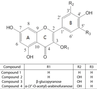
Compound R1 R2 R3
Compound 1 Compound 2 Compound 3 Compound 4
H H β-glucopyranose α-(3’’-O-acetyl)-arabinofuranose
H OH OH OH
H H H H
Fig. 1. Structures of the active compounds 1−4 isolated from Euonymus alatus.
Table 2. 13C (DMSO-d6) NMR spectral data (δ value, ppm).
Carbon Compound
1 2 3 4
2 3 4 5 6 7 8 9 10 1’ 2’ 3’ 4’ 5’ 6’ 1’’
2’’
3’’
4’’
5’’
6’’
COO CH3
147.2 134.0 178.0 161.1 99.1 164.2 93.9 156.7 103.5 122.0 129.9 115.4 159.5 115.4 129.9
147.1 134.1 177.9 161.5 99.3 164.3 94.0 156.6 103.3 122.1 115.4 145.4 149.0 115.4 121.1
147.1 134.0 177.9 161.5 99.2 164.4 94.0 156.7 103.4 122.6 115.4 145.5 148.6 115.4 122.4 104.3 75.6 78.2 71.1 78.0 62.5
147.1 134.0 177.9 161.5 99.3 164.4 94.0 156.6 103.4 122.7 115.4 145.5 148.6 115.3 122.0 108.0 79.2 79.6 85.8 61.5
170.4 20.9
Compound 4는 HRFAB-MS에서m/z 477.1016 [M+H]+ peak와 NMR data에의해분자식 C22H21O12으로확인되었 다. 1H NMR spectrum에서 δ 6.19 (1H, d, J = 2.4 Hz, H- 6), 6.39 (1H, d, J = 2.4 Hz, H-8), 7.48 (1H, d, J = 2.4 Hz, H- 2'), 6.88 (1H, d, J = 8.2 Hz, H-5'), 7.55 (1H, dd, J = 8.4, 1.8 Hz, H-6') peak 등에의해 compound 4 역시 quercetin moiety를포함하고있음을알수있었다. anomeric proton δ 5.27 (1H, d, J = 1.4 Hz, H-1'')과다른 glycosidic protons 4.44 (m, H-2’’) 4.81 (d, J = 3.8, H-3’’), 3.70 (d, J = 3.8, H- 4’’)와 3.42 (2H, m, H-5’’) 이외에 methyl proton signal δ1.88 (s, 3H)과 13C NMR spectrum에서 δ 20.9 (CH3)와 170.4 (COO) signal 등에의해서 glycone moiety가 acetylated arabinofuranoside이며 acetyl group의위치는 H-3’’ (δ 4.81)와 acetoxyl carbonyl carbon (δ 170.4)의 HMBC cross peak 에의해 C-3’’로밝혀졌다. Quercetin의 glycosylation 위치는 anomeric proton δ 5.27 (H-1’’)과 δ 134.0 (C-3) 간의 HMBC long range correlation으로 C-3임을 알수있었고 coupling constant J = 1.4에의해서α-anomeric configuration이 확인되었다. 또한 compound 4을 산 가수분해 후 0.02 N sodium methoxide로처리하여 deacetylation을행한다음 표준당시료와함께실시한 TLC에서도 glycone moiety는 arabinose (rf 0.53)로확인되었다. 따라서 compound 4의구 조는 quercetin-3-O-α-3’’-acetyl-arabinofuranoside로 결정 되었다[13].
본연구에서 activity-based fractionation에의해화살나무 가지로부터α-glucosidase 저해활성물질을분리하였고, 1H,
13C NMR, DEPT, 2D NMR과 MS spectrum 등과문헌 상 의 data을근거로하여 compound 1 (Kaempferol), compound 2 (quercetin), compound 3 (quercetin-3-O-β-glucopyranoside), compound 4 (quercetin-3-O-α-3’’-acetyl-arabinofuranoside) 의구조를결정하였다(Fig. 1). 화살나무의 butanol 용매추 출층에서분리한활성물질 1, 2, 3과 4는모두 5,7-dihydroxy flavonol 물질이다. 다양한천연물소재에서 flavonol 물질의 분리가이루어졌으며, flavonol compound의α-glucosidase 저해활성과항산화작용등에관해다수의연구가보고된 바있다[14]. 그러나화살나무의α-glucosidase 효소저해의 유효성분으로서는보고된바가없다. 또한 compound 4의경 우는 acetylglycosyl 기를포함하는드문 flavonoids로화살 나무성분으로본연구에서처음보고되었다.
Flavonoid 물질의 경우 A ring의 hydroxyl 기가 α- glucosidase enzyme의 tyrosine, tryptophan과수소결합을 형성하여 enzyme의 2차구조의변화를유발시켜 active site 에기질결합을제한함으로써 enzyme 활성을저해한다고알 려졌지만[15, 16], 본연구결과에의하면 A ring 뿐만아니 라 B ring의 OH기수도중요하다. B ring에 1개의 OH기를
갖는 kaempferol 보다 2개 OH기를 포함하는 quercetin이 강한활성을나타냈으며 3개의 OH기를포함하는 myricetin 과비교한결과 myricetin이가장강한활성을나타내어활 성의세기가 myricetin, quecetin, kaempferol 순으로나타 났다. 또한 C ring의 hydroxyl function은활성에영향하지 않는것으로보고된바있으나, kaempferol 활성이 apigenin 에비해크게나타나 C ring의 3-free OH 기는활성을증가 시키나, compound 3와 compound 4와같이 C-3번위치에 당등의 bulky group의치환은 enzyme binding site에의결 합을저해함으로써활성이감소되는것으로추정된다(Table 1).
α-glucosidase 저해제인 acarbose, voglibose 그리고 miglitol 등이당뇨치료제로시판되고있으나복부팽만과설사등의 부작용으로인해사용에제한이있으므로부작용이해결된
새로운치료제개발이시급한실정이다[17]. 본연구에서는
당뇨치료제개발목적으로혈당강하효과가있다고알려져 오랫동안민간요법으로사용되어안전성이확보된화살나무 가지로부터α-glucosidase 저해물질을분리하였다. 분리한 활성물질 Compound 1, 2, 3와 4 모두는 yeast α-glucosidase에 대해 강한 저해 활성을 나타냈으나, β-glucosidase, α- mannosidase, β-galactosidase 등의 다른 glycosidase에는 전혀활성을나타내지않았다(data not shown). 식후혈당 강하작용에의해개발된당뇨병치료제의경우임상적으로 상용되는데있어최대걸림돌은독성이며, 독성문제해결을 위한방안은 glycoprotein processing glycosidase에는활성 이없고, 소장내α-glucosidase에대해특이적저해활성을
갖는물질의선발이다[18]. 이상의결과를종합해보면본연
구에서 분리한α-glucosidase 저해 물질, compound 1-4는 당뇨병치료제로의개발가능성이높다고판단된다.
요 약
화살나무가지로부터 activity based fractionation에의해 α-glucosidase 저해활성물질 compound 1−4을분리하였고, 1H NMR, 13C NMR, 1H−1H COSY와 HMBC 등의 spectral data에 의해 구조를 결정하였다. Compound 1−4는 모두 flavonol 물질로α-glucosidase에대해 IC50값이각각 25.3, 17.1, 47.3과 35.1 μM로 positive control로사용한 acarbose 보 다강한활성을나타내었다. 화살나무의혈당저하기능의 유효성분으로처음동정된 Compound 1−4는α-glucosidase 에만특이적활성을갖는물질로당뇨병치료제로의개발가 능성이높은물질로사료된다.
Acknowledgments
This research was supported by Seokyeong University in 2015.
Conflict of Interest
The author has no conflict of interest to declare.
References
1. Heacock PM, Hertzler SR, Williams JA, Wolf BW. 2005. Effects of a medical food containing an herbal α-glucosidase inhibitor on postprandial glycemia and insulinemia in healthy adults. J. Am.
Diet. Assoc. 105: 65-71.
2. Chapel C, Garcia C, Roingeard P, Zitzmann N, Dubuisson J, Dwek A, et al. 2006. Antiviral effect of α-glucosidase inhibitors on viral morphogenesis and binding properties of the hepatitis C virus- like particles. J. Gen. Virol. 87: 861-871.
3. Goss PE, Baker MA, Carver JP, Dennis JW. 1995. Inhibitors of car- bohydrate processing: a new class of anticancer agents. Clin.
Cancer Res. 1: 935-944.
4. Asano N. 2003. Glycosidase inhibitors: Update and perspectives on practical use. Glycobiology 13: 93R-104R.
5. Yang Z, Wang Y, Wang Y, Zhang Y. 2012. Bioassay-guided screen- ing and isolation of α-glucosidase and tyrosinase inhibitors from leaves of Morus alba. Food Chem. 131: 617-625.
6. Wang SM, Han JJ, Ma K, Jin T, Bao L, Pei Y, et al. 2014. New α- glucosidase inhibitors with p-terphenyl skeleton from the mushroom Hydnellum concrescens. Fitoterapia 98: 149-155.
7. Park SH, Ko SK, Chung SH. 2005. Euonymus alatus prevents the hyperglycemia and hyperlipidemia induced by high-fat diet in ICR mice. J. Ethnopharmacol. 102: 326-335.
8. Markham KR. 1982. Techniques of flavonoid identification, pp. 23-
29. Academic Press, London.
9. Kim S. 2013. α-Glucosidase inhibitor from Buthus martensi Karsch, Food Chem. 136: 297-300.
10. Sakakibara H, Honda Y, Nakagawa S, Ashida H, Kanazawa K.
2003. Simultaneous determination of all polyphenols in vegeta- bles, fruits, and teas. J. Agric. Food Chem. 51: 571-581.
11. Agrawal PK. 1992. NMR spectroscopy in the structural elucida- tion of oligosaccharides and glycosides. Phytochem. 31: 3307- 3330.
12. Harborne JB, Williams CA. 1994. Flavone and flavonol glycosides, pp 290-295. In Harborne JB (ed), The Flavonoids: Advances in research since 1986. Chapman & Hall, London.
13. Madikizela B, Aderogba M, Staden JV. 2013. Isolation and char- acterization of antimicrobial constituents of Searsia chirindensis L. (Anacardiaceae) leaf extracts. J. Ethnopharmacol. 150: 609- 613.
14. Toda M, Kawabata J, Kasai T. 2000. α-Glucosidase inhibitors from clove (Syzgium aromaticum). Biosci. Biotechnol. Biochem. 64: 294- 298.
15. Niwa T, Doi U, Osawa T. 2003. Inhibitory activity of corn-derived bisamide compounds against α-glucosidase. J. Agric. Food Chem. 51: 90-94.
16. Peng X, Zhang G, Liao Y, Gong D. 2016. Inhibitory kinetics and mechanism of Kaemferol on α-glucosidase. Food Chem. 190:
207-215.
17. Asano N. 2003. Glycosidase inhibitors: update and perspectives on practical use. Glycobiology 13: 93R-104R.
18. Jacob GS. 1995. Glycosylation inhibitors in biology and medi- cine. Curr. Opin. Struct. Biol. 5: 605-611.