Journal of Life Science
2009 Vol. 19. No. 5. 680~683 ⓒJLS / ISSN 1225-9918Linoleic Acid from Bamboo (Phyllostachys Bambusoides) Displaying Potent α- Glucosidase Inhibition
Sunin Jung, Su Tae Kang
1, Cheol Yong Choi
1, Kyeong Yeol Oh, Jung Keun Cho, Rajesh Rengasamy and Ki Hun Park*
Division of Applied Life Science (BK21 Program), EB-NCRC, Institute of Agriculture & Life Science, Graduate School of Gyeongsang National University, Jinju 660-701, Korea
1
Cheonghak-Dong Samsundang Food Co., Hadong-Gun 667-883, Korea Received April 12, 2009 /Accepted April 20, 2009
Glycosidase inhibitors are major targets in the treatment of type Ⅱ diabetes, cancer and viral infections. This study was carried out to investigate the glycosidase inhibitory substances from bam- boo (Phyllostachys bambusoides). Bamboo was extracted with methanol and then further fractionated with n-hexane, chloroform, n-BuOH and aqueous to get an active fraction. All extracts were evaluated for α-glucosidase inhibitory activities to identify the n-hexane fraction with 33.5 µg/ml of IC
50value.
Active compound 1 in the n-hexane fraction was identified as linoleic acid, which exhibited inhibitory activity with 12.4 µM of IC
50value. Mechanistic analysis showed that linoleic acid exhibited non- compective inhibition. This is the first study in which bamboo is reported to show α-glucosidase in- hibitory activity.
Key words : Glycosidase, α-glucosidase inhibitor, Phyllostachys bambusoides, linoleic acid
*Corresponding author
*Tel:+82-55-751-5472, Fax:+82-55-757-0178
*E-mail : [email protected]
Introduction
Screening of glycosidase inhibitors is becoming increas- ingly popular because they concern with treatment of nu- merous disease including diabetes mellitus type Ⅱ [6], cancer [5], and HIV [13]. Glycosidase are involved in the biosynthesis and processing of oligosaccharide chains of N-linked glycoproteins in endoplasmic reticulum (ER).
Inhibition of these glycosidases, especially α-glucosidase, has a profound effect on the glycan structure which con- sequently affects the maturation, transport, secretion, and function of glycoproteins, and could therefore alter cell-cell or cell-virus recognition processes [2,4,7]. For instance: by retarding the cleavage of complex carbohydrates, post- prandial glucose absorption in vivo can be attenuated, thus regulating blood sugar levels in diabetics [12], the spread of cancer as well as the structural changes of cell surface glycoconjugates with in neoplasmic cells is proliferated by glycosidases in the sera and interstitial fluid around the tu- mor, thus by effecting glycosidase inhibition, cancer growth may be retarded [3]; finally, cellular signaling rec- ognition is principally orchestrated by glycoproteins. α-
Glucosidases (EC 3. 2. 1. 20; α-D-glucoside glucohydrolase) are a group of exo-acting enzymes that play essential roles in carbohydrate and quality control.
Bamboo is well renowned as a polyphenol plant that is one of the most ubiquitous traditional herbal medi- cines in East Asia. This species, belonging to the family of Gramineae, may be considered to be a nontoxic natu- ral therapeutic agent. The main functional components are flavonoids, lactones, and phenolic acid in this species [9]. Its young leaves have been classified as edible by the KFDA (Korea Food & Drug Administration). Its young leave has also listed in the national standard (i.e.
GB2760) as a kind of food antioxidant in china. Previous workers reported that the antioxidant product derived form bamboo leaves, was capable of blocking chain re- action of lipid autooxidation, chelating metal ions of transition state, and blocking the synthetic reaction of ni- trosamine [11].
In the course of a continuing search for glycosidase in- hibitor from plant source [8,10]. n-Hexane extract of bamboo were found to show significant α-glucosidase inhibitory activity. In this study, we isolated the target α-glucosidase inhibitor from the n-hexane extract of the leaves of bamboo and identified its structure using spectroscopic methods.
Additionally, we carried out kinetic study for the isolated compounds.
- Note -
Journal of Life Science
2009, Vol. 19. No. 5 681Materials and Methods Plant material
The Bamboo (Phyllostachys bambusoides) were collected at Mt. cheongam-myeon in hadog Korea, on June 2008.
Extraction and isolation
The leaves (1 kg) of bamboo were air-dried and extracted with methanol (10 l×2) for 3 days at room temperature and filtered to remove the precipitate. The combined methanol extracts was concentrated in vacuum to yield a dark green gum (21 g). The methanol extract was dissolved in 1.5 l of mixture partitioned with n-hexane, CHCl
3, n-BuOH (each 3 l
×1), yielding n-hexane (4 g), CHCl
3(7 g), n-BuOH (5 g), and H
2O extracts (4 g). The n-hexane phase was chromato- graphed on silica gel (3×30 cm, 230-400 mesh, 130 g) using n-hexane / EtOAc solvent system under gradient condition [20:1 (0.3 l), 10:1 (0.3 l), 5:1 (0.3 l), 2:1 (0.3 l)] to give 4 fractions. The fraction C (230 mg) was subjected to a re- versed-phase column chromatography. Thus, this sample was loaded onto a glass column (1×50 cm), packed with RP-18 (ODS-A, 12 nm, S-150 µm, 40 g). The column was then eluted using MeOH:CH
3CN:H
2O (6:1:1) to afford compound 1 (95 mg). as an oily substance ;
1H NMR (300 MHz, CDCl
3) δ 0.88 (3H, m, H-18), 1.33 (14H, m), 1.65 (2H, m), 2.05 (4H, m, H-8 and H-14), 2.36 (2H, m, H-2), 2.79 (2H, m, H-11), 5.37 (4H, m, H-9, 10, 12, and 13);
13C NMR (75 MHz, CDCl
3) δ 180.2, 130.2, 130.0, 128.0, 127.8, 34.0, 31.5, 29.3, 29.0, 27.1, 25.6, 24.6, 22.5 and 14.0.
Enzyme assay
α-Glucosidase (EC 3.2.1.20, from Baker’s), p-nitrophenyl-α -D-glucopyranoside used for the bioassay were purchased from Sigma Chemical Co. Nitrophenol Methods: The ex- perimental procedure of Lianquan et al [14,15]. for the measurement of α-glucosidase activity was used with some modifications. The α-glucosidase activities were determined using an appropriate p-nitrophenyl-α-D-glucopyranoside as a substrate at the optimum pH of each enzyme. The re- action was stopped by adding 2 M NaOH. The released p-nitrophenol was measured Spectirometrically at 405 nm.
The inhibitory effects of the tested compound were ex- pressed as the concentration that inhibits 50% of the en- zyme activity (IC
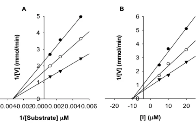
50). Kinetic parameters were determined by the Lineweaver-Burk double-reciprocal-plot method and Dixon plot method at increasing concentration of substrate
and inhibitors.
Results and Discussion
Isolation and structural identification of target compound 1
For investigation of the glycosidase inhibitor, bamboo was extracted with methanol and then further fractionated with n-hexane, chloroform, n-buthanol and aqueous to get active extract. All extracts were tested for their enzymatic in- hibitory activities against α-glucosidase from baker’s yeast.
The enzyme was assayed according to standard procedures by following the hydrolysis of nitrophenyl glycoside spec- trophotometrically [14,15]. As shown in Table 1, n-hexane extract showed a significant degree (>95%) of α-glucosidase inhibition at 500 µg/ml sample concentration, while other extracts did not show.
As the concentrations of n-hexane extract increased, the residual enzyme activity rapidly diminished (Fig. 2A) Activity guided fraction of n-hexane extract gave compound 1 which was purified over octadecyl functionalized silica gel.
The structural elucidation of compound 1 is detailed below.
Compound 1 had the molecular formular C
18H
32O
2and three degrees of unsaturation, as deduced from HREIMS (m/z 280.2402 [M
+]) data. The IR spectrum resembled those of fat- ty acid derivatives. The
1H and
13C NMR data including DEPT experiments showed the presence of eighteen carbon atoms: twelve methylene (sp
3), four methins (sp
2), one meth- yl, and one quaternary carbon. The presence of isolated methylene (H-11) was deduced from connectivity between methylene protons H-11 (δ
H2.79, m) and methin protons (H-10 and H-12, δ
H5.37, m) in the COSY spectrum. The COSY correlation between methylene protons (δ
H2.05, 4H, m) and methins (H-9 and H-13, δ
H5.37) was proved the pres- ence of H-14 and H-8. Esterification of compound 1 with methanol yields methyllinolate that was ascertained by
Table 1. Inhibition percentage of α-glucosidase activity of ex- tracts from Phyllostachys bambusoides
Fraction Inhibition (%)
a)IC
50µg/ml n-Hexane
CHCl
3MeOH Water
>95%
40%
35%
NI
b)33.6 NI
b)NI
b)NI
b)a)
All tests were run at 500 µg/ml sample concentration, values
are means of three experiments
b)NI means no inhibition.
682 생명과학회지 2009, Vol. 19. No. 5
Table 2. α-glucosidase assay results for compound 1 Compound IC
50a(µM) K
ivalue K
m1 Voglibose
12.4 23.4
9.89 NT
b301.41 NT
ba)Compound were examined in a set of experiments repeated
three times; IC
50values of compound represent the concen- tration that caused 50% enzyme activity loss.
b)NT means not test.
OH O 9
10 11
12 13
1 18
Fig. 1. Structure of compound 1.
GC/MS analysis with reference to an authentic sample.
Thus, compound 1 was identified as linoleic acid (Fig. 1).
α-Glucosidase inhibitory activity and kinetic analysis
Isolated compound 1 showed dose-dependent effect on α-glucosidase activity (Fig. 2A). The inhibitory potencies and capacities of compound 1 towards α-glucosidase activity was evaluated as 12.4 µM of IC
50value. The potency of linoleic acid (IC
5012.4 µM) copares with sugar-derived α-glucosi- dase inhibitor currently used for therapeutic purpose such as voglibose (IC
5023.4 µM) [1]. The inhibition mechanisms displayed by the isolated compound 1 were subsequently studied. The compound 1 manifested the same relationship between enzyme activity and enzyme concentration. The in- hibition of α-glucosidase by compound 1 is illustrated (Fig.
[I], µM
0.1 1 10 100 1000
Activity (%)
0 20 40 60 80 100 A
Unit/ml
0.00 0.01 0.02 0.03 0.04 0.05
O.D/min
0.0 0.1 0.2 0.3 B 0.4
Fig. 2. (A) Effect of n-hexane fraction ( ) and compound 1 (●) on the activity of α-glucosidase for the hydrolysis of p-nitirophenyl-α-D-glucopyranoside. (B) Relationship of the hydrolytic activity of α-glucosidase with enzyme concentrations at different concentrations of compound 1. Concentration of compound 1 for cure from top to bottom: 8.9, 17.8, 35.6 and 71.3 µM.
1/[Substrate] µM -0.004-0.0020.0000.0020.0040.006
1/[V] (mmol/min)
0 1 2 3 4 5 A
[I] (µM)
-20 -10 0 10 20
1/[V] (mmol/min)
0 1 2 3 4 5 6 B