한국어병학회지 제20 권 제3 호(2007) J. Fish Pathol., 20(3):299 ~ 306 (2007)
299
�Corresponding Author :
299
�Corresponding Author :
299
Cloning and expression of cDNA for chemokine receptor 9 from Olive flounder Paralichthys olivaceus
Mu Chan Kim, Geun Hee An and Chan Il Park� Institute of Marine Industry, The Colleg of Marine Science, Gyeongsang National University, 455, Tongyeong, 650-160, Korea
Cysteine-cysteine chemokine receptor 9 (CCR9) homologue cDNA was isolated from olive flounder leukocyte cDNA library. Olive flounder CCR9 homologue consisted of 1709 bp encoding 367amino acid residues. When compared with other known CCR peptide sequences, the most conserved region of the olive flounder CCR9 peptide is the seven transmembranes. A phylogenetic analysis based on the deduced amino acid sequence showed the homologous relationship between the olive flounder CCR9 sequence and that of Mouse CCR9. The olive flounder CCR9 gene was predominantly expressed in the Peripheral blood leuko- cytes (PBLs), kidney, spleen, and gills.
Key words: cDNA, Olive flounder, CCR9, Transmembrane
Introduction
Chemokines are cytokines with particular combi- nations of cysteine residues at the amino terminus, and with molecular masses of 5-14 kDa. Like other cytokines, they are secreted by various cell types and are multi-functional. In mammals, they serve not only as chemo-attractants in leukocyte migra- tion during inflammation, but also influence inte- grin activation, angiogenesis, hematopoiesis and homing of lymphocytes in lymphoid tissues (Coillie et al., 1999; Fernandez et al., 2002). To date, more than 50 chemokines have been described. The amino terminal cysteine residues occur in several forms: CXC (αtype), CC (βtype), C (γtype) and CX3C (δtype) (Vaddi et al., 1997; Baggiolini, 1998; Coillie et al., 1999).
Chemokines mediate their effects through G pro- tein coupled 7 transmembrane domain receptors, which are currently divided into 4 families based on
the type of chemokine that they bind; they are CXCR1 to CXCR5, CCR1 to CCR10, XCR1, or CX3CR1. Recent data have identified several chemokines that are constitutively expressed in lymphoid and extralymphoid tissues, indicating that these chemokines might have homeostatic function by regulating lymphocyte trafficking to or within lymphoid organs and in peripheral tissues (Vicari et al., 1997; Willimann et al., 1998; Butcher et al., 1999; Campbell et al., 1999; Morales et al., 1999;
Campbell and Butcher, 2000; Ansel et al., 2000;
Kunkel et al., 2000; Papadakis et al., 2000; Wurbel et al., 2000).
Chemokine receptor 9 (CCR9) is the only known receptor for the chemokine ligand 25, also called thymus-expressed chemokine (Youn et al., 1999;
Zaballos et al., 1999; Norment et al., 2000). CCR9- expressing cells migrate toward chemokine ligand 25, and both molecules colocalize in the thymic cortex and in the small intestinal mucosa (Youn et
�Corresponding Author : Chan Il Park, Tel : 82-55-640-3103 Fax : 82-55-642-4509, E-mail : [email protected]
299
11...
al., 1999; Zaballos et al., 1999; Kunkel et al., 2000;
Norment et al., 2000; Papadakis et al., 2000;
Wurbel et al., 2000). Up-regulation of CCR9 can be induced by pre-TCR signaling (Norment et al., 2000), retinoic acid (Iwata et al., 2004), and den- dritic cells from Peyer’s patches and mesenteric lymph nodes (Lindbom et al., 2003; Mora et al., 2003). Thus, mucosal CCR9 induction may play an important role in the maintenance of CCR9 on gut T cells. Profound down-regulation of CCR9 by TCR cross-linking was shown in persistently acti- vated Th1 cells generated from umbilical cord CD4+ T cells stimulated with IL-2 and IL-12 together with antibodies to IL-4 (Zabel et al., 1999).
Interestingly, this effect was reversible because CCR9 reappeared after removal of the stimulus.
Thus, the level of CCR9 expression on T cells might be modulated by the degree of T-cell activa- tion.
In the present study, we cloned and characterized a novel chemokine chemokine cDNA in Olive flounder, examined its phylogenetic relationship with other chemokine receptors, and detection of its mRNA transcripts in various tissues using RT-PCR.
Materials and Methods
Cloning and sequencing of olive flounder CCR9 cDNA
CCR9 cDNA was identified from the analysis of
expressed sequence tags (ESTs) of olive flounder kidney stimulated with a Con A/PMA cDNA library. Kidney was taken from a single olive floun- der and stimulated with Con A/PMA as previously described by Nam et al. (2003). cDNA clone was sequenced using ThermoSequenase (Amersham) with M13 forward and M13 reverse primers (Table 1) and an automated DNA sequencer LC4200 (Li-Cor).
Phylogeny was inferred using the PHYLIP program (ver. 3.5), and by distance analysis using the neigh- bor joining method. The values supporting each node are derived from 1000 re-samplings.
The determined nucleotide and deduced amino acid sequences, and multiple sequence alignments were analyzed by GENETYX ver. 8.0 (SDC Soft- ware Development).
Tissue distribution of olive flounder CCR9 gene transcript
Total RNA (50 ng) from the brain, heart, intes- tine, kidney, liver and spleen were reverse-tran- scribed into cDNA using an AMV Reverse Tran- scriptase First-Strand cDNA synthesis kit (Life sci- ences). PCR was performed on the resulting cDNA using the CCR9 RT-F / CCR9 RT-R specific primer set (Table 1). β-actin was amplified as a control using the Beta-actin-F and Beta-actin-R primers (Katagiri et al., 1997). The PCR mixtures were denatured at 94℃ for 2 min and then subjected to 25 cycles of 94℃ for 30 s, 57℃ for 30 s and 72℃
Table 1. The oligonucleotide primer sets used in the present study.
Primer name Sequence of oligonucleotide primer
M13 forward 5’-CACGACGTTGTAAAACGAC-3’
M13 reverse 5’-GGATAACAATTTCACACAGG-3’
CCR9 RT-F 5’-GACTGAAGACCATGTCAGATGTG-3’
CCR9 RT-R 5’-GTTGACTCTTGTTGACCAATGGC-3’
Beta actin-F 5’-TTTCCCTCCATTGTTGGTCG-3’
Beta actin-R 5’-GCGACTCTCAGCTCGTTGTA-3’
Fig. 1. Nucleotide and deduced amino acid sequences of the olive flounder CCR9 cDNA. The putative seven transmem- branes are underlined, and the polyadenylation signal is shown in bold print.
for 1 min. The products were visualized by separa- tion on a 1.5% agarose gel.
Results
Cloning and sequencing of olive flounder CCR9 cDNA
The full-length cDNA, designated as OFCCR9, had significant identity to the mammalian CC chemokine receptor group. The OFCCR9 cDNA has a length of 1709 bp, and contains an ORF of 1,104 bp encoding 367 amino acid residues. The 3′UTR contained a polyadenylation signal (AUUAAA) and a polyadenylation site (Fig. 1).
In the phylogenetic analysis, olive flounder CCR9 is grouped with mouse CCR9, and closely
related with mouse and human CCR7 (Fig. 2). This grouping was well supported by bootstrapping.
Alignment of the deduced amino acid sequences of OFCCR9 and other vertebrate chemokines shows that the sequence conserves five cysteine residues at positions 31, 113, 163, 192 and 283 (Fig. 3).
OFCCR9 has also seven hydrophobic regions at positions 45-67, 80-102, 124-143, 164-186, 206- 228, 249-271 and 299-318 that are representing the seven transmembrane domains (TM), which are typical G-protein-coupled receptors (Fig. 3).
Tissue distribution of olive flounder CCR9 gene transcript
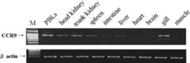
Expression of the OFCCR9 gene in the tissues of olive flounder was detected by RT-PCR. Olive
flounder CCR9 gene transcripts was expressed pre- dominantly in the PBLs, head kidney, trunk kidney, spleen, and gills, but not in liver, brain, and muscle after 25 cycles of PCR (Fig. 4).
Discussion
Chemokine receptor 9 (CCR9) is the only known
receptor for the chemokine ligand 25, also called thymus-expressed chemokine (Norment et al., 2000; Youn et al., 1999; Zaballos et al., 1999).
CCR9-expressing cells migrate toward chemokine ligand 25, and both molecules colocalize in the thymic cortex and in the small intestinal mucosa (Youn et al., 1999; Zaballos et al., 1999; Kunkel et al., 2000 Norment et al., 2000; Papadakis et al., Fig. 2. Phylogenetic tree of amino acid sequences of olive flounder CCR9. The positions of the olive flounder CCR9 was estimated by the neighbor joining method of clustering in the PHYLIP program. Sequences were obtained from DDBJ/EMBL / GenBank.
2000;; Wurbel et al., 2000).
In the phylogenetic analysis (Fig. 2), olive floun- der CCR9 is grouped with mouse CCR9 (Wurbel et al., 2000). This grouping was well supported by bootstrapping. The phylogenetic analysis indicated that known human, cow, and mouse CCRs are more closely related than the olive flounder. This result is possibly due to differences between mammalian
and non-mammalian species. Based on the conser- vation of five cysteine residues and seven trans- membrane regions of the molecule, we predict that the function of olive flounder CCR9 is similar to that in mammals.
The mRNA expression of CCR was detected in leukocytes, kidney, spleen, intestine, liver, and gill of olive flounder. The CCR9 gene was predomi- Fig. 3. Comparison of the derived amino acid sequence of oliveflounder, human, and mouse, CCRs. Asterisks indicate iden- tical amino acid residues, and dashes indicate gaps introduced for maximal alignment. Cysteine residues are shown in bold, and the transmembrane regions are boxed.
nantly expressed in several tissues that contained lymphocytes (PBLs, head kidney, trunk kidney, spleen, and gill) (Fig. 4), thus suggesting that this gene plays important roles in the immune system.
Further studies of recombinant protein of olive flounder CCR9 to understand the roles of this gene should be performed to get a better understanding of the fish immune system.
Acknowledgements
The author thanks Drs. Takashi Aoki and Ikuo Hirono, Tokyo University of Marine Science and Technology, for generously accommodating me for the work on cDNA cloning in their laboratory.
References
Ansel, K.M., Ngo, V.N., Hyman, P.L., Luther, S.A., Forster, R., Sedgwick, J.D., Browning, J.L., Lipp M., Cyster, J.G.: A chemokine-driven positive feedback loop organizes lymphoid follicles. Nature, 406: 309-314, 2000.
Baggiolini, M.: Chemokines and leukocyte traffic.
Nature, 392: 565-568, 1998.
Butcher, E.C., Williams, M., Youngman, K., Rott, L., Briskin, M.: Lymphocyte trafficking and regional immunity. Adv. Immunol., 72: 209- 253, 1999.
Campbell, J.J. and Butcher, E.C.: Chemokines in
tissue-specific and microenvironment spe- cific lymphocyte homing. Curr. Opin.
Immunol., 12: 336-341, 2000.
Campbell, J.J., Haraldsen, G., Pan, J., Rottman, J., Qin, S., Ponath, P., Andrew, D.P., Warnke, R., Ruffing, N., Kassam, N., Wu, L., Butch- er, E.C.: The chemokine receptor CCR4 in vascular recognition by cutaneous but not intestinal memory T cells. Nature, 400: 776- 780, 1999.
Coillie, E.V., Damme, J.V. and Opdenakker, G.:
The MCP/eotaxin subfamily of CC chemokines. Cytokine Growth Factor Rev., 10: 61-86, 1999.
Fernandez, E.J. and Lolis, E.: Structure, function, and inhibition of chemokines. Annu. Rev.
Pharmacol. Toxicol., 42: 469-499, 2002.
Iwata, M., Hirakiyama, A., Eshima, Y., Kagechika, H., Kato, C. and Song, S.Y.: Retinoic acid imprints gut-homing specificity on T cells, Immunity, 21: 527-538, 2004.
Katagiri, T., Hirono, I. and Aoki, T.: Identification of a cDNA for medaka cytoskeletal beta actin and construction for the reverse tran- scriptase-polymerase chain reaction (RTPCR) primer. Fish. Sci., 63: 73-76, 1997.
Kunkel, E.J., Campbell, J.J., Haraldsen, G., Pan, J., Boisvert, J., Roberts, A.I., Ebert, E.C., Vier- ra, M.A., Goodman, S.B., Genovese, M.C., Wardlaw, A.J., Greenberg, H.B., Parker, C.M., Butcher, E.C., Andrew, D.P. andA- gace, W.W.: Lymphocyte CC chemokine receptor 9 and epithelial thymus-expressed chemokine (TECK) expression distinguish the small intestinal immune compartment:
epithelial expression of tissue-specific chemokines as an organizing principle in regional immunity. J. Exp. Med., 192: 761- 768, 2000.
Fig 4. Detection of olive flounder CCR9 mRNA levels from various tissues of healthy olive flounder by RT-PCR.
M indicates a 100-bp ladder marker.
Lindbom, B. J., Svensson, M., Wurbel, M.A., Malissen, B., Marquez, G. and Agace, W.:
Selective generation of gut tropic T cells in gut-associated lymphoid tissue (GALT):
requirement for GALT dendritic cells and adjuvant. J. Exp. Med., 198: 963-969, 2003.
Mora, J.R., Bono, M.R., Manjunath, N., Weninger, W., Cavanagh, L.L., Rosemblatt, M. and Von Andrian, U.H.: Selective imprinting of gut-homing T cells by Peyer’s patch den- dritic cells. Nature, 424: 88-93, 2003.
Morales, J., Homey, B., Vicari, A.P., Hudak, S., Oldham, E., Hedrick, J., Orozco, R., Copeland, N.G., Jenkins, N.A., McEvoy, L.M., Zlotnik, A.: CTACK, a skin-associat- ed chemokine that preferentially attracts skin-homing memory T cells. Proc. Natl.
Acad. Sci. USA, 96: 14470-14475, 1999.
Nam, B.H., Yamamoto, E., Hirono, I. and Aoki, T.:
A survey of expressed genes in the leuko- cytes of Japanese flounder, Paralichthys oli- vaceus, infected with hirame rhabdovirus.
Dev. Comp. Immunol., 24: 13-24, 2000.
Norment, A.M., Bogatzki, L.Y., Gantner, B.N. and Bevan, M.J.: Murine CCR9, a chemokine receptor for thymus-expressed chemokine that is up-regulated following pre-TCR sig- naling. J. Immunol., 164: 639-648, 2000.
Olaussen, R.W., Farstad, I.N., Brandtzaeg, P. and Rugtveit, J., Age-related changes in CCR9+
circulating lymphocytes: are CCR9+ naive T cells recent thymic emigrants? Scand. J.
Immunol., 54: 435-439, 2001.
Papadakis, K.A., Prehn, J., Moreno, S.T., Cheng, L., Kouroumalis, E.A., Deem, R., Breaver- man, T., Ponath, P.D., Andrew, D.P., Green, P.H., Hodge, M.R., Binder, S.W. and Targan, S.R.: CCR9-positive lymphocytes and thy- mus-expressed chemokine distinguish small
bowel from colonic Crohn’s disease. Gas- troenterology, 121: 246-254, 2001.
Papadakis, K.A., Prehn, J., Nelson, V., Cheng, L., Binder, S.W., Ponath, P.D., Andrew, D.P.
and Targan, S.R.: Therole of thymus- expressed chemokine and its receptor CCR9 on lymphocytes in the regional specializa- tion of the mucosal immune system. J.
Immunol., 165: 5069-5076, 2000.
Rossi, D.L., Hardiman, G., Copeland, N.G., Gilbert, D.J., Jenkins, N. and Zlotnik, A.: Cloning and characterization of a new type of mouse chemokine, Genomics, 47: 163-170, 1998.
Vaddi, K., Keller, M. and Newton, R.C.: The chemokine facts book. Academic Press, New York, 1997.
Vicari, A.P., Figueroa, D.J., Hedrick, J.A., Foster, J.S., Singh, K.P., Menon, S., Copeland, N.G., Gilbert, D.J., Jenkins, N.A., Bacon, K.B., Zlotnik, A.: TECK: a novel CC chemokine specifically expressed by thymic dendritic cells and potentially involved in T cell development. Immunity, 7: 291-301, 1997.
Willimann, K., Legler, D.F., Loetscher, M., Roos, R.S., Delgado, M.B., Clark-Lewis, I., Baggi- olini, M., Moser, B.: The chemokine SLC is expressed in T cell areas of lymph nodes and mucosal lymphoid tissues and attracts acti- vated T cells via CCR7. Eur. J. Immunol., 28: 2025-2034, 1998.
Wurbel, M.A., Malissen, M., Grand, D. G., Meffre, E., Nussenzweig, M.C., Richelme, M., Car- rier, A. and Malissen, B.: Mice lacking the CCR9 CC-chemokine receptor show a mild impairment of early T- and B-cell develop- ment and a reduction in T-cell receptor δ+ gut intraepithelial lymphocytes. Blood, 98:
2626-2632, 2001.
Wurbel, M.A., Philippe, J.M., Nguyen, C., Vic- torero, G., Freeman, T., Wooding, P., Miazek, A., Mattei, M.G., Malissen, M., Jor- dan, B.R., Malissen, B., Carrier, A. and Naquet, P.: The chemokine TECK is expressed by thymic and intestinal epithelial cells and attracts double- and single-positive thymocytes expressing the TECK receptor CCR9. Eur. J. Immunol., 30: 262-271, 2000.
Youn, B.S., Kim, C.H., Smith, F.O. and Broxmeyer, H.E.: TECK, an efficacious chemoattractant for human thymocytes, uses GPR-9-6/CCR9 as a specific receptor. Blood, 94: 2533-2536, 1999.
Zaballos, A., Gutierrez, J., Varona, R., Ardavin, C.
and Marquez, G.: Cutting edge: identifica- tion of the orphan chemokine receptor GPR- 9-6 as CCR9, the receptor for the chemokine
TECK, J. Immunol., 162: 5671-5675, 1999.
Zabel, B.A., Agace, W.W., Campbell, J.J., Heath, H.M., Parent, D., Roberts, A.I., Ebert, E.C., Kassam, N., Qin, S., Zovko M., LaRosa, G.J., Yang, L.L., Soler, D., Butcher, E.C., Ponath, P.D., Parker, C.M. and Andrew, D.P.: Human G protein-coupled receptor GPR-9-6/CC chemokine receptor 9 is selec- tively expressed on intestinal homing T lym- phocytes, mucosal lymphocytes, and thymo- cytes and is required for thymus-expressed chemokine-mediated chemotaxis. J. Exp.
Med. 190: 1241-1256, 1999.
Manuscript Received : November 6, 2007 Revision Accepted : November 28, 2007 Responsible Editorial Member : Sung Hee Jung
(NFRDI.)