서 론
어류의 스트레스 반응은 수온(water temperature), 염분(sal
inity), 사육밀도(culture density), 용존산소(dissolved oxygen) 및 기타 화학적인 요소(chemical factors)에 의해 발생하고, 이 중 염분은 생태학적 요인 중 수생환경에서만 해당이 되며, 어류의 생육발달 및 성장단계에 있어 직접적인 영향을 미치 는 환경적 요소이다(Beckmann et al., 1990; Boeuf and Payan, 2001). 급격한 염분변화는 어류에 스트레스 요인으로 작용하 는데, 적정 염분범위를 벗어나게 되면 항상성(homeostasis)
을 유지하기 위하여 삼투압 조절을 하게 된다(Weirich and Tomasso, 1991; Marshall, 2002; Hur et al., 2006).
담수생태계에 있어 해수의 유입은 담수생물의 생존에 위협 적인 것과 마찬가지로 해수생태계 내 담수의 유입 및 기타 환 경적인 영향으로 인한 염분의 변화는 해양생물의 생리 및 생 태에 영향을 미친다(Bervoets et al., 1996). 염분의 변화에 노 출된 생물은 체내 나트륨 및 염화물 등의 불균형으로 인해 생 리적인 스트레스가 유발되고, 체내 기작에 변화가 나타나는 것이 보고되기도 하였다(Hussain et al., 2013; Zhang and Shi, 2013; Deinlein et al., 2014). 이러한 염분의 변화가 장기간 지 속될 경우, 해양생물에게 생리적인 스트레스로 작용하여 유 전자의 발현 및 생존율 변화에 악영향을 미친다(Nikapitiya et
—1 — http://www.fishkorea.or.kr
* Corresponding author: ByungHwa Min Tel: 82336608542, Fax: 82336618514, Email: [email protected]
ISSN: 1225-8598(Print), 2288-3371(Online)
Accepted: March 13, 2018
염분변화에 따른 넙치 (Paralichthys olivaceus)의 스트레스 반응
박형준·민병화1,*
국립수산과학원 양식관리과, 1국립수산과학원 동해수산연구소 양식산업과
Stress Responses of Olive Flounder(Paralichthys olivaceus) to Salinity Changes by Hyung-Jun Park and Byung-Hwa Min1,* (Aquaculture Research Division, National Institute of Fisheries Science, Busan 46083, Republic of Korea; 1Aquaculture Industry Research Division, East Sea Fisheries Research Institute, Gangneung 25435, Republic of Korea)
ABSTRACT We tried to determine the optimum salinity for a cultured of olive flounder(Paralichthys olivaceus) by investigating after exposing the fish at different salinity(10, 15, 20 and 25psu) for 24 and 48 hours compared with control group(fish before transfer to experimental tank). As a control groups, we compared an analyzed with other experimental groups using olive flounder in natural sea water. Hematological parameters including hematocrit(Ht) and hemoglobin(Hb), cortisol and glucose, aspartate aminotransferase(AST) and alanine aminotransferase(ALT), NH3, osmolality, total protein (TP), Na+, K+ and Cl- mostly exhibited significant changes at 10 and 15psu groups compared with control groups for 24 and 48 hours exposed. Plasma SOD(superoxide dismutase) and CAT(catalase) activity also increased with experimental groups(10 and 15psu) compared to the control groups.
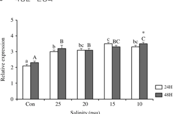
The expression of HSP70 mRNA was also higher at low-salinity(10 and 15psu) than at control group.
In particular, after 24 hours exposed, it expression to 15psu groups showed a significant difference compared to the control group. However, after 48 hours exposed, it expression was higher in the 10psu groups than the control. It is assumed that the changes in the hematological responses and hormone, homeostasis and metabolism were resulted in to protect fish body from stress. Based on these results, we are expected that it will be used as basic data for the culture of olive flounder prepared for low salinity.
Key words: Paralichthys olivaceus, blood physiological response, SOD, CAT, HSP70
al., 2014; Wang et al., 2014; Yang et al., 2016).
넙치(Paralichthys olivaceus) 양식장이 널리 분포하고 있는 제주 연안에서는 여름철에 중국 대륙으로부터 저염수의 유입 및 여름철 집중호우로 인해 양식생물에 피해를 주기도 한다 (Pang and Hyun, 1998). 환경적 요인으로부터 발생하는 저염 분 조건으로 인한 양식생물의 삼투압변화에 대한 생리적인 결 과와 생존에 미치는 영향 및 스트레스 반응이 보고된 바 있고 (Morgan et al., 1997; Sampaio and Bianchini, 2002), 이러한 스 트레스로 인해 어류는 산화적 스트레스를 유발할 수 있으며, 스트레스로 인해 생성된 활성산소종(reactive oxygen species, ROS)을 제거하기 위해 채내 항산화 효소(antioxidant enzyme) 를 활성화시켜 제거함으로써 자가방어를 수행한다(Pandey et al., 2003). 또한 환경(저산소, 수온, 염분) 및 미생물의 감염을 통해 주요하게 발현되는 Heat shock protein(HSP)70은 어류 의 생리 및 생태학적 조건에 영향을 미치며, 세포 내 HSP70 의 합성과 함께 다양한 생리학적 변화가 일어난다(Ryan and Schlesinger, 1992). HSP70은 어류의 스트레스 요인에 의한 충 격으로부터 생체를 보호하기 위한 항상성(homeostasis) 유지 에 중요한 역할을 수행한다(Iwama et al., 1999; Ackerman et al., 2000; Basu et al., 2003).
따라서 본 연구에서는, 광염성 어류로 알려진 넙치(Para
lichthys olivaceus)를 이용하여 급격한 염분변화로 발생되는 스트레스 반응을 스트레스 지표로 잘 알려진 혈액학적 변화 및 HSP70 mRNA 발현분석을 토대로, 저염분의 조건에서 발생 되는 넙치의 스트레스 반응에 대한 기초자료를 제공하고, 넙 치양식에 있어 적정염분조건을 구명하고자 하였다.
재료 및 방법
1. 실험어류 및 조건
넙치(31.8±1.7cm, 308.5±43.3g)는 국립수산과학원의 생 물 사육실에서 2주 동안 유수식 1ton 원형수조에서 상업용 사 료를 급이하며 순화시켰다. 수온은 21±0.2°C를 유지하였다.
이후, 염분별 그룹으로 나누어 대조구로는 자연해수의 조건과 실험구인 25, 20, 15 및 10psu로 5개 수조에 실험어 12마리를 가온 및 냉각이 가능한 50L 사각수조에 각각 수용한 뒤, 24 및 48시간 동안 노출시켰다. 모든 실험은 2반복으로 수행하였다.
2. 혈액분석
24시간 및 48시간 급격한 염분조건에 노출 후, 모든 실험 어는 150mg/L의 tricaine methan sulphonate(MS222, Sigma, USA)로 마취시킨 다음, heparin sodium(Sigma, USA) 처리 된 3mL syringe(50IU)를 이용하여 대조구 및 실험구마다 각
각 12마리씩 넙치의 미부정맥으로부터 혈액을 샘플하였다.
샘플 후, 혈액의 일부를 hematocrit(Ht) 및 hemoglobin(Hb) 분석에 사용하고 나머지 혈액은 실온에서 20분간 방치한 다 음 원심분리(4°C, 10,000rpm, 15분)하여 분석 전까지 deep
freezer(-80°C)에 보관하였다. Ht는 모세관을 원심분리(RT, 10,000rpm, 15분)하여 Htlog chip(Microhaematocrit reader, Hawksley Co, UK)을 이용하여 측정하고, Hb, 혈장 글루코 스, 총 단백질(TP), aspartate aminotransferase(AST), alanine aminotransferase(ALT) 및 NH3는 자동 생화학 분석기(Fuji dry-chem 4000i, Fujifilm Co., Japan)로 분석하였다. 삼투질농 도는 삼투압 측정기(Vapro 5520, WESCOR Co., USA)로 측정 하였다. 혈장 코티졸 농도는 Fish ELISA Cortisol kit(Cusabio Biotech, China), 혈장 내 SOD 및 CAT 활성은 Superoxide Dismutase Assay Kit, Catalase Assay Kit(Cayman Chemical, USA)로 각각 상법에 따라 분석하였다.
3. Quantitative real-time PCR(q-PCR)
24시간 및 48시간 동안 염분별로 노출시킨 넙치의 HSP70 mRNA 발현을 분석하기 위해 각 실험구의 넙치로부터 간 조 직을 적출하여 액체질소에서 급속 동결시킨 후, -80°C에서 실험 전까지 보관하였다. Total RNA는 TRizol Reagent(Gibco/
BRL, USA)법을 이용하여, 각 시료에서 RNA를 추출한 후, 1 μg의 total RNA는 Transcriptor First Strand cDNA synthesis kit(Roche, USA)를 이용하여 cDNA를 합성하였다.
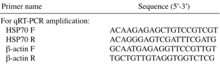
PCR 분석에 앞서, 이전에 알려진 넙치 HSP70 nucleotide sequence(accession no. AAC33859)에 기초하여 primer를 제 작하였다(Table 1). HSP70 mRNA 발현은 CFX96TM Realtime System(BioRad, USA)과 iQTMSYBR green Supermix(Bio
Rad, USA)를 이용하여 분석하였다. 대조구로써 housekeeping gene은 넙치에서 알려진 β-actin(accession no. HQ386788)을 사용하였으며, 모든 분석값은 β-actin에 대한 cycle(Ct) level 과의 차이로 계산하였다. Ct값은 다음과 같은 식으로 결정지 었다: 2-ΔΔCt Method, [ΔΔCt=2^-(ΔCtsample-ΔCtinternalcontrol)]
(Livak and Schmittgen, 2001).
4. 통계분석
결과값은 평균 표준오차±(S.E.)로 나타내었으며, 실험구
Table 1. Primers used in this study
Primer name Sequence (5′3′)
For qRT-PCR amplification:
HSP70 F ACAAGAGAGCTGTCCGTCGT
HSP70 R ACAGGGAGTCGATTTCGATG
β-actin F GCAATGAGAGGTTCCGTTGT
β-actin R TGCTGTTGTAGGTGGTCTCG
별 유의차 유무는 SPSS 통계처리프로그램(version 10.0; SPSS inc., USA)에 의한 Oneway ANOVA 및 Duncan’s multiple range test를 실시하였고, 24시간 및 48시간 사이의 유의차는 ttest를 이용하여 평균간의 유의성(P<0.05)을 검정하였다.
결과 및 고찰
1. 혈액 생리학적 분석
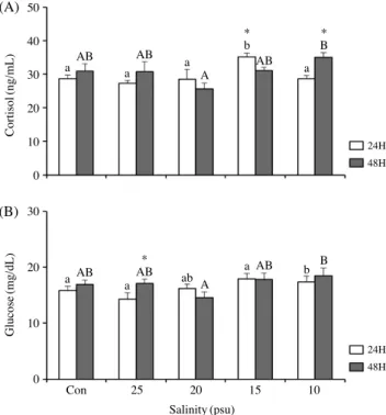
코티졸 농도는 24시간 노출 후, 대조구인 28.6±1.2ng/mL 과 실험구 간의 유의적인 차이를 보이지 않았지만, 48시간 노 출 후에서는 10psu에서 35.0±1ng/mL로 대조구인 30.9±2.2 ng/mL에 비해 유의적으로 높은 값이 나타났다(Fig. 1A). 글루 코스의 변화값은 17.4±1.0, 18.5±1.4mg/dL로 10psu에서 대 조구 15.8±0.8, 16.9±0.8mg/dL에 비해 유의적인 차이를 나 타내었다(Fig. 1B).
Ht는 24시간 노출 후, 대조구 및 실험구 간의 유의적인 차이 를 보이지 않았지만, 48시간 노출 후에 10psu에서 23.0±0.8%
로 대조구 20.0±1.1%에 비해 유의하게 높았다(Fig. 2A). 또한 Hb에서는 24시간 및 48시간 후, 저염분구인 10psu에서 6.6±
0.4, 6.7±0.4g/dL로 대조구 5.6±0.4, 5.3±0.4g/dL에 비해 유 의한 차이를 보였다(Fig. 2B).
24시간 노출 후의 AST는 실험구와 대조구 간의 유의적인 차이를 보이지 않았으나, 48시간 노출 후에는 대조구 22.1±
1.0U/L에 비해 10psu 및 15psu에서 각각 28.9±2.7 및 25.2±
1.9U/L로 유의적인 차이를 나타낸 반면(Fig. 3A), ALT는 24 시간 노출 후에는 10psu 및 15psu에서 각각 15.2±1.3, 12.3±
0.8U/L로 대조구 10.8±0.6U/L에 비해 유의하게 증가하였지 만 48시간 노출 후에는 실험구와 대조구 간의 유의적인 차이 를 보이지 않았다(Fig. 3B).
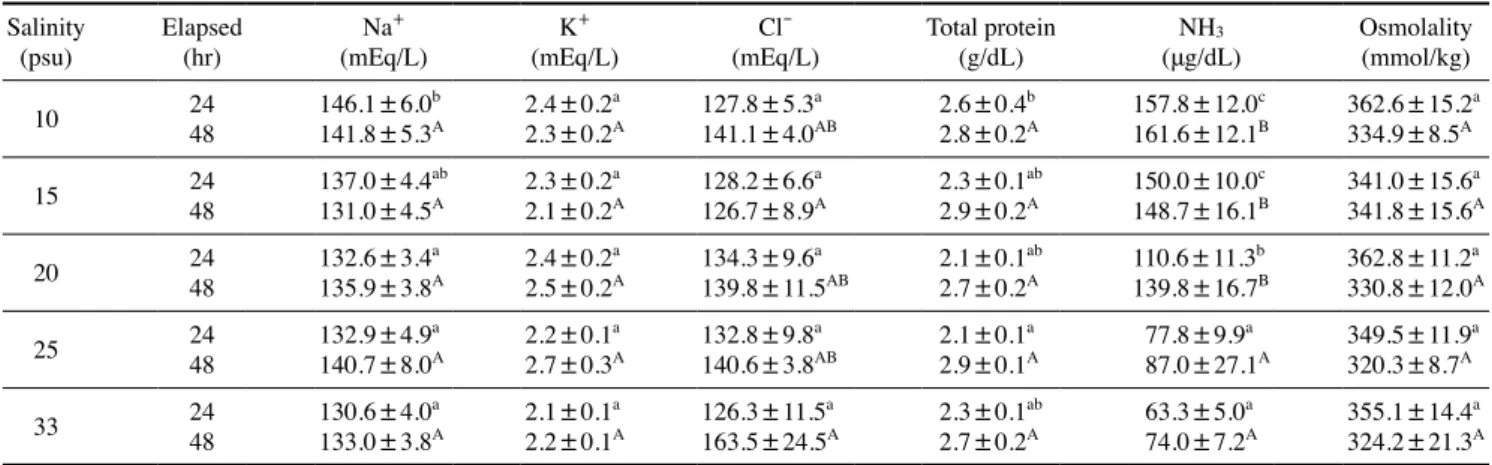
24시간 및 48시간 노출 후의 총 단백질은 24시간 노출 후, 대조구에 비해 10psu에서 2.6±0.4g/d로 유의적인 차이를 보 였지만 48시간 후에는 실험구 간의 유의적인 차이가 나타나 지 않았다. NH3는 24시간 및 48시간 노출 후, 10psu에서 각 각 157.8±12.0, 161.6±12.1, 331.8±13.5μg/dL로 대조구에 비해 유의적으로 높았으나, 삼투질 농도에서는 24시간 및 48 시간 노출 후, 모든 실험구 간 유의적인 차이가 나타나지 않았 다. Na+는 24시간 노출 후, 10psu에서 146.1±6.0mEq/L로 대 조구에 비해 유의적으로 높았으나, 48시간 노출 후에는 실험 구 간에 유의적인 차이가 나타나지 않았다. 또한 K+는 24시간 및 48시간 염분별 노출 후, 모든 실험구에서 유의적인 차이를 보이지 않았으나, Cl-은 48시간 노출 후에, 10psu에서 127.8±
5.3mEq/L로 대조구에 비해 낮은 값을 보이며 유의적인 차이 가 나타났다(Table 2).
(A)
(B)
Fig. 1. Plasma cortisol(A) and glucose(B) of olive flounder, Para- lichthys olivaceus in different salinity at the conclusion of this exper
iment. Data were expressed as mean±SE(n=12). Different letters denote significant difference among each group(P<0.05). * : signifi
cance between 24 and 48 hours(P<0.05)
Cortisol(ng/mL)
24H 48H
24H 48H
Glucose(mg/dL)
Salinity(psu)
Con 25 20 15 10 a
a
a
a
a
ab
b
a
a
b AB
AB
AB
AB
A
A
AB
AB
B
B 50
40 30 20 10 0 30 20 10 0
*
* *
Fig. 2. Hematocrit(A) and hemoglobin(B) of olive flounder, Para- lichthys olivaceus in different salinity at the conclusion of this exper
iment. Data were expressed as mean±SE(n=12). Different letters denote significant difference among each group(P<0.05). * : signifi
cance between 24 and 48 hours(P<0.05) (A)
(B)
Hematocrit(%)
24H 48H
24H 48H
Hematocrit(%)
Salinity(psu)
Con 25 20 15 10 a
a
ab
a
b
ab
b
ab
b
b A
A
AB
A
AB
B
AB
B
B
B 30
20 10
0 10 8 6 4 2 0
* *
어류에 있어 대사나 스트레스 반응을 확인하기 위한 여러 가지 지표 중 주요 분석항목으로써 코티졸 및 글루코스의 변 화가 있다(Barton et al., 1986; Flos et al., 1988). 1차적으로 급 격한 스트레스에 따른 어류의 코티졸이 증가 또는 감소하여, 2차적인 스트레스 반응으로 글루코스 값의 변화가 일어난다 (Perry and Reid, 1993; Chang and Hur, 1999). 따라서 10psu 에서 높아진 글루코스 값은 염분변화에 따라 넙치가 스트레 스를 받을 때, 분비되는 코티졸의 영향으로 글루코스가 신생
합성을 위한 효소 활성이 높아져 값이 증가되는 것으로 생각 되며, 염분변화에 노출된 넙치가 누적된 스트레스로 인해 에 너지의 소비가 증가하는 것으로 여겨진다(Barton and Iwama, 1991). 염분의 변화에 따른 코티졸 및 글루코스 변화에 관 한 연구가 모잠비크 틸라피아, Oreochromis mossambicus (Morgan et al., 1997)와 페헤레이, Odontesthes bonariensis (Tsuzuki et al., 2001) 등의 보고가 있다. 이와 비슷한 결과로, 농어, Lateolabrax japonicus 유어에서 저염분에 24시간 노출 후, 코티졸 및 글루코스가 증가하는 경향을 보였고(Han et al., 2003), 감성돔, Acanthopagrus schlegeli에서도 마찬가지로 10 psu의 염분에 24시간 노출시켰을 때 유의적으로 증가하였다 (An et al., 2010), 이러한 결과는 넙치가 적정염분조건을 벗어 났을 때, 급격한 스트레스의 영향으로 인해 혈장 내 코티졸 및 글루코스의 변화가 일어나는 것으로 여겨진다.
Hematocrit(ht)는 혈구의 용적비율을 나타내며 어류의 건 강도와 질병의 진단에 필수적인 지표이고, hemoglobin(Hb) 은 그 농도에 따라 어류의 생리 및 병리학적 상태를 결정짓 는 데 있어 주요 혈액학적 분석항목이다(Houston and Rupert, 1976; Lukianenko, 1984). 특히, Ht 및 Hb는 어체 내 생리학 적인 측면에 있어 pH, 산소운반능력의 저하, 용존산소의 감 소에 따라 그 변화가 나타나는데, 일부 연구에서는 어류의 유 영활동 및 연령, 계절변화, 염분, 수온 및 수질오염을 통해 그 값의 변화에 대한 보고가 있다(Houston, 1997). 무지개송어 (Onchorhynchus mykiss)의 연구에서 염분 7psu 및 11psu에 노출시킨 후, 시간의 증가에 따라 Hb 농도가 유의적으로 증 가하였지만(Hosseini et al., 2011), 페헤레이(O. bonariensis) 에서는 24시간 및 128시간 저염분에 노출시킨 후, 0, 5psu 및 20psu 간 유의적인 차이를 보이지 않았으며(Tsuzuki et al., 2001), 본 연구의 결과와 마찬가지로, 염분별 노출에 따른 넙 치의 Ht 및 Hb의 변화를 확인할 수 있었다.
AST 및 ALT는 대부분의 척추동물의 간 세포에 존재하며,
Table 2. Plasma Na+, K+, Cl-, total protein, NH3 and osmolality of olive flounder, Paralichthys olivaceus at different salinity Salinity
(psu) Elapsed
(hr) Na+
(mEq/L) K+
(mEq/L) Cl-
(mEq/L) Total protein
(g/dL) NH3
(μg/dL) Osmolality (mmol/kg)
10 24 146.1±6.0b 2.4±0.2a 127.8±5.3a 2.6±0.4b 157.8±12.0c 362.6±15.2a
48 141.8±5.3A 2.3±0.2A 141.1±4.0AB 2.8±0.2A 161.6±12.1B 334.9±8.5A
15 24 137.0±4.4ab 2.3±0.2a 128.2±6.6a 2.3±0.1ab 150.0±10.0c 341.0±15.6a
48 131.0±4.5A 2.1±0.2A 126.7±8.9A 2.9±0.2A 148.7±16.1B 341.8±15.6A
20 24 132.6±3.4a 2.4±0.2a 134.3±9.6a 2.1±0.1ab 110.6±11.3b 362.8±11.2a
48 135.9±3.8A 2.5±0.2A 139.8±11.5AB 2.7±0.2A 139.8±16.7B 330.8±12.0A
25 24 132.9±4.9a 2.2±0.1a 132.8±9.8a 2.1±0.1a 77.8±9.9a 349.5±11.9a
48 140.7±8.0A 2.7±0.3A 140.6±3.8AB 2.9±0.1A 87.0±27.1A 320.3±8.7A
33 24 130.6±4.0a 2.1±0.1a 126.3±11.5a 2.3±0.1ab 63.3±5.0a 355.1±14.4a
48 133.0±3.8A 2.2±0.1A 163.5±24.5A 2.7±0.2A 74.0±7.2A 324.2±21.3A Data were expressed as mean±SEM(n=10). Different letters denote significant difference among each group(P<0.05).
Fig. 3. Plasma AST(A) and ALT(B) of olive flounder, Paralichthys olivaceus in different salinity at the conclusion of this experiment.
Data were expressed as mean±SE(n=12). Different letters denote significant difference among each group(P<0.05). * : significance between 24 and 48 hours(P<0.05)
(A)
(B)
AST(U/L)
24H 48H
24H 48H
ALT(U/L)
Salinity(psu)
Con 25 20 15 10 a
a
a
a
a
a
a
a
a
b A
A
A
A
A
A
B
A
B
A 50
40 30 20 10 0 30 20 10 0
*
* *
*
간 기능의 검사지표로 사용된다. 다양한 스트레스 요인에 의 해 간 세포가 단기간 및 장기간에 걸쳐 파괴되면 혈액 중에 서 이들 효소의 수치가 증가하게 된다(Kang et al., 2007). 특 히 어류에서는 주로 간 및 비장세포에 분포하며, 급격한 수온 및 염분의 변화, 저산소(hypoxia), pH, 암모니아 및 중금속 오 염에 의한 스트레스 반응으로 수치가 증가 또는 감소하는 경 향을 나타낸다(Pan et al., 2003). 또한 경골어류는 어체 내에 적정 염분을 유지함으로써 세포 내외의 이온 경사를 유지하 며 끊임없이 삼투압 조절작용을 하게 되는데, 이는 해수와 담 수에서 많은 차이를 보인다(Min et al., 2006). 수온 및 염분의 변화 등 많은 환경적인 요인에 따라 삼투압 조절에 있어 많은 영향을 미치는데, 이에 따라 삼투질농도, Na+, K+ 및 Cl- 이온 의 농도변화가 발생한다(Kim et al., 2009). 따라서, 본 연구에 서도 Na+ 및 Cl- 값의 변화가 나타남에 따라, 급격한 염분변 화에 따른 스트레스로 인해 유의적인 차이를 보인 것으로 생 각된다. 혈장 내 총 단백질과 NH3는 급격한 염분변화에 따른 스트레스로 인해 염분이 낮아질수록 다른 실험구에 비해 유 의적인 차이를 보였다. 어류의 총 단백질량은 건강도, 영양상 태 및 질병진단의 지표로 사용되어지고 있고(Nakagawa et al., 1977; Siddiqui, 1977; Byrne et al., 1989), 수온, 수질 및 염분 등 환경변화에 따른 스트레스에 의한 영향이 보고되어진 바 있다(McLeay and Brown, 1979; Ishioka, 1980). NH3는 간 조 직 기능의 저하로 인해 혈액 내에서 증가하게 된다(Randall, 2002). 조피볼락, Sebastes schlegeli(Do et al., 2016), 붉바리, Epinephelus akaara(Lim et al., 2016), 숭어, Mugil cephalus 및 나일틸라피아, Oreochromis niloticus(Chang and Hur, 1999)로 부터 수온 및 염분변화에 따른 스트레스 반응과 저산소증에 따른 삼투질농도의 변화가 보고된 바 있다.
2. 혈장 내 SOD 및 CAT 활성
염분변화에 따른 스트레스 요인이 넙치의 혈장 내 SOD 및 CAT 활성에 어떠한 영향을 미치는지에 대한 분석을 수행하 였다. 염분별 SOD 및 CAT는 모두 24시간 및 48시간 노출 후 에 각각 131.1±10.0, 127.5±4.5U/mL, 14.3±0.6, 11.0±0.5 nmol/min/mL으로 대조구 및 다른 실험구에 비해 유의적인 차 이를 나타내었다(Fig. 4).
특정 유해물질이나 독소, 환경적 요인에 의한 스트레스로 인해 과도하게 생성되는 활성산소종은 어류의 조직이나 세 포에 치명적인 손상을 줄 수 있다(Goldberg and Sterm, 1977;
Simon et al., 1981). 어체 내에서 생성된 ROS에 의해 단백질, 핵산 및 생물학적 손상은 SOD(superoxide dismutase), CAT (catalase) 등의 항산화 효소에 의해 보호되며(Moody and Hassan, 1982), 이들 SOD 및 CAT는 ROS를 무해하고 안정 된 분자로 전환시켜 어체를 보호한다(Forman and Fridovich,
1973). 이러한 항산화 효소의 활성은 어종 및 조직에 따라 차 이가 나타나고, 계절적인 변화에 의해 나타나는 환경적 요소 로 인한 항산화 효소의 활성이 증가 또는 감소하는 것이 보 고된 바 있다(Aksnes and Njaa, 1981; Gabryelak et al., 1983).
담수어인 철갑상어(Acipenser naccarii)에서는 20일 동안 35 psu에 노출시켰을 때, SOD 및 CAT 활성이 유의적으로 증가 하였고(Álvarez et al., 2002), 병어(Pampus argenteus)에서도 마찬가지로 10psu에서 시간이 증가함에 따라 SOD 및 CAT 의 활성도가 25psu에 비해 유의적인 차이를 보였다(Fei et al., 2011). 따라서 본 연구의 결과에서도 마찬가지로, 급격한 염분 변화에 따른 스트레스 반응으로 인해 넙치의 SOD 및 CAT 활 성이 증가한 것으로 보인다.
3. HSP70 mRNA 발현
염분별 넙치의 스트레스의 영향을 알아보기 위하여 스트레 스 관련 단백질인 HSP70 mRNA 발현을 QPCR법을 통해 확 인하였다. 24시간 및 48시간 염분별 노출 후, 저염분그룹인 10 psu 및 15psu에서 각각 3.3±0.1 및 3.5±0.1fold, 3.5±0.1 및 3.3±0.1fold로 대조구에 비해 높은 발현을 보임에 따라 유의 적인 차이를 확인할 수 있었다(Fig. 5).
Heat shock proteins(Hsps)는 세포의 수송 및 분해를 돕는 Fig. 4. The activities of superoxide dismutase(SOD)(A) and cata
lase(CAT)(B) in olive flounder, Paralichthys olivaceus after salinity changes. Values are significantly different from control in the same time of sampling(P<0.05). Values are means±SE(n=12). * : sig
nificance between 24 and 48 hours(P<0.05) (A)
(B)
SOD activity(U/mL)
24H 48H
24H CAT activity(nmol/min/mL) 48H
Salinity(psu)
Con 25 20 15 10 a
a
ab
b
bc
c
c
d
c
d B
A
A
B
BC
BC
BC
C
C
C 200
150 100 50
0 20 15 10 5 0
* *
*
분자 샤페론(molecular chaperone)의 역할과 손상된 세포나 단백질의 회복을 돕는다(Becker and Craig, 1994; Currie et al., 2000). 일반적으로 Hsps는 열충격으로 인해 발생하여 유도되 는 단백질로 알려져 있지만, 미생물의 감염, 독소, 저산소 및 기타 환경 스트레스 요인에 따른 노출에도 높은 발현을 유발 한다(Basu et al., 2002). 그중, HSP70은 어류의 스트레스 반응 에 따른 분자생물학적인 기능과 발현에 관한 연구가 활발히 이루어지고 있다. 해양무척추동물인 오만둥이, Styela plicata (Pineda et al., 2012)와 참담치, Mytilus coruscus(Kim and Kang, 2015)에서 HSP70 mRNA 발현의 증가가 시간의 흐름에 따른 염분변화에 의해 유도되는 것으로 잘 알려져 있고, 어류 에서는 무지개송어, O. mykiss(Hosseini et al., 2011)를 해수에 노출시켜 급격한 염분의 변화가 HSP70 mRNA 발현을 증가시 킨다는 것이 보고된 바 있지만, 아직까지 염분변화에 따른 스 트레스가 HSP70에 미치는 영향에 관한 연구가 미흡하여 많은 연구가 필요할 것으로 생각된다.
이상의 결과, 넙치의 염분에 의한 스트레스가 미치는 영향 을 생리학적 관점에서 조사하였다. 염분변화에 따른 분석의 경우 저염분구인 10psu 및 15psu로 갈수록 코티졸 및 글루코 스를 비롯한 혈액생리학적 변화를 확인할 수 있었고, HSP70 mRNA의 발현에서도 대조구와 유의적인 차이가 나타난 것을 알 수 있었으며, 이는 적어도 양식장의 저염분 대비를 위해서 는 적정 염분을 유지하여 넙치양식이 이루어져야만 건강도 측 면에서 유리하다는 것을 보여주고 있다. 이러한 염분의 변화 가 장기간 지속될 경우, 넙치의 생존율에도 영향을 미칠 것으 로 생각되고, 적어도 20~25psu를 유지해 주어야만 혈액생리 학적인 인자를 비롯한 스트레스 반응에 영향을 미치지 않으 며, 넙치에 있어 적정 염분조건이 건강도에도 영향을 미치지 않는다는 것을 보여주고 있다. 본 연구의 결과들을 바탕으로 염분변화에 따른 넙치의 혈액생리학적 반응 및 HSP70 mRNA
발현을 밝힘에 따라 기초적인 분자 및 생리학적 메커니즘을 밝히는데 있어 유용한 연구보고가 될 것으로 판단되며, 저염 분에 대비한 넙치양식에 있어 참고적인 자료로 활용될 수 있 을 것이 기대된다.
요 약
스트레스를 유발하는 환경적인 요소 중, 염분의 변화는 어 류의 성장과 생존에 있어 많은 영향을 미친다. 본 연구에서 는 한국의 양식대상 어종인 넙치(P. olivaceus)를 대상으로 넙 치양식에 있어 적정 염분범위를 구명하기 위하여 각 염분별 (25, 20, 15psu 및 10psu) 조건을 통해 24시간 및 48시간 동안 노출시킨 후, 혈액생리학적 분석 및 HSP70 mRNA 발현을 조 사하였다. 혈액학적 분석에서 hematocrit(Ht) 및 hemoglobin (Hb), 혈장 코티졸(cortisol) 및 글루코스(glucose)의 변화, aspartate aminotransferase(AST) 및 alanine aminotransferase (ALT), Na+, K+, Cl-, NH3, 삼투질농도(osmolality) 및 총 단백 질(total protein, TP)는 저염분 조건인 10psu 및 15psu에서 다 른 염분별 실험구에 비해 대부분의 항목에서 유의적인 차이를 보였다. 혈장 내 SOD(superoxide dismutase) 및 CAT(catalase) 활성에서도 마찬가지로 대조구인 자연해수의 조건보다 저염 분(10psu 및 15psu)으로 갈수록 증가하는 경향을 보였다. 스 트레스성 단백질로 알려진 HSP 70 mRNA 발현에서도 대조구 보다 저염분에서 유의적인 차이를 확인하였다. 이러한 결과로, 염분변화에 의한 스트레스가 넙치의 생체방어메커니즘과 항 상성 유지를 위한 결과로 혈액학적 반응, 호르몬의 분비 및 단 백질 발현의 증가가 나타나는 것으로 여겨지며, 이를 바탕으 로 저염분에 대비한 넙치양식에 있어 기초적인 자료로 활용될 것이 기대된다.
사 사
이 논문은 2018년도 국립수산과학원 수산과학연구사업 동 해특성품종 양식기술개발(R2018012)의 지원으로 수행된 연 구입니다.
REFERENCES
Ackerman, P.A., R.B. Forsyth, C.F. Mazur and G.K. Iwama. 2000.
Stress hormones and the cellular stress response in salmo
nids. Fish. Physiol. Biochem., 23: 327336.
Aksnes, A. and L.R. Njaa. 1981. Catalase, glutathione peroxide and superoxide dismutase in different fish species. Comp. Bio
Fig. 5. The HSP70 mRNA expression during salinity changes to after 24 and 48(9, 12, 15, 18 and 21°C) hours in the olive flounder (Paralichthys olivaceus) of liver. Data were expressed as mean±SE (n=10). Different letters denote significant difference among each group(P<0.05). * : significance between 24 and 48 hours(P<0.05)
24H 48H
Relative expression
Salinity(psu)
Con 25 20 15 10 a
b bc c bc
A
B B BC C
5 4 3 2 1 0
*
chem. Physiol., 69: 357358.
Álvarez, R.M.M., M.C. Hidalgo, A. Domezain, A.E. Morales, M.G.
Gallego and A. Sanz. 2002. Physiological changes of stur
geon Acipenser naccarii caused by increasing environmen
tal salinity. J. Exp. Biol., 205: 3699-3706.
An, K.W., H.S. Shin, B.H. Min, G.S. Kil and C.Y. Choi. 2010.
Physiological changes in response to osmotic and thermal stress conditions in black porgy(Acanthopagrus Schlegeli).
Korean J. Icthyol., 22: 17-24. (in Korean)
Barton, B.A., C.B. Schreck and L.A. Sigismondi. 1986. Multiple acute disturbances evoke cumulative physiological stress responses in juvenile Chinook salmon. T. Am. Fish. Soc., 115: 245251.
Barton, B.A. and G.K. Iwama. 1991. Physiological changes in fish from stress in aquaculture with emphasis on the response and effects of corticosteroids. Annu. Rev. Fish. Dis., 1:
326.
Basu, A., A.E. Todgham, P.A. Ackerman, M.R. Bibeau, K, Nakano, P.M. Schulte and G.K. Iwama. 2002. Heat shock protein genes and their functional significance in fish. Gene, 295:
173183.
Basu, N., C.J. Kennedy and G.K. Iwama. 2003. The effects of stress on the association between hsp70 and the glucocorticoid receptor in rainbow trout. Comp. Biochem. Phys. A, 134:
655663.
Becker, J. and E.A. Craig. 1994. Heat-shock proteins as molecular chaperones. Eur. J. Biochem., 219: 11-23.
Beckmann, R.P., L.E. Mizzen and W.J. Welch. 1990. Interaction of HSP70 with newly synthesized proteins: implications for protein folding and assembly. Science, 248: 850854.
Bervoets, L., R. Verheyen and R. Blust. 1996. Uptake of zinc by the midge larvae Chironomus riparius at different salinities:
Role of speciation acclimation, and calcium. Envrion. Toxi
col. Chem., 15: 14231428.
Boeuf, G. and P. Payan. 2001. How should salinity influence fish growth? Comp. Biochem. Physiol. Part C, 130: 411423.
Byrne, P., D.J. Speare and H.W. Ferguson. 1989. Effects of cationic detergent on the gills and blood chemistry of rainbow trout (Salmo gairdneri). Dis. Aquat. Organ, 6: 195196.
Chang, Y.J. and J.W. Hur. 1999. Physiological responses of grey mullet(Mugil cepjalus) and Nile tilapia(Oreochromis nilo
ticus) by rapid changes in salinity of rearing water. Korean J.
Fish. Soc., 32: 310316.(in Korean)
Currie, S., C.D. Moyes and B.L. Tufts. 2000. The effects of heat shock and acclimation temperature on Hsp70 and Hsp30 mRNA expression in rainbow trout: in vivo and in vitro comparisons. J. Fish. Biol., 56: 398-408.
Deinlein, U., A.B. Stephan, T. Horie, W. Luo, G, Xu and J.I. Schro
eder. 2014. Plant salttolerance mechanisms. Trends Plant Sci., 19: 371379.
Do, Y.H., B.H. Min, Y.D. Kim and M.S. Park. 2016. Changes on hematological factors and oxygen consumption of Korea rockfish Sebastes schlegeli in high water temperature. J.
Kor. Soc. Fish. Mar. Edu., 28: 738745.(in Korean) Fei, Y., P. Shiming, S. Peng and S. Zhaohong. 2011. Effects of low
salinity on antioxidant enzymes activities in kidney and muscle of juvenile silver pomfret Pampus argenteus. Acta.
Ecologica Sinca, 31: 5560.
Flos, R., L. Reig, P. Torres and L. Tort. 1988. Primary and sec
ondary stress responses to grading and hauling in rainbow trout, Salmo gairdneri. Aquac., 71: 99106.
Forman, H.J. and I. Fridovich. 1973. Superoxide dismutase: A com
parison of rate constant. Arch. Biochem. Biophys., 158:
369p.
Gabryelak, T., M. Piatkowska, W. Leyko and G. Pérès. 1983. Sea
sonal variation in the activities of peroxide metabolism enzymes in erythrocytes of freshwater fish species. Comp.
Biochem. Physiol., 75: 383385.
Goldberg, B. and A. Stern. 1977. The role of the superoxide anion as a toxic species in the erythrocyte. Arch. Biochem. Bio
phys., 178: 218225.
Han, H.K., D.Y. Kang, C.Y. Jun and Y.J. Chang. 2003. Effect of salinity change on physiological response and growth of yearling sea bass, Lateolabrax japonicas. Aquaculture, 16:
3136.(in Korean)
Hosseini, P., H. Vahabzade, M.S. Bourani and R. Kazemi. 2011.
The effects of salinity stress on hematocrit and hemoglobin in fingerling rainbow trout(Oncorhynchus mykiss). Int.
Con. Med. Biol. Pharma. Sci.,(ICMBP ’2011).
Houston, A.H. 1997. Review: Are the classical hematological vari
ables acceptable indicators of fish health? T. Am. Fish. Soc., 126: 879894.
Houston, A.H. and R. Rupert. 1976. Immediate response of the hemoglobin system of the goldfish, Carassius auratus, to temperature change. Can. J. Zoolog., 54: 1737-1741.
Hur, J.W., J.Y. Lee, Y.H. Kim, I.S. Park and Y.J. Chang. 2006. Ef
fects of salinity on hematological changes and survival of cultured olive flounder, Paralichthys olivaceus. J. Kor. En
viron. Biol., 24: 380386.(in Korean)
Hussain, M., H.W. Park, M. Farooq, K. Jabran and D.J. Lee. 2013.
Morphological and physiological basis of salt resistance in different rice genotypes. J. Int. Agri. Biol., 15: 113-118.
Ishioka, H. 1980. Stress reactions in the marine fish, 1: Stress re
actions induced by temperature change. B. Jpn. Soc. Sci.
Fish., 46: 523532.
Iwama, G.K., M.M. Vijayan, R.B. Forsyth and P.A. Ackerman.
1999. Heat shock proteins and physiological stress in fish.
Amer. Zool., 39: 901909.
Kang, D.Y., H.W. Kang, G.H. Kim, K.C. Jo and H.C. Kim. 2007.
Effect of cold shock on the physiological response of the cultured mullet, Mugil haematocheilus in winter. Korean J.
Fish. Soc., 40: 226233.(in Korean)
Kim, C.W. and H.S. Kang. 2015. The expression of Hsp70 and GST genes in Mytilus coruscus exposed to water temperature and salinity. Korean J. Environ. Biol., 33: 450-458.(in Korean) Kim, Y.S., Y.H. Do, B.H. Min, H.K. Lim, B.K. Lee and Y.J. Chang.
2009. Physiological responses of starry flounder Platich- thys stellatus during freshwater acclimation with different speeds in salinity change. J. of Aquaculture, 22: 28-33.(in Korean)
Lim, S.G., S.B. Han and H.K. Lim. 2016. Effects of salinity on the growth, survival and stress responses of red spotted grouper Epinephelus akaara and hybrid grouper E. akaara ♀×E.
lanceolatus ♂. Korean J. Sci., 49: 612-619(in Korean).
Livak, K.J. and T.D. Schmittgen. 2001. Analysis of relative gene expression data using realtime quantitative PCR and the 2-ΔΔCT metheod. Methods, 25: 402408.
Lukianenko, V. 1984. Hemoglobin polymorphism and monomor
physm in two Acipenseridae populations. Translate by:
Younos Adeli. Iran. Fish. Res. Ins., 86p.
Marshall, W.S. 2002. Na+, Cl-, Ca2+ and Zn2+ transport by fish gills: retrospective review and prospective synthesis. J.
Exp. Zool., 293: 264283.
McLeay, D.J. and D.J. Brown. 1979. Stress and chronic effects of untreated and treated bleched keaft pulpmill effluent on the biochemistry and stamina of juvenile coho salmon(Onco- rhynchus kisutch). J. Fish. Res. Board Can., 36: 1049-1059.
Min, BH., G.A. Noh, M.H. Jeong, D.Y. Kang, C.Y. Choi, I.C. Bang and Y.J. Chang. 2006. Effects of oral administration of thy
roid hormone on physiological activity and growth of black porgy reared in fresh water or seawater. J. of Aquaculture, 19: 149156.(in Korean)
Moody, C.S. and H.M. Hassan. 1982. Mutagenicity of oxygen free radicals. Proc. Natl. Acad. Sci. USA, 79: 28552859.
Morgan, J.D., T. Sakamoto, E.G. Grau and G.K. Iwama. 1997.
Physiological and respiratory Responses of the Mozam
bique tilapia(Orepchromis mossambicus) to salinity accli
mation. Comp. Biochem. Physiol., 3: 391398.
Nakagawa, H., M. Kayama and K. Ikuta. 1977. Electrophoretic evi
dence of seasonal variation of carp plasma albumin. J. Fac.
Fish. Anim. Husb. Hiroshima Univ., 16: 99106.
Nikapitiya, C., W.S. Kim, K. Park and I.S. Kwak. 2014. Identifi
cation of potential markers and sensitive tissues for low or high salinity stress in an intertidal mud crab(Macrophthal- mus japonicas). Fish Shellfish Immun., 41: 407416.
Pan, C.H., Y.H. Chien and B. Hunter. 2003. The resistance to am
monia stress of Penaeus monodon Fabricus juvenile fed di
ets supplemented with astaxanthin. J. Exp. Mar. Biol. Ecol., 297: 107118.
Pandey, S., S. Parvez, I. Sayeed, R. Haques, B. BinHafeez and S.
Raosuddin. 2003. Biomarkers of oxidative stress: a compar
ative study of river Yammuna fish Wallagattu(Bi. & Schn.).
Sci. Total Environ., 309: 105115.
Pang, I.C. and K.H. Hyun. 1998. Seasonal variation of water mass distributions in the Eastern yellow sea and the yellow sea warm current. Korean J. Soc. Oceanogr., 33: 41-52.
Perry, S.F. and S.D. Reid. 1993. β-adrenergic signal transduction in fish: interactive effects of catecholamines and cortisol. Fish.
Physiol. Biochem., 11: 195203.
Pineda, M.C., X. Turon and S.L. Legentil. 2012. Stress levels over time in the introduced ascidian Styela plicata: the effects of temperature and salinity variations on Hsp70 gene expres
sion. Cell Stress Chaperon., 17: 435444.
Randall, D.J. 2002. Ammonia toxicity in fish. Mar. Pollut. Bull., 45:
112.
Ryan, C. and M.J. Schlesinger. 1992. Inhibitory effects of HSP70 chaperones on nascent polypeptide. Protein Sci., 1: 980
Sampaio, L.A. and A. Bianchini. 2002. Salinity effects on osmoreg985.
ulation and growth of the euryhaline flounder Paralichthys orbignyanus. J. Exp. Mar. Biol. Ecol., 269: 187-196.
Siddiqui, N. 1977. Seasonal, size and comparative study of plasma proteins of four air breathing freshwater fishes. P. Indian Acad. Sci. B, 85: 384390.
Simon, R.H., C.H. Scoggin and D. Patterson. 1981. Hydrogen per
oxide causes the fatal injury to human fibroblasts exposed to oxygen radicals. J. Biol. Chem., 256: 7181-7186.
Tsuzuki, M.Y., K. Ogawa, C.A. Strüssmann, M. Maita, F. Takashi
ma. 2001. Physiological responses during stress and sub
sequent recovery at different salinities in adult pejerrey Odontesthes bonariensis. Aquaculture, 200: 349362.
Wang, Q.L., S.S. Yu, C.X. Qin, S.L. Dong and Y.W. Dong. 2014.
Combined effects of acute thermal and hypoosmotic stress
es on osmolality and hsp70, hsp90 and sod expression in the sea cucumber Apostichopus japonicas Selenka. Aqua
cult. Int., 22: 11491161.
Weirich, C.R. and J.R. Tomasso. 1991. Confinement- and transport- induced stress on red drum juveniles: effect of salinity.
Prog. Fish. Cult., 53: 146149.
Yang, S.J., J.Y. Lee, Y.K. Shin, H.K. Hwang and J.I. Myeong. 2016.
Effects temperature and salinity on survival, metabolism and histological change of the rockfish, Sebastes schlegeli. J.
Kor. Soc. Fish. Mar. Edu., 28: 10681075.(in Korean) Zhang, J.L. and H. Shi. 2013. Physiological and molecular mecha
nisms of plant salt tolerance. Photo. Res., 115: 122.