Korean J Gastroenterol Vol. 62 No. 3, 182-184 http://dx.doi.org/10.4166/kjg.2013.62.3.182 pISSN 1598-9992 eISSN 2233-6869
IMAGE OF THE MONTH
Korean J Gastroenterol, Vol. 62 No. 3, September 2013 www.kjg.or.kr
간내담관-십이지장 문합부위 담관에 발생한 난치성 담관결석
김민정, 차상우, 조영덕
순천향대학교 의과대학 내과학교실 소화기병센터
Refractory Bile Duct Stones Occurring at Hepaticoduodenostomy Site
Min Jeong Kim, Sang-Woo Cha and Young Deok Cho
Digestive Disease Center, Department of Internal Medicine, Soonchunhyang Uinversity College of Medicine, Seoul, Korea
CC This is an open access article distributed under the terms of the Creative Commons Attribution Non-Commercial License (http://creativecommons.org/licenses/
by-nc/3.0) which permits unrestricted non-commercial use, distribution, and reproduction in any medium, provided the original work is properly cited.
교신저자: 차상우, 140-743, 서울시 용산구 대사관로 59, 순천향대학교 의과대학 내과학교실 소화기병센터
Correspondence to: Sang-Woo Cha, Digestive Disease Center, Department of Internal Medicine, Soonchunhyang Uinversity College of Medicine, 59 Daesagwan-ro, Yongsan-gu, Seoul 140-743, Korea. Tel: +82-2-709-9494, Fax: +82-2-709-9696, E-mail: [email protected]
Financial support: None. Conflict of interest: None.
Fig. 1. Computed tomography images show large stones (black arrow) in distal portion of hepaticoduodenostomy site with dilatation of both intrahepatic duct.
증례: 36세 여자가 내원 4개월 전부터 발생하고 1주일 전 부터 악화된 우상복부 통증을 주소로 내원하였다. 환자는 14 년 전 교통사고로 간의 우후엽절제술(right posterior seg- mentectomy)를 타 병원에서 시행하였고 그로부터 3년 뒤 총 담관 협착으로 right hepaticoduodenostomy를 본원에서 시 행받은 과거력이 있었다. 내원 당시 신체활력징후는 안정적이 었고, 심한 우상복부 통증을 호소하고 있었으며, 검사실 소견 으로 말초혈액검사에서 백혈구 8,900/mm3, 혈색소 12.9 g/dL, 혈소판 267,000/mm3, AST 28 IU/L, ALT 43 IU/L, 총 빌리루빈 1.4 mg/dL, ALP 619 IU/L, GGT 502 IU/L이었 고, CRP 0.92 mg/dL로 상승되어 있었다. 내원 당일 시행한 복부 CT에서 간내담관-십이지장 문합부(hepaticoduodenos- tomy site)의 원위부 담관에 두 개의 큰 결석이 관찰되며 양 측 간내담관 확장이 관찰되었다(Fig. 1).
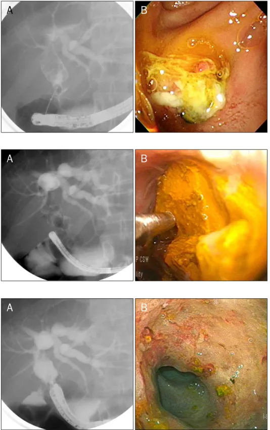
입원 2일째 췌담도 내시경을 시행하여 십이지장을 관찰하 였을 때 간내담관-십이지장 문합부 개구부가 관찰되었으며 이 부위로 카테터 삽관 후 조영제 주입시 관내 충만결손이 보여 결석이 있음을 확인하였다. 이후 basket을 이용하여 담석을 제거하려 하였으나 관강 내에 박혀있고 크기도 커서 담석이 제거되지 않았다(Fig. 2). 내시경적 경비 담도 배액술(endo- scopic naso-biliary drainage) 시행 후에도 복통은 지속되었 다. 입원 5일째 초슬림 위장관내시경(ultra-slim gastroscope, EG-1690k; Pentax, Tokyo, Japan)을 이용하여 간내담관-십
이지장 문합부를 통해 직접 관강 내로 접근하여 내시경 직시 하 전기수압쇄석술(electrohydraulic lithotripy, EHL)을 시행 하였다(Fig. 3). 이후 분쇄조각들은 일반 내시경으로 basket 을 이용하여 제거하였다. 환자는 EHL을 통한 1차 쇄석술 이 후 복통은 호전되기 시작하였고, 입원 7일째와 9일째에도 같
Kim MJ, et al. Refractory Bile Duct Stones Occurring at Hepaticoduodenostomy Site
183
Vol. 62 No. 3, September 2013
Fig. 3. (A) Direct cholangioscope was inserted into the bile duct. (B) Elec- trohydraulic lithotripy probe is seen adjacent to the stone.
Fig. 4. (A) Fluoroscopic image obtain- ed during follow-up endoscopic retro- grade cholangiopancreatography shows no residual filling defects in the bile duct. (B) Direct cholangioscope i-scan image shows red colored irregular in- trahepatic bile duct mucosa.
Fig. 2. (A) Fluoroscopic image taken during endoscopic retrograde chol- angiopancreatography demonstrates large bile duct stones at hepaticodu- odenostomy site. (B) Duodenoscopic image shows impacted stone at the orifice of hepaticoduodenostomy site.
은 방식으로 초슬림 위장관내시경을 통해 내시경직시하 EHL 시행 후 돌을 여러 차례 제거하였으며 담관 내에 더 이상의 잔존 분쇄조각이 없음을 확인하였다(Fig. 4A). 또한 초슬림 위장관 내시경 i-scan (Pentax, Tokyo, Japan)으로 담관 내를 관찰하였을 때 표면의 염증 소견을 보여 조직검사 시행 후 시술
을 종료하였다(Fig. 4B). 조직검사에서도 역시 염증 소견 외 이상소견 없었다. 이후 더 이상의 복통은 호소하지 않았으며 환자는 퇴원 후 4개월 뒤 추적 관찰로 시행한 복부초음파에서 결석이 없음을 확인하였다.
184
김민정 등. 간내담관-십이지장 문합부위 담관에 발생한 난치성 담관결석The Korean Journal of Gastroenterology
진단: 간내담관-십이지장 문합부위 담관에 발생한 난치성 담관결석
담관결석 환자의 대부분은 총담관 결석으로 1 cm 이내의 결 석의 경우 내시경 유두괄약근 절개술 후 바스켓이나 풍선카테터 를 이용하여 결석을 제거한다. 하지만 크기가 큰 결석(≥2 cm) 이나 담관 내에 박혀있어 위의 방법만으로 결석 제거가 되지 않는 난치성 담관결석의 경우 쇄석술을 통하여 결석을 제거한다. 가장 흔 하게 쓰이는 방법으로는 기계적 쇄석술(mechanical lithotriopsy)이 며 이를 통하여 2 cm 이상의 큰 담관 결석에 대하여 80% 정도 결석 제거 성공률을 보인다.1 이 외에도 체외 충격파 쇄석술 (estracorporeal shock wave lithotripsy) 및 레이저 쇄석술 (laser lithotripsy)이나 EHL을 이용한 체내 결석 쇄석술(in- tracorporeal lithotripsy)이 있다.2
EHL은 액체용매 내에서 고전압 방전(spark)을 일으켜 발 생한 충격파가 결석을 분쇄시키는 효과를 이용한 것으로 내시 경 직시하에 쇄석 탐침을 정확하게 결석의 표면에 접근시켜 쇄석술을 시행하는 방법이다. EHL은 결석이 기계식 쇄석 바 스켓 내에 포획되지 못할 정도로 크거나 결석의 크기에 비해 담관이 확장되지 않아 바스켓에 의한 결석의 포착이 곤란할 경우에 유용하다.3 EHL은 대부분 경피경간 담관내시경(per- cutaneous transhepatic cholangioscopy, PTCS)을 이용하 거나 motherscope과 babyscope이 필요한 경구용 담도내시 경하 쇄석법(peroral cholangioscopci lithotripsy, POC-L)을 시행한다. 그러나 PTCS를 시행하기 위해서는 간내담관을 천 자하여 삽입한 담도배액관의 누공을 확장하는데 시간이 걸리 며, POC-L의 경우 내시경적 역행성 담췌관 조영술(endosco- pic regtrograde cholangiopancreatography)에 익숙한 두 사람의 시술자를 필요로 하며 수기가 비교적 복잡하다는 단점 이 있다.
최근 초슬림 위장관 내시경을 이용하여 직접 담관계를 관 찰할 수 있게 되었다. 초슬림 위장관 내시경은 한 명의 시술자 가 시행 가능하며 형상 화질도 좋으나 담관의 입구에 삽입하 기 어려운 단점이 있다.4 여러 액세서리를 이용하여 담관 내로 초슬림 위장관 내시경을 위치시켜 담관결석 제거술도 성공적 으로 시행한 사례가 늘고 있다.5 초슬림 위장관 내시경의 두께
는 약 5-6 mm 정도로 총담관 직경이 10 mm 이상인 경우 내시경을 총담관으로 직접 삽입시킬 수 있다. 또한 이 초슬림 위장관 내시경은 겸자공의 직경이 2 mm로 기존의 경구용 담 도내시경(babyscope)의 직경 1.2 mm에 비해 훨씬 커서, EHL 또는 레이저 쇄석술을 시행하기가 더 용이하다. 또한 이 러한 초슬림 위장관 내시경은 최근 이번 증례의 환자에서 사 용한 i-scan이나 협대역(narrow band imaging) 내시경과 같 은 영상증강(image-enhanced) 기법 또한 개발되어 담관 종 양에 대한 보다 정확한 진단에도 많은 도움을 줄 것으로 예상 된다.4
이번 증례의 경우 일반적인 담관에서 발생한 결석이 아니 라 간내담관과 십이지장을 연결한 문합부 주위 담관에서 발생 하였고 크기가 큰 결석이어서 제거에 어려움을 겪었다. 또한 환자 증상이 매우 심하여 경피경간 담도배액관의 누공을 확장 하는데 시간을 지체할 수 없었다. 다행히 담관 문합부의 크기 가 약 1 cm 가량 되고 결석이 문합부를 통하여 관찰될 정도로 가까이 있어 직경 5.6 mm 크기의 초슬림 위장관 내시경은 통과가 가능할 것이라는 점에 착안, 이를 이용한 초슬림 위장 관 내시경하 EHL을 성공적으로 시행할 수 있었다.
REFERENCES
1. Garg PK, Tandon RK, Ahuja V, Makharia GK, Batra Y. Predictors of unsuccessful mechanical lithotripsy and endoscopic clearance of large bile duct stones. Gastrointest Endosc 2004;59:601-605.
2. Stefanidis G, Christodoulou C, Manolakopoulos S, Chuttani R.
Endoscopic extraction of large common bile duct stones: A review article. World J Gastrointest Endosc 2012;4:167-179.
3. Arya N, Nelles SE, Haber GB, Kim YI, Kortan PK. Electrohydraulic lithotripsy in 111 patients: a safe and effective therapy for difficult bile duct stones. Am J Gastroenterol 2004;99:2330-2334.
4. Moon JH, Terheggen G, Choi HJ, Neuhaus H. Peroral cholangio- scopy: diagnostic and therapeutic applications. Gastroenterology 2013;144:276-282.
5. Moon JH, Ko BM, Choi HJ, et al. Intraductal balloon-guided direct peroral cholangioscopy with an ultraslim upper endoscope (with videos). Gastrointest Endosc 2009;70:297-302.