Ⅰ. INTRODUCTION
Many clinical studies have documented high success rates of dental implant therapy.1,2 When success has been evaluated in these studies, following specific criteria have been included; lack of mobility, ab- sence of persistent infection or discomfort, lack of pain, and absence of continuous peri- apical radiolucency.3 Most of these criteria are designed to evaluate osseointegration of the implant and provide little information related to the soft tissue.1)
Recently, many studies documented the soft tissues around dental implants.4-11
In a series of studies of the dog model, Berglundh et al. found that the structure and function of the keratinized, non-mobile gingiva and the corresponding periimplant
mucosa were observed around implants .4,6,12 The gingiva around natural teeth and periimplant mucosa were found to have many features in common. But, the majority of the fibers at the implant sites occurred in an avascular compartment and were appa- rently anchored in the periosteum of the bone crest.
In another experiment in the dog,7 it was observed that the mucosal barrier that formed following successful 1-and 2-stage implant installations had similar composition.
It was comprised of zone of junctional epi- thelium and zone of connective tissue. They suggested that a certain width of the peri- implant mucosa is required to enable a prop- er epithelial - connective tissue attachment.
And, if this soft tissue dimension is not sat- isfied, bone-resorption will occur to ensure
* Corresponding author:In-Chul Rhyu, Department of Periodontology, College of Dentistry, Seoul National University, 28 Yongon-Dong, Chongno-Ku, Seoul, 110-744, South Korea. Fax:82-2-744-0051,
E-mail:icrhyu@snu.ac.kr
대한치주과학회지 : Vol. 36, No. 1, 2006
Soft tissue responses to differential shapes of the implant abutment
Soo-Yong Ahn1, Chong-Hyun Han2, Seong-Joo Heo3, Tae-Il Kim1, Yang-Jo Seol1, Yong-Moo Lee1, Young Ku1, Hae-Jun Lee1, Chong-Pyoung Chung1, Soo-Boo Han1, In Chul Rhyu1
1Department of Periodontology, College of Dentistry, Seoul National University,
2Department of Prosthodontics, Yong Dong Sevrance Hospital, Yonsei University,
3Department of Prosthodontics, College of Dentistry, Seoul National University
the establishment of attachment with a ap- propriate "biologic width".
Cochran et al.13 documented the soft tis- sue dimensions and described the Biologic Width around non-submerged, one-piece dental implants. This study showed that an area of Connective Tissue Contact was found between the apical extension of the Junctional Epithelium and the alveolar bone comprising the first bone-to-implant contact.
The dimensions of these tissues, the Biologic Width, for non-submerged, one-piece im- plants were demonstrated to be similar to the dimensions for the same tissues de- scribed for natural teeth.14,15
Earlier studies about soft tissue around transmucosal implant surface were com- monly reported that the similarity of soft tissue compared to that of the natural teeth.
They usually used conventional external connection implant system, i.e. straight form abutment. Recently successful use of internal connection implant system has been reported. The internal connection type had wedge shape abutments. Around this type of abutment, soft tissue inserted following the curvature form. The study about internal connection implant system has focused the Morse taper for prevention of abutment loosening, difference of geometry7,16 or mi- crogap-free implant neck.17,18 Little studies about soft tissue around curvature form abutment.
The objective of this study was to eval- uate the soft tissue responses to the differ- ential shapes of the implant abutment, and evaluate the advantage of curvature form abutment.
Ⅱ. MATERIALS AND METHODS
Two beagle dogs, 1-year old, were used for the present experiment. All mandibular premolars were extracted. After 3 months of healing, 3 implants (∅8.5 × 3.3 mm, Warantes.
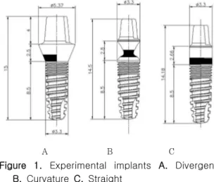
co. Seoul, Korea) were installed in the right and the left edentulous, mandibular pre- molar region of the 2 dogs bilaterally. In each dog the following types of abutments were used: divergent form (Figure 1A), cur- vature form (Figure 1B), straight form (Figure 1C), and the fixture and abutment part were connected to the one body. In the lower abutment part of all implants, there were about 1 mm of microgroove area .
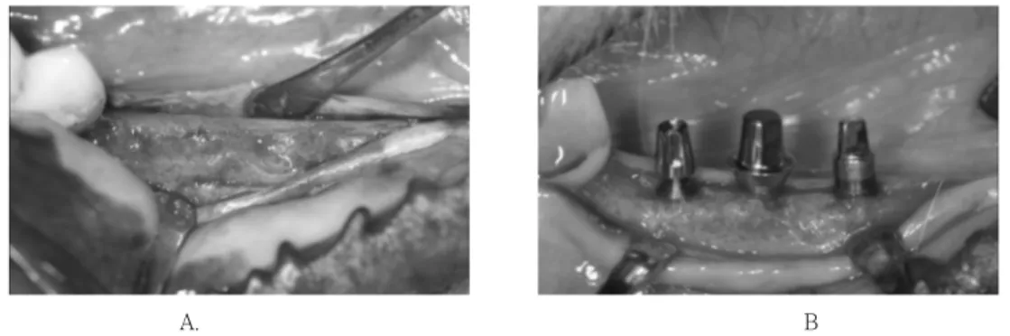
An incision was made through the mucosa at the crest of the alveolar ridge. Buccal and lingual full thickness flaps were ele- vated (Figure 2A), and self-tapping fixtures were placed in accordance with the recom- mendations given in the manual for this particular implant system. The implants were margin coincided with the bone crest (Figure 2B). The mucoperiosteal flaps were resutured by resorbable suture material.
A B C
Figure 1. Experimental implants A. Divergent B. Curvature C. Straight
Three months later, the animals were sac- rificed with an overdose of sodium-pentotal and perfused with a fixative through the carotid arteries. The fixative consisted of a mixture of 5% glutaraldehyde and 4% form- aldehyde buffered to pH 7.2. The mandibles were removed and placed in the fixative.
Each implant region was dissected using a diamond saw. The biopsies selected for the
"fracture technique"6 were placed in EDTA.
Before the hard tissue was fully decalcified, implants were removed by counter-clockwise rotating. And, incisions were placed at the buccal and lingual aspects of the curvature form abutment implants only. The cuts pe-
netrated the superior part of peri-implant tissue and the implants were removed by counter-clockwise rotating. The specimen was not divided entirely. Decalcification was completed in EDTA and dehydration per- formed in serial steps of ethanol concentrations. The units were finally em- bedded in paraffin. Sections were produced from each tissue unit with the microtome set at 3㎛. The sections were stained with H&E.
Each section was examined histologically and histomorphometrically. The following distances (Figure 3) were measured in a millimeter using a light microscope (magnification X 40) under a digitizing pad(TDI Scope Eye 3.0 Techsan Co. Ltd).
(a) Epithelium = Gingival margin to ap- ical margin of junctional epithelium
(b) Connective tissue = apical margin of junctional epithelium to bone crest
(c) Soft tissue height = Gingival margin to bone crest
A one-way analysis of variance (ANOVA;
p = 0.05) for each variable (soft tissue height, epithelium, connective tissue) was Figure 3. The distance measured for
histometric analysis. (a) Epithelium (b) Connective tissue (c) Soft tissue height
A. B
Figure 2. Clinical photos of implant installation surgery. A, After flap reflection. B, Implants in- stalled in a crest level.
employed to test the hypothesis that there was no difference between curvature form, straight form, divergent form abutment. The SPSS 10.0 software was used for all stat- istical procedures.
III. RESULTS
Histometric analysis
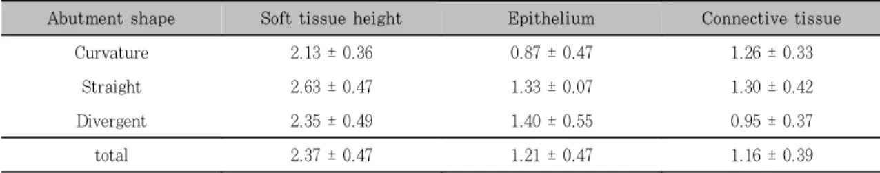
Due to the technically difficulty, some of the bucco-lingual sections of 12 implants re- vealed processing artifacts in the implant surface. Thus, the localization of the most apical epithelial cells was only accurately detectable in 6 implants, 2 of each type. The results with calculated means and standard deviations for each variable and surface are listed for buccal and lingual sites separately in Table 1. The hypothesis that there is no difference between curvature, divergent and straight form abutment showed to be gen- erally valid for the location of the gingival margin in relation to the soft tissue height, epithelium and connective tissue. But they were not statistically significant.
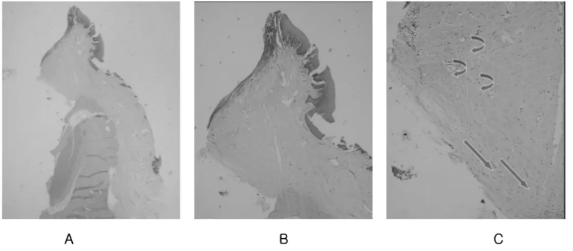
Histologic observation Straight form (Figure 5)
In the specimen, it was observed that a connective tissue invaded into the micro- groove of the abutment. In the interior part of connective tissue, connective tissue fiber ran paralleled to the abutment surface. By the higher magnification, in contact to the abutment surface, very densely arranged connective tissue oriented abutment surface.
There were no inserting fiber to the implant surface. Additionally more fibroblast was observed in close to the abutment surface.
Divergent form (Figure 6)
Similar histologic characteristics were ob- served in the divergent form abutment. The soft tissue around abutment were formed to the abutment shape; divergent. The hori- zontal soft tissue dimension was smaller than the curvature form. And, connective tissue fiber ran parallel to the abutment surface.
Curvature form (Figure 7)
The connective tissue invaded into the concave space of the curvature form abutment. As a result, horizontally thicker soft tissue dimension than the straight and divergent form were observed. Additionally, more circular form fiber than the straight and divergent form.
Table 1. Results from the histometric analysis (mm)
Abutment shape Soft tissue height Epithelium Connective tissue
Curvature 2.13 ± 0.36 0.87 ± 0.47 1.26 ± 0.33
Straight 2.63 ± 0.47 1.33 ± 0.07 1.30 ± 0.42
Divergent 2.35 ± 0.49 1.40 ± 0.55 0.95 ± 0.37
total 2.37 ± 0.47 1.21 ± 0.47 1.16 ± 0.39
A B C
Figure 5. The histologic section of straight form. A, X 40 magnification. B, X100. C, X 200.
Connective tissue fiber paralleled to the abutment surface(arrow). Connective tissue inserted into the microgroove(circle).
A B C
Figure 6. The histologic section of divergent form. A, X 12.5 magnification. B, X 40. C, X 100.
A B C
Figure 7. The histologic section of curvature form. A, X 12.5 magnification. B, X 40. C, X 100. The multiple orientation of connective tissue fiber
IV. DISCUSSION
Gingival tissue around implant
Dental implants are surrounded by 3 dif- ferent tissues: epithelium, fibrocollagenous soft connective tissue and bone. Although the success rate of dental implants is very high today, there are still failures, which may be related to an absence of attachment of the gingival connective tissue to the implant.
One of the main differences between the gingival tissues surrounding a natural tooth and a dental implant is the collagen fiber orientation around the collar of the device just below the gingival surface. In a natural tooth, dentogingival collagen fibers are in- serted into the cementum and the bone, and oriented perpendicular or oblique to the tooth surface.14 In contrast, around a dental implant they are mainly parallel to the im- plant surface.4,5
Berglundh et al.4 demonstrated that the peri-implant mucosa which formed at tita- nium implants following abutment
connection had many features in common with gingival tissue at teeth. Thus, like the gingiva, the peri-implant mucosa established a cuff-like barrier which adhered to the sur- face of the titanium abutment. Further, both the gingiva and the peri-implant muco- sa had a well-keratinized oral epithelium which was continuous with a junctional epi- thelium that faced the enamel or the tita- nium surface. In the peri-implant mucosa, the collagen fibers appeared to commence at
the marginal bone and were paralleled with the abutment surface.
In the same study,4 no inflammatory cell infiltrates were found in the peri-implant region despite the parallel arrangement of collagen fibers around the implant in a ca- nine model. The suggestion was that the col- lagen fibers, while parallel to the surface.
provided a cuff-like barrier to bacterial invasion.
From Hermann's study,19 while changes occur in the dimensions of the sulcus depth, junctional epithelium, and connective tissue contact, the overall dimension was not al- tered whether unloaded or loaded for 1 year around dental implants. Futhermore, these results were similar to those same di- mensions around natural teeth.14. And they suggested that dental implants have physio- logically formed and stable soft tissue units.
In this study, all of 3 different abutment shapes had the same gingival structure.
None of sections revealed an epithelial downgrowth to the alveolar crest. It might be assumed that the epithelial downgrowth was prevented by a direct connective tissue contact to the implant surface. The observed connective tissue close to the implant sur- face had no similarities with that around natural teeth, since inserting perpendicular fibers were not found. The inner zone of connective tissue resembled most likely an inflammation-free scar tissue formation into which collagen fibers from different direc- tions inserted. The surrounding connective tissue was well organized, dominated by col- lagen bundles running in different direc- tions, and forming a 3-dimensional network
around the implant abutment. This network of collagen fibers in the outer zone is sim- ilar to that found around natural teeth.
Connective tissue(Fiber) orientation
In the study for evaluation the orientation of collagen in the canine gingival connective tissue around dental implants,20 vertical col- lagen fibers ran from bone to the epi- thelium, and horizontal fibers ran perpen- dicularly towards the implant surface and then, when close to the surface, became vertical. These findings were in accordance with the fiber orientation reported in prior studies of titanium dental implants in the canine5 and in implants retrieved from hu- man subjects11 that showed that only near the crestal margin of bone were fibers found obliquely inserting in the bone surface.
The histologic section of this study re- vealed the similar connective fiber orientation. But, in the narrow neck abut- ment, more circular oriented fiber was found than other implant of different abutment shape. This circular oriented fiber is ex- pected to prevent mechanical force to soft tissue around implant. Schierano et al.21 suggested that the presence of a dominant circular system of collagen fibers around the abutment is in accordance with the concept of peri-implant 'circular ligament' proposed by Ruggeri,22 and confirmed by Piattelli.11 And their three-dimensional graphic repre- sentation of the microscopic data suggested the presence around titanium implant abut- ments of a differentiated network of fibers, which might be of clinical relevance as a
mechanical protection for the underlying bone-implant interface.
Microgroove
The role of microgroove around abutment surface is suggested proliferation and migra- tion of fibroblast, formation of thick con- nective tissue, adaptation effect of con- nective tissue and finally maintenance of soft tissue around abutment. But the study about this theme has not been performed, the author reviewed about the studies of rough surface around abutment surface.
In studies about the effect of rough sur- face to the soft tissue formation,5 when com- paring three different surfaces (sandblasted, find sandblasted, polished) at the trans- mucosal level, no significant differences con- cerning soft tissue reactions were found be- tween the 3 implant surfaces. In particular, the length of direct connective tissue contact was similar. It is concluded that the differ- ent surface textures did not influence the healing pattern of the soft tissues.
And in other study23 similar opinions were suggested. They did not find any such dif- ference when comparing soft tissue healing with acid-etched and machined abutments.
The attachment comprised a barrier epi- thelium and a zone of connective tissue at- tachment of similar dimension at both surfaces. It was demonstrated that the soft tissue attachment that formed to implants made of c.p. titanium was not influenced by the roughness of the titanium surface.
But, Glauser et al.24 recently demon- strated contrast results. They reported that
the periimplant soft tissue formed at the ex- perimental one-piece mini-implants in hu- mans was of a character similar to that de- scribed in animal studies. The oxidized and acid-etched implants revealed less epithelial downgrowth and longer connective tissue seal than machined implants. They sug- gested the hypothesis of this results; a rough surface has a certain "conductive" ef- fect on the connective tissue adhesion dur- ing healing, thereby inhibiting epithelial downgrowth. On the other hand, a smooth surface may allow for pronounced epithelial downgrowth compared with rougher surfaces.
In conclusion, the curvature form abut- ment with microgroove may result thicker soft tissue and more circular connective tis- sue fiber. This thick soft tissue and circular fiber may strengthen the function of soft tis- sue barrier. Finally, using this form abut- ment, the clinicians can get additional ad- vantage to prevent the gingival recession around dental implant in esthetic area.
V. REFERENCES
1. Adell R, Lockhole U, Rockier B, Branemark PI. A 15-year study of os- seointegrated implants in the treatment of the edentulous jaw. Int J Oral Surg.
1981 Dec;10(6):387-416.
2. Buser D, Mericske-Stern R, Bernard JP, Behneke A, Behneke N, Hirt HP, Belser UC, Lang NP. Long-term evaluation of non-submerged ITI implants. Part 1:
8-year life table analysis of a pro- spective multi-center study with 2359
implants. Clin Oral Implants Res. 1997 Jun;8(3):161-72.
3. Albrektsson T, Zarb G, Worthington P, Eriksson AR. The long-term efficacy of currently used dental implants: a re- view and proposed criteria of success.
Int J Oral Maxillofac Implants. 1986 Summer;1(1):11-25.
4. Berglundh T, Lindhe J, Ericsson I, Marinello CP, Liljenberg B, Thomsen P.
The soft tissue barrier at implants and teeth. Clin Oral Implants Res. 1991 Apr-Jun;2(2):81-90.
5. Buser D, Weber HP, Donath K, Fiorellini JP, Paquette DW, Williams RC. Soft tissue reactions to non-sub- merged unloaded titanium implants in beagle dogs. J Periodontol. 1992 Mar;
63(3):225-35.
6. Berglundh T, Lindhe J, Jonsson K, Ericsson I. The topography of the vas- cular systems in the periodontal and peri-implant tissues in the dog. J Clin Periodontol. 1994 Mar;21(3):189-93.
7. Abrahamsson I, Berglundh T, Wennstrom J, Lindhe J. The peri-implant hard and soft tissues at different implant systems.
A comparative study in the dog. Clin Oral Implants Res. 1996 Sep; 7(3):212-9.
8. Berglundh T, Lindhe J. Dimension of the periimplant mucosa. Biological width revisited. J Clin Periodontol. 1996 Oct;23(10):971-3.
9. Hammerle CH, Bragger U, Burgin W, Lang NP. The effect of subcrestal place- ment of the polished surface of ITI im- plants on marginal soft and hard tissues. Clin Oral Implants Res. 1996
Jun;7(2):111-9.
10. Weber HP, Buser D, Donath K, Fiorellini JP, Doppalapudi V, Paquette DW, Williams RC. Comparison of healed tissues adjacent to submerged and non-submerged unloaded titanium dental implants. A histometric study in beagle dogs. Clin Oral Implants Res. 1996 Mar;7(1):11-9.
11. Piattelli A, Scarano A, Piattelli M, Bertolai R, Panzoni E. Histologic as- pects of the bone and soft tissues sur- rounding three titanium non-submerged plasma-sprayed implants retrieved at autopsy: a case report. J Periodontol.
1997 Jul;68(7):694-700.
12. Ericsson I, Berglundh T, Marinello C, Liljenberg B, Lindhe J. Long-standing plaque and gingivitis at implants and teeth in the dog. Clin Oral Implants Res. 1992 Sep;3(3):99-103.
13. Cochran DL, Hermann JS, Schenk RK, Higginbottom FL, Buser D. Biologic width around titanium implants. A his- tometric analysis of the implanto-gin- gival junction around unloaded and loaded nonsubmerged implants in the canine mandible. J Periodontol. 1997 Feb;68(2):186-98.
14. Gargiulo AW, Wentz FM, Orban B.
Dimensions and relations of the dento- gingival junction in humans. J Periodontol 1961;32:261-267
15. Vacek JS, Gher ME, Assad DA, Richardson AC, Giambarresi LI. The di- mensions of the human dentogingival junction. Int J Periodontics Restorative Dent. 1994 Apr;14(2):154-65.
16. Abrahamsson I, Berglundh T, Moon IS, Lindhe J. Peri-implant tissues at sub- merged and non-submerged titanium implants. J Clin Periodontol. 1999 Sep;
26(9):600-7.
17. Moon IS, Berglundh T, Abrahamsson I, Linder E, Lindhe J. The barrier between the keratinized mucosa and the dental implant. An experimental study in the dog. J Clin Periodontol. 1999 Oct;26(10) :658-63.
18. Tenenbaum H, Schaaf JF, Cuisinier FJ.
Histological analysis of the Ankylos peri-implant soft tissues in a dog model.
Implant Dent. 2003;12(3):259-65.
19. Hermann JS, Buser D, Schenk RK, Higginbottom FL, Cochran DL. Biologic width around titanium implants. A physiologically formed and stable di- mension over time. Clin Oral Implants Res. 2000 Feb;11(1):1-11.
20. Comut AA, Weber HP, Shortkroff S, Cui FZ, Spector M. Connective tissue ori- entation around dental implants in a ca- nine model. Clin Oral Implants Res.
2001 Oct;12(5):433-40.
21. Schierano G, Ramieri G, Cortese M, Aimetti M, Preti G. Organization of the connective tissue barrier around long-term loaded implant abutments in man. Clin Oral Implants Res. 2002 Oct;
13(5):460-4.
22. Ruggeri A, Franchi M, Marini N, Trisi P, Piatelli A. Supracrestal circular col- lagen fiber network around osseointe- grated nonsubmerged titanium implants.
Clin Oral Implants Res. 1992 Dec;3(4) :169-75.
23. Abrahamsson I, Berglundh T, Glantz PO, Lindhe J. The mucosal attachment at different abutments. An experimental study in dogs. J Clin Periodontol. 1998 Sep;25(9):721-7.
24. Glauser R, Schupbach P, Gottlow J, Hammerle CH. Periimplant soft tissue
barrier at experimental one-piece mini- implants with different surface top- ography in humans: A light-microscopic overview and histometric analysis. Clin Implant Dent Relat Res. 2005;7 Suppl 1:S44-51.
-Abstract-
임플란트 지대주 모양에 따른 주위 연조직 반응에 관한 연구
안수용1, 한종현2, 허성주3, 김태일1, 설양조1, 이용무1, 구영1, 이해준1, 정종평1, 한수부1, 류인철1
서울대학교 치과대학 치주과학교실1,연세대학교 영동세브란스병원 보철학교실2
서울대학교 치과대학 보철학교실3
연구배경
임플란트에 관한 전통적인 연구들은 주로 임플란트 매식체와 골조직간의 결합에 중점을 두어왔다. 최근 임 플란트의 심미적 관점에 대한 관심이 높아지면서 임플란트 주위 연조직의 재건 및 유지에 대한 연구들이 많이 이루어지고 있다. 이번 연구는 임플란트 주위 연조직이 임플란트 지대주의 모양에 따라 어떻게 반응하는지 알 아보고자 한다.
연구방법 및 재료
2 마리의 성견을 대상으로, 먼저 하악의 모든 소구치를 발치하고, 3개월의 치유 기간 후에 각 4분악에 실험 에 사용된 3개의 다른 모양의 지대주를 가진 임플란트를 식립하였다: (1) 위로 벌어진 모양의 지대주 (2) 안 쪽으로 오목하게 좁아진 지대주 (3) 평행한 모양의 지대주. 식립 순서는 무작위로 하였으며, 3개월 후 실험동 물을 희생하고 조직 표본을 얻었다. 조직 표본은 광학 현미경을 통해 관찰하고, 상피, 결합조직, 전체 연조직의 수직적 거리를 측정하여 비교하였다.
연구 결과
이번 연구에서 임플란트 주위의 연조직을 측정한 결과, 접합 상피는 1.21 ± 0.47 mm, 결합조직은 1.16 ± 0.39 mm, 전체 연조직 두께는 2.37 ± 0.47 mm로 이전의 연구들과 비슷한 결과를 보였다. 지대주 주위의 연 조직 중 결합조직이 많은 부위에서는 여러 주행 방향의 교원 섬유들이 관찰되었다. 그 중에서 결합조직이 차단 막으로써의 역할을 할 수 있도록 하는 원형으로 주행하는 교원 섬유들이 모든 지대주 모양에서 관찰되었다. 특 히 오목하게 좁아진 모양을 가진 지대주에서는 오목하게 파인 부분으로 많은 원형으로 주행하는 교원 섬유들이 관찰되었다. 오목한 모양의 지대주는 다른 모양의 지대주에 비해서 측면 방향의 연조직 두께가 두꺼웠다. 특히 위로 벌어진 모양의 지대주에 비해서 두꺼운 연조직을 확보할 수 있었으며, 내부에 많은 결합조직 교원 섬유들 을 관찰할 수 있었다.
결론
이번 연구에서 오목한 모양의 지대주가 연조직을 두껍게 유지하고, 많은 원형으로 주행하는 교원 섬유들을 확보할 수 있었다. 이를 통해 오목한 형태의 지대주가 연조직 유지에 더 유리하고, 따라서 심미적인 부위에서 연조직의 퇴축을 예방하는데 더 유리하다고 할 수 있다.2)
주요어:임플란트, 연조직, 오목한 모양의 지대주, 교원 섬유