권고안 개발 배경
갑상선 결절은 전 세계적으로 상당히 흔한 질환으로, 촉진 시 4~8%, 초음파에서 19~67%, 부검 시 50%의 인구에서 갑상
선 결절이 발견된다고 보고되어 있다(1-3). 갑상선 결절의 5~15%는 최종적으로 갑상선 암으로 진단되고 있으며 다양한 유전적 환경적 요인 및 의료 기기의 발달, 건강검진 프로그램의 보급 등으로 전 세계적으로 갑상선암 발생은 지속적으로 증가
Guidelines for Primary Imaging Test and Biopsy Methods
in the Diagnosis of Thyroid Nodules: Joint Report by the Korean Society of Radiology and National Evidence-Based Healthcare Collaborating Agency
갑상선 결절 진단에 있어 일차적인 영상검사 및 조직검사 방법에 대한 권고안: 대한영상의학회와 한국보건의료연구원 공동보고서
Hyun Kyung Lim, MD
1, Eun Ju Ha, MD
2*, In Young Youn, MD
3, Jung Hyun Yoon, MD
4, Jung Hwan Baek, MD
5, Kyung Hyun Do, MD
5, Miyoung Choi, RN
6, Jin A Choi, MPH
5, Min Lee, MPH
5, Dong Gyu Na, MD
71Department of Radiology, Soonchunhyang University Seoul Hospital, Seoul, Korea
2Department of Radiology, Ajou University School of Medicine, Suwon, Korea
3Department of Radiology, Sungkyunkwan University School of Medicine, Kangbuk Samsung Hospital, Seoul, Korea
4Department of Radiology, Severance Hospital, Research Institute of Radiological Science, Yonsei University College of Medicine, Seoul, Korea
5Department of Radiology and Research Institute of Radiology, Asan Medical Center, University of Ulsan College of Medicine, Seoul, Korea
6Division for Healthcare Technology Assessment Research, National Evidence-Based Healthcare Collaborating Agency, Seoul, Korea
7Department of Radiology, Gangneung Asan Hospital, Gangneung, Korea
The Korean Society of Radiology and the National Evidence-based Healthcare Col- laborating Agency developed the guideline for primary imaging and biopsy methods in the diagnosis of thyroid nodules. The development committee, the working com- mittee, and the advisory committee were formed to develop the recommendation.
The development committee mainly plays a role of methodological consulting and overall planning and management of the advisory development stage. The working committee was composed of experts recommended by the Korean Society of Thy- roid Radiology and conducted a practical adaptation process from the selection of core questions to the final recommendation. The Advisory Committee consisted of clinical experts recommended by the Korean Thyroid Association and reviewed core questions and draft recommendations and participated in the Expert Panel Survey.
This guideline recommends cervical ultrasound as the first imaging modality for di- agnosis of suspected thyroid nodules and recommends ultrasound-guided fine nee- dle aspiration for histologic diagnosis of thyroid nodules. This guideline is expected to be of significant benefit to clinicians treating thyroid nodules.
Index terms Thyroid Nodule Thyroid Neoplasm Ultrasonography Biopsy, Fine-Needle
Received November 8, 2017 Revised December 5, 2017 Accepted December 7, 2017
*Corresponding author: Eun Ju Ha, MD Department of Radiology, Ajou University School of Medicine, 164 Worldcup-ro, Yeongtong-gu, Suwon 16499, Korea.
Tel. 82-31-219-4057 Fax. 82-31-219-5852 E-mail: [email protected]
This is an Open Access article distributed under the terms of the Creative Commons Attribution Non-Commercial License (http://creativecommons.org/licenses/by-nc/4.0) which permits unrestricted non-commercial use, distri- bution, and reproduction in any medium, provided the original work is properly cited.
J Korean Soc Radiol 2018;79(1):1-10 https://doi.org/10.3348/jksr.2018.79.1.1
추세에 있다(4). 2014년 보건복지부 암 발생률 통계에 따르면 국내에서 갑상선암은 암 발생률 1위로 전체 암의 14.2%를 차 지하고 10만 명당 60.7명의 조발생률(crude rate)을 보이고 있 어 사회적인 문제가 되고 있다(5).
갑상선 암의 발생이 증가하면서 갑상선 결절이 의심되는 환 자의 진단을 위한 다양한 영상 장치 및 확진을 위한 조직학적 진단 방법이 대두되었다. 초음파(US), 컴퓨터단층촬영(CT), 자 기공명영상(MRI), 양전자방사단층촬영(PET) 등과 같은 많은 영상검사가 결절의 진단에 사용되게 되었고, 결절의 조직학적 진단에도 촉진 하 세침흡인생검, 초음파 유도 하 세침흡인생검, 초음파 유도 하 중심바늘생검, 진단적 수술 등 다양한 방법이 보 고되었다. 따라서 결절 진단의 전반적인 과정에 있어 검사자 및 기관에 상관없이 통일된 근거기반 임상 영상 가이드라인 개발의 필요성이 제기되게 되었으며, 이에 개발위원회, 실무위원회 및 유관학회의 자문위원단이 모여 갑상선 결절의 진단을 위한 권 고안 개발이 시작되었다.
권고안 개발을 위한 위원회의 구성
권고안 개발을 위해 개발위원회와 실무위원회 및 자문위원 회를 구성하였다. 개발위원회는 대한영상의학회 진료지침위원 회에서 추천한 임상 전문가, 연구방법론 전문가, 한국보건의료 연구원 연구진으로 구성하였다. 개발위원회는 주로 방법론적 컨설팅 역할을 수행하고, 권고안 개발 단계를 전반적으로 기획 및 관리하였다. 실무위원회는 대한갑상선영상의학회에서 추천 한 전문가로 구성되어 핵심질문 선정부터 최종 권고안 도출까 지 실질적인 수용개작(adaptation) 과정을 수행하였다. 자문위 원회는 핵심질문별 임상 영상검사를 의뢰하거나 시행한 최종사 용자(end-user)로 예상되는 유관 외부 전문학회(대한갑상선 학회)에서 추천을 받은 임상 전문가들로 구성되었으며, 핵심질 문의 검토 및 권고안 초안에 대한 검토와 전문가 패널조사에 참여하였다.
권고안 수용개작 과정
권고안 개발절차는 개발위원회에서 개발한 근거기반 임상 영상 권고안 수용개작 방법론에 근거하여 이루어졌다(6).
핵심질문선정
실무위원회에서 선정한 핵심질문은 개발위원회 및 최종사용 자로 예상되는 임상 전문학회(대한갑상선학회)로부터 추천받 은 자문위원회의 검토 및 수정사항 반영 후 최종 확정되었다.
최종 확정된 2가지 문장형 핵심질문은 아래와 같다.
1) 갑상선 결절이 의심되는 환자에서 진단을 위한 일차적인 영상 검사는 무엇인가?
2) 갑상선 결절 진단에 적절한 조직검사 방법은 무엇인가?
진료지침 검색 전략
검색 데이터베이스는 국외 주요 문헌검색데이터베이스(Ovid- Medline, Ovid-Embase) 및 해외진료지침관련 주요 사이트 (National Guideline Clearinghouse, Guideline International Network)와 국내 데이터베이스(KoreaMed, KMbase, KoMGI, KGC)를 모두 포함하여 검색하였다. 또한, 주요 학회사이트 및 주요 권고안이 누락되지 않도록 수기검색을 통해 보완하였다.
진료지침 선별 및 선정
핵심질문 1에 대한 검색결과는 중복 배제 후 213건 검색, 제 목과 초록으로 1차 선별 시 37건이 선택되었다. 원문을 확보하 여 2차 선정과정을 통해 최종 9건이 선택되었다. 핵심질문 2에 대한 검색결과는 중복 배제 후 100건 검색, 제목과 초록으로 1 차 선별 시 8건이 선택되었다. 원문을 확보하여 2차 선정과정을 통해 최종 5건이 선택되었다(Figs. 1, 2).
진료지침의 질 평가
개발위원회에서 선택한 진료지침에 대한 appraisal of guide- lines for research & evaluation (이하 AGREE) II 질 평가 결과 를 회신하였고, 실무위원회에서는 진료지침의 질을 고려하여 핵심질문 1에서 최종 5편의 지침을, 핵심질문 2에서 최종 5편 의 지침을 선택했다. 2011년 대한갑상선영상의학회에서 발표된
“Ultrasonography and the Ultrasound-Based Management of Thyroid Nodules: Consensus Statement and Recommendations”
의 경우 AGREE II 질 평가에서는 41점으로 개발위원회의 추천 을 받지 않았으나 갑상선 영상 전문가들에 의해 제정된 국내 진 료지침이라는 점에서 실무위원 만장일치로 채택되었다(4).
근거 검토 및 권고안 초안 작성
최종 선택된 지침들의 권고 및 권고등급을 비교하고(Tables 1, 2), 각 권고를 지지하는 근거 문헌을 다시 검토하여 Korean Clinical Imaging Guideline (KCIG)의 근거수준 결정 방법에 따라 근거의 내용 및 질 평가 결과를 근거표(evidence table) 형 태로 정리했다. 근거표를 바탕으로 실무위원회의 최종 논의를 거쳐 권고안 초안이 도출되었다.
Fig. 1. Flow diagram of guideline selection (Key Question 1).
갑상선 KQ1
국외 데이터베이스 검색을 통해 확인된 지침 수
• Ovid-MEDLINE (n = 53)
• Ovid-EMBASE (n = 182)
• NGC (n = 104)
• GIN (n = 2)
(total n = 341)
국내 데이터베이스 검색을 통해 확인된 지침 수
• KoreaMed (n = 2)
• KMBASE (n = 9)
• KGC (n = 0)
• KoMGI (n = 0)
(total n = 11)
수기 검색을 통해 확인된 지침 수(n = 0)
중복제거 후 지침 수 (국내 n = 9/국외 n = 204)
(total n = 213)
1차 선택 지침 수 (국내 n = 0/국외 n = 37)
(total n = 37)
최종 선택된 지침 수 (국내 n = 0/국외 n = 9)
(total n = 9)
1차 배제된 지침 수 (total n = 176)
2차 배제된 지침 수 및 배제사유(n = 28) 1. P: 핵심질문의 관심환자를 대상으로 하지 않은 경우(n = 7) 2. I&C: 핵심질문 관련 영상검사가 포함되지 않은 경우(n = 3) 3. O: 적절한 결과(진단정확성, 유효성, 안전성, 비교효과성,
환 자선호도 등)를 보고하지 않은 경우(n = 0) 4. 진료지침(practical guideline)이 아닌 경우(n = 7) 5. 권고(recommendation)가 제시되지 않은 경우(n = 2) 6. 근거기반 방법으로 작성되지 않은 경우(즉, 체계적
근거검색 없이 합의만으로 자성한 지침)(n = 0) 7. 영어 또는 한국어로 보고되지 않은 지침(n = 5) 8. 중복으로 게재된 경우(즉, 동일 내용으로 다른 저널에
게재 혹은 출판형태만 차이가 있는 경우(n = 1) 9. 원문확보가 불가능한 경우(n = 3)
Fig. 2. Flow diagram of guideline selection (Key Question 2).
갑상선 KQ2
KQ1과 동일 국외 데이터베이스
검색을 통해 확인된 지침 수
• Ovid-MEDLINE (n = 61)
• Ovid-EMBASE (n = 29)
• NGC (n = 104)
• GIN (n = 2)
(total n = 196)
중복제거 후 지침 수 (국내 n = 9/국외 n = 91)
(total n = 100)
1차 선택 지침 수 (국내 n = 0/국외 n = 8)
(total n = 8)
최종 선택된 지침 수 (국내 n = 0/국외 n = 5)
(total n = 5)
1차 배제된 지침 수 (total n = 92)
2차 배제된 지침 수 및 배제사유(n = 3) 1. P: 핵심질문의 관심환자를 대상으로 하지 않은 경우(n = 1) 2. I&C: 핵심질문 관련 영상검사가 포함되지 않은 경우(n = 1) 3. O: 적절한 결과(진단정확성, 유효성, 안전성, 비교효과성,
환자선호도 등)를 보고하지 않은 경우(n = 0) 4. 진료지침(practical guideline)이 아닌 경우(n = 0) 5. 권고(recommendation)가 제시되지 않은 경우(n = 0) 6. 근거기반 방법으로 작성되지 않은 경우(즉, 체계적 근거검색
없이 합의만으로 자성한 지침)(n = 0) 7. 영어 또는 한국어로 보고되지 않은 지침(n = 1) 8. 중복으로 게재된 경우(즉, 동일 내용으로 다른 저널에
게재 혹은 출판형태만 차이가 있는 경우(n = 0) 9. 원문확보가 불가능한 경우(n = 0)
국내 데이터베이스 검색을 통해 확인된 지침 수
• KoreaMed (n = 2)
• KMBASE (n = 9)
• KGC (n = 0)
• KoMGI (n = 0) (total n = 11)
수기 검색을 통해 확인된 지침 수(n = 0)
권고안 합의 및 권고등급 결정
실무위원회에서 작성한 초안에 대해서는 개발위원회와의 논 의를 거쳐 최종 근거 수준과 권고등급을 결정했다.
권고안의 합의
갑상선 학회 등 외부 관련 학회 전문가가 포함된 자문위원회 의 권고안 동의 정도 설문조사(9점 척도) 결과, 핵심질문 1에 대한 동의 정도는 1차에서 평균(표준편차) 7.80(1.25)에서 2 차는 8.00(0.53)이었고, 핵심질문 2에 대한 동의 정도는 1차 에서 7.60(1.19), 2차에서 7.88(0.35)로 향상되었고, 동의 정 도는 매우 높았다.
외부 검토
최종 권고안에 대한 검토는 권고안 개발에 참여하지 않은 영 상의학 전문의 검토와 지침의 최종사용자가 될 유관학회의 검
토를 받았다.
권고안
핵심질문 1. 갑상선 결절이 의심되는 환자에서 진단을 위한 일차적인 영상 검사는 무엇인가?
권고 1. 갑상선 결절이 의심되거나 초음파 이외의 영상 기법으로 발견된 갑상선 결절의 세부 진단에 경부 초음파 검사를 권고한다(권고등급 A, 근거수준 II).
근거요약
갑상선 결절의 진단 및 치료에 관한 권고안을 검색 후 최종 5개의 진료지침을 선택했다(4, 7-10). 갑상선 결절은 흔한 질 Table 1. Recommendations Matrix of Existing Guidelines (Key Question 1)
Source Guidelines (Publication Year)
AACE/AME/ETA Medical Guidelines for Clinical Practice for the Diagnosis and
Management of Thyroid Nodules
(2010)
Ultrasonography and the US-Based Management of Thyroid
Nodules: Consensus Statement and Recom-
mendations (2011)
British Thyroid Association Guidelines
for the Management of Thyroid Cancer
(2014)
Thyroid Carcinoma, Version 2. 2015 (NCCN Guideline)
(2015)
2015 American Thyroid Association Manage-
ment Guidelines for Adult Patients with Thyroid Nodules and Differentiated Thyroid
Cancer (2016) AGREE II (domain 3.
rigour of development)
69 41 84 74 63
Recommendation US evaluation is recommended for
• Patients at risk for thyroid malignancy
• Patients with palpable thyroid nodules or MNGs
• Patients with lymphadenopathy suggestive of a malignant lesion
Among the modern imaging modalities, high-resolution US is the most sensitive diagnostic modality for the detection of the thyroid nodules and it is necessary to perform US for the nodules found after palpation
US is an extremely sensitive examination for thyroid nodules. It can be specific for the diagnosis of thyroid carcinoma (particularly papillary carcinoma), and aids decision making about which nodules to perform FNAC
1) All patients being investigated for possible thyroid cancer should undergo an US of the neck in second- ary care by an appro- priate, competent practitioner
For thyroid nodules known or suspected on clinical or imaging findings, US recommended
Thyroid sonography with survey of the cervical lymph nodes should be performed in all patients with known or suspected thyroid nodules
Grading of recommendation
Grade B, BEL 3 Not available Good practice point Category 2A Strong recommendation, High-quality evidence AACE = American Association of Clinical Endocrinologists, AGREE = Appraisal of Guidelines for Research & Evaluation, AME = Associazione Medici Endo- crinologi, BEL = best evidence level, ETA = European Thyroid Association, FNAC = fine needle aspiration cytology, MNG = multinodular goiter, NCCN = Na- tional Comprehensive Cancer Network, US = ultrasound
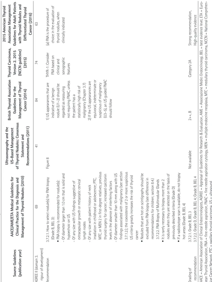
Table 2. Recommendations Matrix of Existing Guidelines (Key Question 2) Source Guidelines (publication year) AACE/AME/ETA Medical Guidelines for Clinical Practice for the Diagnosis and Management of Thyroid Nodules (2010) Ultrasonography and the US-Based Management of Thyroid Nodules: Consensus Statement and Recommendations (2011)
British Thyroid Association Guidelines for the Management of Thyroid Cancer (2014) Thyroid Carcinoma, Version 2. 2015 (NCCN guideline) (2015)
2015 American Thyroid Association Management Guidelines for Adult Patients with Thyroid Nodules and Differentiated Thyroid Cancer (2016) AGREE II (domain 3. rigour of development)6941847463 Recommendation3.7.2.1. How to select nodule(s) for FNA biopsy (Grade B, BEL 3): • FNA biopsy is recommended for nodule(s): - Of diameter larger than 1.0 cm that is solid and hypoechoic on US - Of any size with US findings suggestive of extracapsular growth or metastatic cervical lymph nodes - Of any size with patient history of neck irradiation in childhood or adolescence; PTC, MTC, or MEN 2 in first-degree relatives; previous thyroid surgery for cancer; increased calcitonin levels in the absence of interfering factors - Of diameter smaller than 10 mm along with US findings associated with malignancy (see section 3.7.1.2.); the coexistence of 2 or more suspicious US criteria greatly increases the risk of thyroid cancer • Nodules that are hot on scintigraphy should be excluded from FNA biopsy (see difference in recommendations for children; section 8.4.) 3.7.2.2. FNA Biopsy of Multinodular Glands • It is rarely necessary to biopsy more than 2 nodules when they are selected on the basis of previously described criteria (Grade D) • If a radioisotope scan is available, do not biopsy hot areas (Grade B, BEL 4)
Figure 81) US appearances that are indicative of a benign nodule (U1–2) should be regarded as reassuring not requiring FNAC, unless the patient has a statistically high risk of malignancy (Chapter 3.7) 2) If the US appearances are equivocal, indeterminate or suspicious of malignancy (U3–5), an US guided FNAC should follow THYR-1: Consider FNA based on clinical and sonographic features
(a) FNA is the procedure of choice in the evaluation of thyroid nodules, when clinically indicated Grading of recommendation3.7.2.1. Grade B, BEL 3 3.7.2.2. Grade D, Grade B, BEL 4; Grade B, BEL 4Not available2++, BCategory 2AStrong recommendation, High-quality evidence AACE = American Association of Clinical Endocrinologists, AGREE = Appraisal of Guidelines for Research & Evaluation, AME = Associazione Medici Endocrinologi, BEL = best evidence level, ETA = Euro- pean Thyroid Association, FNA = fine-needle aspiration, FNAC = fine needle aspiration cytology, MEN = multiple endocrine neoplasia, MTC = medullary thyroid carcinoma, NCCN = National Comprehen- sive Cancer Network, PTC = papillary thyroid carcinoma, US = ultrasound
환으로, 무증상 성인 인구의 약 19~67%에서 발견된다고 보고 된 바 있으며(1-3), 5개의 권고안에서 공통적으로 갑상선 결절 이 의심될 때는 진단을 위하여 경부 초음파 검사를 권고한다.
초음파 검사는 갑상선 결절에 대한 진단 예민도가 매우 높은 방법으로, 결절의 진단 및 이를 바탕으로 세침흡인 검사의 필요 유무를 결정할 수 있다.
2011년 대한갑상선영상의학회에서 발표된 “Ultrasonography and the Ultrasound-Based Management of Thyroid Nodules:
Consensus Statement and Recommendations”에서는 갑상선 결절을 발견하는 데 있어 가장 민감한 검사법은 고해상도 경부 초음파 검사이며, 갑상선 결절이 의심되는 경우 진단을 위해 경 부 초음파 검사를 시행할 것을 권고하고 있다. 초음파 검사를 통해 결절의 진단뿐만 아니라 크기와 형태학적 특징을 확인할 수 있고, 주변 경부 림프절 전이 여부를 판단할 수 있으며, 이를 바탕으로 초음파 유도 하 세침흡인생검의 필요성 및 가능 여부 를 결정할 수 있다고 보고하였다(4).
2015년 American Thyroid Association (이하 ATA)에서 발표 된 “ATA Management Guidelines for Adult Patients with Thyroid Nodules and Differentiated Thyroid Cancer”에서는 갑상선 결절이 의심되거나, 결절성 갑상선종, CT/MRI/PET 등 의 영상 검사에서 우연히 발견된 갑상선 결절의 진단을 위해 갑 상선과 경부 림프절에 대한 초음파 검사를 권고하고 있다 (Grade of recommendation: strong, level of evidence: high- quality). 경부 초음파 검사를 통해 증상을 유발하는 병변이 갑 상선 결절과 연관성이 있는지 확인할 필요가 있으며 결절의 위 치, 크기, 형태학적 특징에 대한 분석 및 경부 림프절 전이 여부 에 대한 검사가 필요하다고 권고한다. 또한, 이러한 소견을 바 탕으로 초음파 유도 하 세침흡인생검의 필요성 및 가능 여부를 판단할 수 있다고 보고하였다(7, 11, 12).
2014년 British Thyroid Association (이하 BTA)에서 발표된
“BTA Guidelines for the Management of Thyroid Cancer”에 서는 경부 초음파 검사를 갑상선 결절의 진단에 매우 민감한 검 사 기법으로 정의하였고, 특히 유두상 갑상선암의 감별 진단에 사용을 권고하고 있다(Good Practice Point √; Important practical points for which there is not, nor is there likely to be, any research evidence are shown in the guidelines as Good Practice Points, and are marked with a tick box √).
또한, 초음파 검사는 결절의 형태학적 소견에 따른 초음파 유 도 하 세침흡인생검 여부를 결정할 수 있고, 이의 진단률을 높이 는 데 유용하다고 하였다(Level of Evidence: 2++, Grade of Recommendation: B)(9, 13, 14).
2010년에 American Association of Clinical Endocrinologists
(이하 AACE), Associazione Medici Endocrinologi (이하 AME), and European Thyroid Association (이하 ETA)에서 발표된
“AACE/AME/ETA Medical Guidelines for Clinical Practice for the Diagnosis and Management of Thyroid Nodules”에서 는 경부 초음파 검사를 갑상선 결절의 발견 및 진단, 결절 이외 의 갑상선 실질 변화를 판단하는 데 있어 가장 유용한 검사로 정의하였고, 만져지는 결절이 있거나 갑상선 병증이 의심되는 경우 우선적으로 초음파 검사를 시행할 것을 권고하였다. 또한, 만져지는 경부 림프절이 있을 경우 무증상 갑상선암으로 인한 경부 림프절 전이를 배제할 수 없으므로 경부 초음파 검사를 통 한 갑상선 병증의 확인이 필요함을 권고하고 있다(Grade of Recommendations: C, Level of Evidence: 3)(7, 15).
2015년 National Comprehensive Cancer Network (이하 NCCN)에 발표된 “NCCN Clinical Practice Guidelines for Thyroid Carcinoma, Version 2. 2015”에서는 경부 초음파 검 사를 갑상선 결절이 의심되는 환자에서 일차적인 진단 검사로 시행할 것을 권고하였다(NCCN Categories of Evidence and Consensus: Category 2A). 발견된 갑상선 결절의 초음파 소견 과 함께 환자의 임상 소견, thyroid stimulating hormone, thy- roglobulin 수치 등을 종합하여 초음파 유도 하 세침흡인검사 또는 초음파 추적 관찰을 결정하도록 권고하고 있다(10).
권고 고려사항
이득과 위해
초음파 검사는 갑상선 결절의 발견 및 진단에 있어 매우 민감 한 검사 방법으로 방사선 노출에 대한 위험이 없고, 갑상선 결 절의 진단을 비롯하여 갑상선 실질 변화에 대한 평가, 주변 경 부 림프절에 대한 검사 등이 가능하다. 또한, 갑상선 결절의 초 음파 영상 소견을 분석하여 초음파 유도 하 세침흡인생검의 필 요성을 판단하고, 진단의 정확도를 높일 수 있다(13, 14).
그러나 무증상 성인 인구에서 갑상선 결절은 매우 흔하게 발 견되는 질환이며, 여러 초음파 소견 중 단독으로 악성 갑상선 결절에서 특이적으로 보이는 소견은 아직까지 밝혀지지 않았고 (15), 양성 및 악성 갑상선 결절 모두에서 여러 초음파 소견이 중복되어 나타날 수 있다(1, 14, 16). 이로 인해 악성 결절의 진 단을 위한 양성 결절의 불필요한 세침흡인생검이 시행될 수 있 으며, 필요 이상의 의료비 지출 증가 및 검사로 인한 합병증 등 의 위해가 초래될 수 있다. 단, 의료비 지출 증가에 있어서 비용 효과적인 측면에 대해서는 경부 초음파의 의료보험 적용 여부 에 따라 달라질 수 있으므로 추후 의료보험 적용 시 재평가가 필요하다. 마지막으로 초음파를 통한 결절의 진단이 환자에게
불필요한 걱정과 불안을 야기시킬 수 있다.
국내 수용성과 적용성
갑상선 결절이 의심되거나 진단된 환자의 일차적인 영상 검사 방법으로 5개의 진료지침에서 동일하게 경부 초음파 검사를 선 택하였다. 이들 5개 진료지침에 대한 국내 수용성 및 적용성 평 가 결과, 갑상선 결절의 발견과 진단에 있어 경부 초음파 검사 를 적용하는 것은 국내 수용성과 적용성에 무리가 없는 것으로 판단되었다.
검사 별 방사선량 경부 초음파 검사: 0
핵심질문 2. 갑상선 결절의 진단에 적절한 조직검사 방법은 무엇인가?
권고 2. 갑상선 결절의 조직검사를 위한 방법으로는 초 음파 유도 하 세침흡인생검을 권고한다(권고등급 A, 근거 수준 II).
근거요약
갑상선 결절의 조직학적 진단을 위해 적절한 검사 방법에 대 해 최종 5개의 진료지침이 선택되었다(4, 7-10). 상기 5개 진 료지침은 모두 갑상선 결절의 조직검사 방법으로 세침흡인생검 을 권고하였으며, 촉진에 의한 세침흡인생검과 초음파 유도 하 세침흡인생검을 비교한 연구들을 바탕으로 표본의 적절성(비진 단결과와 표본오류)과 위음성률의 측면에서 초음파 유도 하 세 침흡인생검이 촉진에 의한 세침흡인생검보다 우수함을 보고하 였다.
2014년에 발표된 “BTA Guidelines for the Management of Thyroid Cancer”에서는 세침흡인생검이 가치 있고, 비용대비 효과적인 수술 전 검사방법이라고 하였으며, 초음파 유도 하 세 침흡인생검은 진단의 정확도를 높이고, 비적절한 표본을 얻을 확률을 줄인다고 하였다. 215명을 대상으로 한 전향적 연구 결 과, 초음파 유도 하 세침흡인생검의 비진단결과율(21.4%)은 촉진에 의한 비진단결과율(32.4%)보다 유의하게 낮았으며, 위음성률 역시 초음파 유도 하 세침흡인생검(5.6%)이 촉진에 의한 경우(15.8%)보다 유의하게 낮았다(9, 13).
2015년에 발표된 “ATA Management Guidelines for Adult Patients with Thyroid Nodules and Differentiated Thyroid Cancer”에서는 세침흡인생검이 갑상선 결절을 평가하는 데 있어
가장 정확하고 비용대비 효과적인 검사라고 강력히 권고하였 다. 후향적 연구에서 초음파 유도 하 세침흡인생검의 비진단결 과율(3.5%)과 위음성률(1%)이 촉진에 의한 비진단율(8.7%) 과 위음성률(2.3%)보다 유의하게 낮았다(17). 특히, 낭성 결절 이나 만져지지 않는 결절, 깊은 곳에 위치하는 결절의 경우 비 진단결과와 표본오류가 나올 확률이 높으므로 초음파 유도 하 세침흡인생검이 더욱 권고된다고 하였다. 단, 촉진으로 확인되 는 결절이 초음파에서 고형 결절인 경우에는 초음파 유도 하 세 침흡인생검과 촉진에 의한 세침흡인생검을 모두 사용할 수 있 다고 하였다. 초음파 유도 하 세침흡인생검의 진단 민감도는 97.1~100%, 특이도는 70.9~100%, 정확도는 75.9%로 보고 되었다(8, 15, 18).
2011년에 대한갑상선영상의학회에서 발표된 “Ultrasonography and the Ultrasound-Based Management of Thyroid Nodules:
Consensus Statement and Recommendations”의 경우 발견된 갑상선 결절의 초음파 소견에 따라 특정 적응증에 해당하면 조 직학적 진단을 위해 초음파 유도 하 세침흡인생검을 권고하였 으나 구체적인 근거는 제시하지 않았다(4).
2010년에 발표된 “AACE/AME/ETA Medical Guidelines for Clinical Practice for the Diagnosis and Management of Thyroid Nodules”에서는 갑상선 결절의 진료는 초음파검사 및 세침흡인생검의 결과에 의해야 한다고 권고하였으며, 세침흡인 생검은 초음파 유도 하에 이루어져야 좀 더 신뢰할 만하고 비진 단결과율을 낮출 수 있다고 권고하였다. 특히 결절이 만져지지 않거나, 환자가 뚱뚱하거나, 경부 근육이 매우 발달하여 있거 나, 다결절성인 경우 초음파 유도 하 세침검사를 강력히 권고하 였다. 386명을 대상으로 한 전향적 연구 결과 초음파 유도 하 세침흡인생검의 비진단율(12.5%)은 촉진에 의한 세침흡인생 검의 비진단율(27.2%)보다 유의하게 낮았다(7, 17, 19-23).
2015년에 발표된 “NCCN Clinical Practice Guidelines for Thyroid Carcinoma, Version 2. 2015”에서는 갑상선 결절의 초 음파 소견에 따라 적응증에 해당하면 조직학적 진단을 위해 세 침흡인생검을 시행할 것을 권고하였다. 특히, 이전 세침흡인생 검에서 고형 결절이면서 부적절한 검체를 얻었던 경우 또는, 비 진단결과인 경우에는 반드시 초음파 유도 하 세침흡인생검을 시 행할 것을 권고하였으나 구체적인 근거는 제시하지 않았다(10).
권고 고려사항
이득과 위해
갑상선 결절의 초음파 유도 하 세침흡인생검은 비교적 쉽고, 촉진에 의한 세침흡인생검보다 정확하고 합병증이 적은 안전한
검사로, 적절한 교육을 받은 갑상선 진료를 전문으로 하는 의 사라면 누구나 시행할 수 있는 검사법이다.
그러나 시술자의 기술적 숙련도와 검체 처리 오류, 결절 자체 의 내인적 요인들에 의해 다양한 빈도의 비진단결과가 나올 수 있어 이를 최대한 줄이려는 노력이 필요하다(4). 이에 최근에는 이전 초음파 유도 하 세침흡인생검에서 비진단결과가 나온 결 절의 경우, 초음파 유도 하 중심바늘생검이 세침흡인생검의 대 안적 검사법이 될 수 있다는 보고들이 있다. 초음파 유도 하 세 침흡인생검의 보고된 합병증은 0~8.6%이며, 대부분이 갑상선 주변의 혈종, 갑상선의 부종, 일시적인 목소리 변화 등이고, 입 원이 필요한 정도의 중증 합병증은 거의 보고되지 않았다. 출혈 성향이나 이에 대한 기왕력이 있는 경우 사전 준비와 합병증에 대한 적절한 예방 및 처치방법 등에 대해 잘 알고 있어야 한다.
양성 결절에 대한 불필요한 초음파 유도 하 세침흡인 세포검사 는 환자의 걱정과 불안을 증가시키고, 불필요한 의료비 지출을 유발할 수 있다.
국내 수용성과 적용성
갑상선 결절의 적절한 조직검사 방법으로 5개의 진료지침에 서 모두 초음파 유도 하 세침흡인생검을 권고하였다. 이들 5개 진료지침에 대한 국내 수용성 및 적용성 평가 결과, 수용성 및 적용성에서 모두 무리가 없는 것으로 판단되었다.
검사 별 방사선량 경부 초음파 검사: 0
요약
이번 권고안은 갑상선 결절 진단에 있어 한국에서 최초로 개 발된 근거기반의 임상 진료 권고안으로 수용개작 방법을 통해 개발되었다. 본 권고안은 갑상선 결절이 의심되는 환자의 진단 에 있어 일차적으로 경부 초음파를 시행하는 것을 권고하며, 결 절의 조직학적 진단을 위해 초음파 유도 하 세침흡인생검을 권 고한다. 이번 권고안은 갑상선 결절을 진료하는 임상의에게 많 은 도움이 될 것으로 기대된다.
Acknowledgments
This study was co-supported by the National Evidence- based Collaborating Agency (NECA-C-15-003) and the Ko- rean Society of Radiology (NECA-S-15-002).
RefeRences
1. Frates MC, Benson CB, Charboneau JW, Cibas ES, Clark OH, Coleman BG, et al. Management of thyroid nodules detect- ed at US: Society of Radiologists in Ultrasound consensus conference statement. Radiology 2005;237:794-800 2. Guth S, Theune U, Aberle J, Galach A, Bamberger CM. Very
high prevalence of thyroid nodules detected by high fre- quency (13 MHz) ultrasound examination. Eur J Clin Invest 2009;39:699-706
3. Tan GH, Gharib H. Thyroid incidentalomas: management approaches to nonpalpable nodules discovered incidental- ly on thyroid imaging. Ann Intern Med 1997;126:226-231 4. Moon WJ, Baek JH, Jung SL, Kim DW, Kim EK, Kim JY, et al.
Ultrasonography and the ultrasound-based management of thyroid nodules: consensus statement and recommen- dations. Korean J Radiol 2011;12:1-14
5. 2014 Statistics of the Ministry of Health and Welfare. Avail- able at. https://www.data.go.kr/dataset/15003005/fileDa- ta.do. Published 2017. Accessed Nov 13, 2017
6. Choi SJ, Jeong WK, Jo AJ, Choi JA, Kim MJ, Lee M, et al.
Methodology for developing evidence-based clinical imag- ing guidelines: joint recommendations by Korean Society of Radiology and National Evidence-Based Healthcare Col- laborating Agency. Korean J Radiol 2017;18:208-216 7. Gharib H, Papini E, Garber JR, Duick DS, Harrell RM, Hegedüs
L, et al. American Association of Clinical Endocrinologists, American College of Endocrinology, and Associazione Medici Endocrinologi Medical Guidelines for clinical practice for the diagnosis and management of thyroid nodules--2016 update. Endocr Pract 2016;22:622-639
8. Haugen BR, Alexander EK, Bible KC, Doherty GM, Mandel SJ, Nikiforov YE, et al. 2015 American Thyroid Association man- agement guidelines for adult patients with thyroid nod- ules and differentiated thyroid cancer: The American Thy- roid Association Guidelines Task Force on thyroid nodules and differentiated thyroid cancer. Thyroid 2016;26:1-133 9. Perros P, Boelaert K, Colley S, Evans C, Evans RM, Gerrard Ba
G, et al. Guidelines for the management of thyroid cancer.
Clin Endocrinol (Oxf) 2014;81 Suppl 1:1-122
10. National Comprehensive Cancer Network (NCCN). NCCN Guidelines for Thyroid Carcinoma. Available at. https://www.
nccn.org/professionals/physician_gls/pdf/thyroid.pdf. Pub- lished 2015. Accessed Aug 14, 2015
11. Smith-Bindman R, Lebda P, Feldstein VA, Sellami D, Gold- stein RB, Brasic N, et al. Risk of thyroid cancer based on thy- roid ultrasound imaging characteristics: results of a popula- tion-based study. JAMA Intern Med 2013;173:1788-1796 12. Brito JP, Gionfriddo MR, Al Nofal A, Boehmer KR, Leppin AL,
Reading C, et al. The accuracy of thyroid nodule ultrasound to predict thyroid cancer: systematic review and meta-anal- ysis. J Clin Endocrinol Metab 2014;99:1253-1263
13. Cesur M, Corapcioglu D, Bulut S, Gursoy A, Yilmaz AE, Erdo- gan N, et al. Comparison of palpation-guided fine-needle aspiration biopsy to ultrasound-guided fine-needle aspi- ration biopsy in the evaluation of thyroid nodules. Thyroid 2006;16:555-561
14. Hambly NM, Gonen M, Gerst SR, Li D, Jia X, Mironov S, et al.
Implementation of evidence-based guidelines for thyroid nodule biopsy: a model for establishment of practice stan- dards. AJR Am J Roentgenol 2011;196:655-660
15. Solbiati L, Osti V, Cova L, Tonolini M. Ultrasound of thyroid, parathyroid glands and neck lymph nodes. Eur Radiol 2001;
11:2411-2424
16. Lee YH, Kim DW, In HS, Park JS, Kim SH, Eom JW, et al. Dif- ferentiation between benign and malignant solid thyroid nodules using an US classification system. Korean J Radiol
2011;12:559-567
17. Danese D, Sciacchitano S, Farsetti A, Andreoli M, Pontecor- vi A. Diagnostic accuracy of conventional versus sonogra- phy-guided fine-needle aspiration biopsy of thyroid nod- ules. Thyroid 1998;8:15-21
18. Carmeci C, Jeffrey RB, McDougall IR, Nowels KW, Weigel RJ.
Ultrasound-guided fine-needle aspiration biopsy of thyroid masses. Thyroid 1998;8:283-289
19. Gharib H, Papini E. Thyroid nodules: clinical importance, as- sessment, and treatment. Endocrinol Metab Clin North Am 2007;36:707-35, vi
20. Wu HH, Jones JN, Osman J. Fine-needle aspiration cytology of the thyroid: ten years experience in a community teach- ing hospital. Diagn Cytopathol 2006;34:93-96
21. Yang J, Schnadig V, Logrono R, Wasserman PG. Fine-needle aspiration of thyroid nodules: a study of 4703 patients with histologic and clinical correlations. Cancer 2007;111:306-315 22. Deandrea M, Mormile A, Veglio M, Motta M, Pellerito R, Gal-
lone G, et al. Fine-needle aspiration biopsy of the thyroid:
comparison between thyroid palpation and ultrasonography.
Endocr Pract 2002;8:282-286
23. Can AS, Peker K. Comparison of palpation-versus ultra- sound-guided fine-needle aspiration biopsies in the evalu- ation of thyroid nodules. BMC Res Notes 2008;1:12
갑상선 결절 진단에 있어 일차적인 영상검사 및 조직검사 방법에 대한 권고안: 대한영상의학회와 한국보건의료연구원 공동보고서
임현경
1· 하은주
2* · 윤인영
3· 윤정현
4· 백정환
5· 도경현
5· 최미영
6· 최진아
5· 이 민
5· 나동규
7대한영상의학회와 한국보건의료연구원은 갑상선 결절 진단에 있어 일차적인 영상검사 및 조직검사 방법에 대한 권고안을 개발하였다. 권고안 개발을 위해 개발위원회와 실무위원회, 자문위원회를 구성하였으며 개발위원회는 주로 방법론적 컨 설팅 역할을 수행하고, 권고안 개발 단계를 전반적으로 기획 및 관리하였으며, 실무위원회는 대한갑상선영상의학회에서 추천한 영상의학 전문가로 구성되어 핵심질문 선정부터 최종 권고안 도출까지 실질적인 수용개작 과정을 수행하였다. 자 문위원회는 대한갑상선학회의 추천을 받은 임상 전문가들로 구성되었으며 핵심질문의 검토 및 권고안 초안에 대한 검토 와 전문가 패널조사에 참여하였다. 본 권고안은 갑상선 결절이 의심되는 환자의 진단에 있어 일차적으로 경부 초음파를 시 행하는 것을 권고하며, 결절의 조직학적 진단을 위해 초음파 유도 하 세침흡인생검을 권고한다. 이번 권고안은 갑상선 결 절을 진료하는 임상의에게 많은 도움이 될 것으로 기대된다.
1순천향대학교 서울병원 영상의학과, 2아주대학교 의과대학 영상의학교실, 3성균관대학교 의과대학 강북삼성병원 영상의학과,
4연세대학교 의과대학 세브란스병원 방사선의과학연구소 영상의학과,
5울산대학교 의과대학 서울아산병원 영상의학과, 영상의학과 연구소, 6한국보건의료연구원 보건의료근거연구본부,
7강릉아산병원 영상의학과