ABSTRACT
Background: Post-transplant lymphoproliferative disease (PTLD) is one of the major complications of organ transplantation, especially in children with Epstein-Barr virus (EBV) viremia (EV). We performed a retrospective study to evaluate risk factors for PTLD in children with EV.
Methods: Among 199 pediatric kidney transplantation (KT) recipients at our center from January 2001 to October 2015, records of those with EBV viral loads of > 1,000 copies/mL and/
or PTLD were reviewed.
Results: Diagnosis of PTLD was made in seven patients (PTLD group), and 39 patients had EV only (EV only group). The median time from KT to EV and PTLD diagnosis was 6.7 (range 0.4–47.8) months and 8.2 (range, 2.8–98.9) months, respectively. There were no significant differences between the groups in terms of sex, age at transplantation, donor type, EBV viral load, or EV-free duration after KT. Higher tacrolimus level before EV (hazard ratio, 44.5;
P = 0.003) was an independent risk factor for PTLD in multivariate Cox regression analysis.
Six patients with a high EBV load (median 171,639 copies/mL) were treated with preemptive rituximab (RTX) therapy, resulting in transient reduction of EBV load. None of these patients developed PTLD (median follow-up 51.5 months); however, two had neutropenia and two developed infection requiring hospital admission.
Conclusion: In pediatric KT recipients, higher tacrolimus levels were associated with a higher incidence of PTLD. Conversely, those who received preemptive RTX for EV did not develop PTLD.
Keywords: Post-Transplant Lymphoproliferative Disease; Kidney Transplantation;
Epstein-Barr Virus; Rituximab
Original Article
Hyesun Hyun ,1 Eujin Park ,2 Myunghyun Cho ,3 Sang-Il Min ,4 Jongwon Ha ,4,5 Hyoung Jin Kang ,6 Hee Young Shin ,6 Il-Soo Ha ,3 Hae Il Cheong ,3 Yo Han Ahn ,7 and Hee Gyung Kang 3
1 Department of Pediatrics, St. Vincent's Hospital, College of Medicine, The Catholic University of Korea, Seoul, Korea
2 Department of Pediatrics, Kangnam Sacred Heart Hospital, Hallym University College of Medicine, Seoul, Korea
3 Department of Pediatrics, Seoul National University Children's Hospital, Seoul National University College of Medicine, Seoul, Korea
4 Department of Surgery, Seoul National University Hospital, Seoul National University College of Medicine, Seoul, Korea
5 Transplantation Research Institute, Seoul National University College of Medicine, Seoul, Korea
6 Department of Pediatrics, Seoul National University College of Medicine, Seoul National University Cancer Research Institute, Seoul, Korea
7Department of Pediatrics, Seoul National University Bundang Hospital, Seongnam, Korea
Post-Transplant Lymphoproliferative Diseases in Pediatric Kidney Allograft Recipients with Epstein-Barr Virus Viremia
Received: Nov 7, 2018 Accepted: Jul 7, 2019 Address for Correspondence:
Hee Gyung Kang, MD, PhD
Department of Pediatrics, Seoul National University Children's Hospital, Seoul National University College of Medicine, 101 Daehak-ro, Jongno-gu, Seoul 03080, Korea.
E-mail: [email protected] Yo Han Ahn, MD
Department of Pediatrics, Seoul National University Bundang Hospital, 82 Gumi-ro 173-beon-gil, Bundang-gu, Seongnam 13620, Korea.
E-mail: [email protected]
© 2019 The Korean Academy of Medical Sciences.
This is an Open Access article distributed under the terms of the Creative Commons Attribution Non-Commercial License (https://
creativecommons.org/licenses/by-nc/4.0/) which permits unrestricted non-commercial use, distribution, and reproduction in any medium, provided the original work is properly cited.
ORCID iDs Hyesun Hyun
https://orcid.org/0000-0001-8525-1471 Eujin Park
https://orcid.org/0000-0002-4413-468X Myunghyun Cho
https://orcid.org/0000-0002-3237-3173 Sang-Il Min
https://orcid.org/0000-0002-0688-0278 Jongwon Ha
https://orcid.org/0000-0003-2285-3517 Hyoung Jin Kang
https://orcid.org/0000-0003-1009-6002
Pediatrics
Hee Young Shin
https://orcid.org/0000-0003-2091-1947 Il-Soo Ha
https://orcid.org/0000-0001-5428-6209 Hae Il Cheong
https://orcid.org/0000-0001-7556-1265 Yo Han Ahn
https://orcid.org/0000-0002-8185-4408 Hee Gyung Kang
https://orcid.org/0000-0001-8323-5320 Funding
This study was supported by a grant from the Korean Health Technology R&D Project, Ministry of Health & Welfare, Republic of Korea (HI12C0014).
Disclosure
The authors have no potential conflicts of interest to disclose.
Author Contributions
Conceptualization: Hyun H, Ahn YH, Kang HG.
Data curation: Hyun H, Park E, Cho M, Min SI, Ha J, Kang HJ, Shin HY, Ha IS, Cheong HI, Ahn YH, Kang HG. Formal analysis: Hyun H, Ahn YH. Investigation: Hyun H, Park E, Cho M, Min SI, Ha J, Kang HJ, Shin HY, Ha IS, Cheong HI, Ahn YH, Kang HG. Methodology: Hyun H, Park E, Cho M, Ha IS, Cheong HI, Ahn YH, Kang HG. Validation: Min S, Ha J, Kang HJ, Shin HY. Writing - original draft: Hyun H, Kang HG.
Writing - review & editing: Hyun H, Ahn YH, Kang HG.
INTRODUCTION
Post-transplant lymphoproliferative disease (PTLD) is one of the major complications of organ transplantation, with an incidence ranging from 1% to 16% depending on the allograft organ.1 This incidence is 0.2%–2.5% in liver and kidney transplantation cases, which is relatively lower than that in other organ transplant cases.2,3 Incidence rates of PTLD are higher in pediatric kidney transplant recipients than in adult kidney transplant recipients, ranging from 1.2% to 10%.4-6 More than 90% of pediatric PTLDs are due to Epstein-Barr virus (EBV)-positive B-cell proliferation. Previously reported risk factors for EBV-associated PTLD include recipient EBV seronegativity, degree of immunosuppression, acute rejection episode, use of OKT3 or tacrolimus, recipient age and race, allograft type, host genetic variations, and especially Epstein-Barr virus viremia (EV).7-12 While adult allograft recipients usually have acquired-immunity to EBV at the time of transplantation, pediatric allograft recipients often experience primary EBV infection after transplantation. Although EBV- infected transformed B cells are highly immunogenic and rapidly eliminated by EBV-specific T cells in healthy hosts, if immunosuppressed pediatric patients have primary EBV infection, then an inadequate immune response may result in massive infection of B cells. Primary EBV infection increases the chance of developing PTLD 6–76 fold.13-16
EBV infection and/or reactivation, which can be detected as increasing copy numbers of EBV DNA in the peripheral blood, usually precede PTLD. Therefore, it is recommended that EBV titer be regularly monitored after solid-organ transplantation in patients at a higher risk for PTLD. The Kidney Disease Improving Global Outcomes clinical practice guideline for the care of kidney transplant recipients recommended the following monitoring regimen for EBV viral load monitoring in EBV-seronegative patients who received an allograft from a seropositive donor: every 1 week in the first 3 months after transplantation, at least monthly for 3–6 months, and then every 3 months for the rest of the first year.17 Once EBV infection and/or reactivation is noted, transplantation physicians reduce immunosuppression to prevent EBV-associated PTLD. However, EBV infection often persists and progresses despite reduction in immunosuppressive drug regimen, and there is no anti-viral agent with proven efficacy against EBV.8 In contrast, during stem cell transplantation, pre-emptive treatment with rituximab is often used to eradicate B lymphocytes, the reservoir of EBV, thereby to prevent PTLD.18,19 A similar approach has been attempted in solid organ transplantation in patients at high-risk for EBV-associated PTLD.20
In this study, the risk factors for PTLD were assessed in pediatric kidney allograft recipients with EV, including the effect of pre-emptive rituximab treatment to prevent PTLD.
METHODS
Patients and data collection
We performed a retrospective study that included all patients aged 0–19 years who underwent kidney transplantation in Seoul National University Children's Hospital from January 2001 to October 2015. Patients were enrolled if their EBV load in whole blood was greater than 1,000 copies/mL for 2 consecutive tests. The EBV Q-PCR Alert kit (ELITech Group, Puteaux, France) was used to quantify the amount of Epstein-Barr virus nuclear antigen (EBNA)-1 in whole blood. The diagnosis of PTLD was made histologically after biopsy, and the association with EBV was assessed in tissue specimens by in-situ hybridization of Epstein-Barr virus-encoded
RNA (EBER). Patients who received another solid organ transplantation before or after kidney transplantation were excluded. Data were obtained from electronic medical records.
Data reviewed included underlying disease, sex, age at transplantation, EBV serologic status of the donor and the recipient at transplantation, donor source, type of induction therapy, maintenance immunosuppressive medication, rejection episodes, and tacrolimus level at EV onset and median levels before and after onset of EV. The median levels of tacrolimus before the onset of EV were calculated using all values measured from transplantation to the appearance of EV. EBV serostatus at the time of transplant was determined by viral capsid antigen (VCA)-IgM, VCA-IgG, and EBNA. Cytomegalovirus (CMV) viremia was defined as positive antigenemia or detection of CMV DNA determined by whole-blood PCR.
Immunosuppression
In our center, induction immunosuppression therapy for kidney transplantation consisted of methylprednisolone with or without basiliximab or antithymocyte globulin. Steroids, tacrolimus, and mycophenolate mofetil were started perioperatively and continued as maintenance therapy. Methylprednisolone was administered as 10 mg/kg intravenous bolus dose at the time of surgery and was tapered gradually to a maintenance dose of prednisolone 0.3 mg/kg by 1 month after transplantation. The target tacrolimus trough level was 8–12 ng/mL for up to 3 months, 6–8 ng/mL between 3 and 6 months, and 4–6 ng/mL thereafter.
Tacrolimus levels were monitored weekly for a month after discharge from operation, biweekly for the next 3 months, and monthly thereafter. From 2001 to 2008, basiliximab was used as induction therapy for high-risk patients with deceased-donor kidney transplant or higher number of human leukocyte antigen (HLA) mismatches. After 2008, most patients received basiliximab induction therapy.
EV monitoring
Principally, EBV was monitored every month for the first three months following transplant, then every 3 months for the rest of the first year, and then yearly. For recipients who were positive for EBV VCA IgG, EBV was monitored every three months. When EV was detected, EBV was monitored again in 2 weeks, and if the titer was still high, immunosuppression was reduced, usually by reducing antimetabolites and then tacrolimus according to the judgment of the physicians.
Pre-emptive rituximab therapy
Some of the patients who exhibited persistent high titers of EV (more than 1 × 10⁴ copies/
mL in whole blood for two consecutive weeks) despite immunosuppression reduction were treated with rituximab (RTX). These patients had prolonged high viremia over 1 year, a 3-fold or greater increase in EBV titer, or higher risk of malignancy (WT1 mutation). After confirming that the patients did not have active infection or neutropenia, a single dose of RTX therapy of 375 mg/m2 body surface area was administered.
Statistical analysis
To determine statistical differences between groups, we used the chi-square test or Fischer's exact test for categorical variables and the t-test or Mann-Whitney test for continuous variables.
Cox regression analysis was performed to identify risk factors for PTLD following EV. We performed the univariate Cox regression test to identify significant independent variables, and used independent variables with a univariate P value < 0.2 for multivariate Cox regression analysis. A P value < 0.05 was considered statistically significant. The statistical analysis was performed using IBM SPSS Statistics version 22.0 (IBM cooperation, Armonk, NY, USA).
Ethics statement
The study was approved by the Institutional Review Board (IRB) of our center (IRB No.
H-1312-068-541). The informed consent requirement was waived by the board.
RESULTS
During the study period, 199 children underwent kidney transplantation in our center. Of these, 46 (23.1%) had viremia defined as an EBV load greater than 1,000 copies/mL in whole blood for 2 consecutive tests during a median follow-up period of 5.3 years (Fig. 1). Viremia of all patients (EBV > 1,000 copies/mL) was first detected at a median of 6.7 months (range, 0.4–47.8 months) after kidney transplantation. The diagnosis of PTLD was made in seven patients (PTLD group) at a median of 8.2 months (2.8–98.9 months) after transplantation.
The other 39 patients had EV only (EV only group).
Clinical course of PTLD
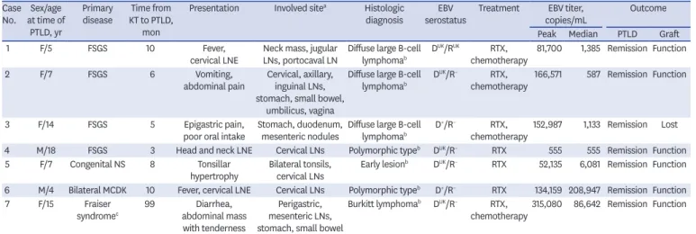
Patients with PTLD presented with fever, lymph node enlargement, or gastrointestinal symptoms such as abdominal pain, vomiting, and diarrhea (Table 1). Any gastrointestinal symptoms and/or lymph node enlargement raised suspicion for PTLD and prompted the clinician to perform further work-up to rule out PTLD. The majority of patients (n = 4) had gastrointestinal organ involvement, including small bowel and intraperitoneal lymph nodes.
There was no extranodal PTLD. Pathologic diagnosis of PTLD revealed one case of early lesion, two cases of polymorphic PTLD, one case of Burkitt lymphoma, and three cases of diffuse large B-cell lymphoma. Upon diagnosis of PTLD, immunosuppressive medications
All patients n = 199
Excluded
EBV ≤ 1,000 copies/mL n = 153 (76.9%)
Only EBV viremia n = 39 (84.8%) EBV related PTLD
n = 7 (15.2%)
EBV > 1,000 copies/mL n = 46 (23.1%)
EBV ≤ 10,000 copies/mL n = 25 (64.1%) EBV > 10,000 copies/mL
n = 14 (35.9%)
No RTX n = 8 (57.1%) Preemptive RTX
n = 6 (42.9%)
Fig. 1. Distribution of patients with kidney transplantation by EBV status and PTLD.EBV = Epstein-Barr virus, PTLD = post-transplant lymphoproliferative disease, RTX = rituximab.
were reduced, and RTX and/or chemotherapy were administered as appropriate. All patients achieved complete remission of PTLD after treatments. While one patient lost her allograft kidney due to complications of chemotherapy, six patients retained renal function after follow-up for 2.5–10.5 years.
Risk factors for PTLD
Table 2 shows the comparison of clinical variables between the PTLD and EV only group by univariate analysis. There were no significant differences between the two groups in terms of sex, age at transplantation, donor type, interval between transplantation, and first appearance of EV. Although the peak median EBV titer was higher in the PTLD group (152,987 EBV copies/mL whole blood) than the EV only group (17,305 copies/mL whole blood), there was no statistical significance. There were also no significant differences between groups in terms of median EBV viral load and EV-free duration after kidney transplant. At the time of transplantation, six patients (85.7%) in the PTLD group and 14 patients (35.9%) in the EV only group were seronegative for EBV (P = 0.009). Data of donor EBV status before transplantation were available only in a few cases, with no statistically significant difference observed between the groups.
Tacrolimus levels before EV tended to be higher in the PTLD group (9.5 ng/mL) than in the EV only group (7.7 ng/mL, P = 0.039). Maintenance immunosuppression regimen or history of rejection was not significantly different between the two groups. Six patients were treated with pre-emptive RTX, none of whom developed PTLD, while the number of RTX-treated patients was too small to be statistically significant.
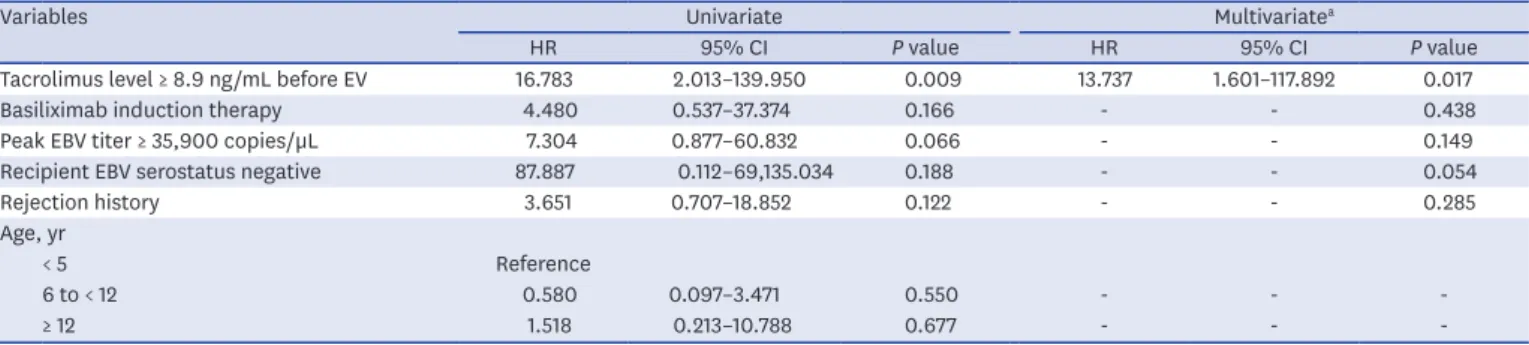
The Cox proportional-hazard model was used to identify factors associated with an increased risk of developing PTLD after EV (Table 3). Values of 8.9 ng/mL for tacrolimus level and 35,900 copies/μL for peak EBV titer were determined as cutoff values based on the receiver operating characteristic curve analysis. The areas under curve of tacrolimus and peak EBV titer were 0.745 (95% confidence interval [CI] 0.522–0.969, sensitivity 85.7%, and specificity 79.5%) and 0.634 (95% CI 0.399–0.869, sensitivity 85.7%, and specificity Table 1. Characteristics of patients with post-transplant lymphoproliferative disease
Case No. Sex/age
at time of PTLD, yr
Primary
disease Time from KT to PTLD,
mon
Presentation Involved sitea Histologic
diagnosis EBV
serostatus Treatment EBV titer,
copies/mL Outcome
Peak Median PTLD Graft
1 F/5 FSGS 10 Fever,
cervical LNE Neck mass, jugular
LNs, portocaval LN Diffuse large B-cell
lymphomab DUK/RUK RTX,
chemotherapy 81,700 1,385 Remission Function
2 F/7 FSGS 6 Vomiting,
abdominal pain Cervical, axillary, inguinal LNs, stomach, small bowel,
umbilicus, vagina
Diffuse large B-cell
lymphomab DUK/R− RTX,
chemotherapy 166,571 587 Remission Function
3 F/14 FSGS 5 Epigastric pain,
poor oral intake Stomach, duodenum,
mesenteric nodules Diffuse large B-cell
lymphomab D+/R− RTX,
chemotherapy152,987 1,133 Remission Lost 4 M/18 FSGS 3 Head and neck LNE Cervical LNs Polymorphic typeb DUK/R− RTX 555 555 Remission Function
5 F/7 Congenital NS 8 Tonsillar
hypertrophy Bilateral tonsils,
cervical LNs Early lesionb DUK/R− RTX 52,135 6,081 Remission Function 6 M/4 Bilateral MCDK 10 Fever, cervical LNE Cervical LNs Polymorphic typeb D+/R− RTX 134,159 208,947 Remission Function
7 F/15 Fraiser
syndromec 99 Diarrhea, abdominal mass with tenderness
Perigastric, mesenteric LNs, stomach, small bowel
Burkitt lymphomab DUK/R− RTX,
chemotherapy315,080 86,642 Remission Function PTLD = post-transplant lymphoproliferative disease, KT = kidney transplantation, EBV = Epstein-Barr virus, F = female, FSGS = focal segmental
glomerulosclerosis, LNE = lymph node enlargement, LN = lymph node, D/R = donor/recipient, UK = unknown, RTX = rituximab, M = male, NS = nephrotic syndrome, MCDK = multicystic dysplastic kidney.
aLesion detected by imaging study (computed tomography or positron emission tomography); bEBV in-situ hybridization positive; cGenetic disorder caused by WT1 mutation.
59.0%), respectively. A higher tacrolimus level (hazard ratio [HR], 13.7; 95% CI, 1.6–117.9;
P = 0.017) was associated with PTLD. Basiliximab induction therapy, higher EBV titer, EBV seronegativity of recipients, and rejection history were not significant in multivariate Cox regression analysis.
Table 2. Characteristics of patients
Characteristics PTLD (n = 7) EV only (n = 39) P value
Sex, M:F 2:5 18:21 0.446
Age at transplantation, yr 6 (3–18) 7 (1–16) 0.811
Donor type 1.000
Deceased 4 (57.1) 21 (53.8)
Living related 3 (42.9) 18 (46.2)
EBV recipient serostatus 0.009
Positive 0 23 (59.0)
Negative 6 (85.7) 14 (35.9)
Unknown 1 (14.3) 2 (5.1)
EBV donor serostatus 1.000
Positive 2 (28.6) 10 (25.6)
Negative 0 3 (7.7)
Unknown 5 (71.4) 26 (66.7)
Time to EV, mon 5 (1–48) 8 (0–47) 0.946
Peak EBV level, copies/mL 152,987 (555–1,341,159) 17,305 (1,198–1,279,841) 0.278 Median EBV level, copies/mL 6,081 (555–208,947) 4,250 (485–326,880) 0.834
CMV viremia 2 (28.6) 12 (30.8) 1.000
Induction therapy
Basiliximab 6 (85.7) 22 (56.4) 0.220
Thymoglobulin 0 2 (5) 1.000
Maintenance medication 0.496
Steroid + tacrolimus + MMF 6 (85.7) 36 (92.3)
Steroid + tacrolimus + AZA 1 (14.3) 1 (2.6)
Steroid + tacrolimus 0 2 (5.1)
Immunosuppressant after EV 0.423
Monotherapy 2 (28.6) 5 (12.8)
Double immunotherapy 3 (42.9) 26 (66.7)
Triple immunotherapy 2 (28.6) 8 (20.5)
Tacrolimus level, ng/mL
Pre EV diagnosis 9.5 (6.2–10.3) 7.7 (5.4–12.7) 0.039
At EV diagnosis 6.5 (2.5–9.6) 5.9 (2.2–14.3) 0.549
Post EV diagnosis 4.1 (0–5.8) 4.5 (2.4–7.2) 0.278
Rejection history 5 (71.4) 15 (38.5) 0.213
Preemptive RTX 0 6 (15.4) 0.266
Values are expressed as numbers (%) and median (range).
PTLD = post-transplant lymphoproliferative disease, EV = Epstein-Barr virus viremia, EBV = Epstein-Barr virus, CMV = cytomegalovirus, MMF = mycophenolate, AZA = azathioprine, RTX = rituximab.
Table 3. Risk factors for post-transplant lymphoproliferative disease in patients with EV
Variables Univariate Multivariatea
HR 95% CI P value HR 95% CI P value
Tacrolimus level ≥ 8.9 ng/mL before EV 16.783 2.013–139.950 0.009 13.737 1.601–117.892 0.017
Basiliximab induction therapy 4.480 0.537–37.374 0.166 - - 0.438
Peak EBV titer ≥ 35,900 copies/µL 7.304 0.877–60.832 0.066 - - 0.149
Recipient EBV serostatus negative 87.887 0.112–69,135.034 0.188 - - 0.054
Rejection history 3.651 0.707–18.852 0.122 - - 0.285
Age, yr
< 5 Reference
6 to < 12 0.580 0.097–3.471 0.550 - - -
≥ 12 1.518 0.213–10.788 0.677 - - -
HR = hazard ratio, CI = confidence interval, EV = Epstein-Barr virus viremia, EBV = Epstein-Barr virus.
aFactors with a value of P < 0.2 in the univariate Cox regression analysis were included in the multivariate analysis.
Efficacy and safety of pre-emptive rituximab treatment in pediatric kidney transplant
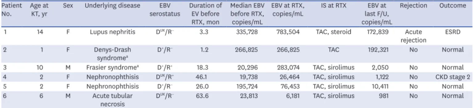
Six patients in the EV only group (male: female, 2:4) who had a high EBV load received preemptive RTX therapy. Their median age at transplant was 4 years (1–14 years), and EBV infection was first detected at 4.3 months (3.9–9.9 months) after kidney transplantation.
Administration of RTX was carried out at a median of 29.2 months (5.1–69.6 months) after transplantation. These six patients did not exhibit any symptoms such as fever, lymph node enlargement, or gastrointestinal problems; and no imaging studies were performed.
Two patients were EBV seropositive and four patients were EBV seronegative (Table 4). The two EBV-seropositive patients were at low risk for the development of PTLD, but RTX was administered to these patients based on the clinician's decision; one patient had a mutation of WT1, and was therefore prone to tumor development, while the other had persistently high EBV load for 46 months despite the reduction of immunosuppression. Median EBV viral loads at the time of RTX treatment were 171,639 copies/mL (6,181–783,504 copies/mL).
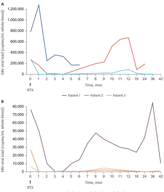
After a single dose of RTX therapy, a concordant decrease in EBV load and B lymphocytes was observed (Fig. 2). In five patients, EV disappeared within months; the other patient showed reduction of EBV titer but persistence of EV despite RTX treatment. Unfortunately, the patient lost the allograft due to rejection and concomitant infection within 8 months after RTX therapy, and EBV titer was not monitored after this adverse event. In the remaining five patients, EBV load rebounded along with recovery of B cells in a median 8 months. However, none of these five patients developed PTLD over a median follow-up of 51.5 months.
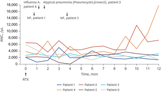
Regarding the safety of RTX therapy, only one patient complained of chest discomfort during RTX infusion. Two patients experienced neutropenia at 4 months and 1 month after RTX treatment (Fig. 3). Two patients were admitted for viral or bacterial infections at 1 month and 2 months after RTX treatment. In comparison, of 8 patients with high EV (EBV
> 10,000 copies/mL) but without RTX treatment, three patients experienced 6 infection episodes and one patient developed neutropenia during 12 months after the occurrence of high EV. Infectious complications included Citrobacter freundii, CMV, influenza A, adenovirus and Pneumocystis jirovecii infections. There were no significant differences in the infectious complications and neutropenia between the RTX group and the non-RTX group in patients with high EV.
Table 4. Description of six patients with preemptive rituximab therapy Patient
No. Age at
KT, yr Sex Underlying disease EBV
serostatus Duration of EV before RTX, mon
Median EBV before RTX, copies/mL
EBV at RTX,
copies/mL IS at RTX EBV at last F/U, copies/mL
Rejection Outcome
1 14 F Lupus nephritis DUK/R− 3.3 335,728 783,504 TAC, steroid 172,839 Acute
rejection ESRD
2 1 F Denys-Drash
syndromea D+/R− 1.2 266,825 266,825 TAC 192,321 No Normal
3 10 M Frasier syndromea D+/R+ 18.3 20,296 283,074 TAC, sirolimus 2,050 No Normal
4 2 F Nephronophthisis DUK/R+ 46.1 19,738 26,464 TAC, sirolimus 1,122 No CKD stage 2
5 2 F Nephronophthisis D+/R− 26.0 195,724 76,453 TAC, sirolimus 10,411 No Normal
6 6 M Acute tubular
necrosis DUK/R− 63.6 23,813 6,181 TAC, sirolimus 981 No Normal
KT = kidney transplantation, EBV = Epstein-Barr virus, EV = Epstein-Barr virus viremia, RTX = rituximab, IS = immunosuppressant, F/U = follow up, F = female, D/R = donor/recipient, UK = unknown, TAC = tacrolimus, ESRD = end stage renal disease, CKD = chronic kidney disease, M = male.
aGenetic disorder caused by WT1 mutation.
DISCUSSION
During the last 15 years, among the almost 200 pediatric kidney transplantation recipients at our center, seven developed EBV-associated PTLD (3.5%). In our study, the risk factor for PTLD in pediatric kidney transplant recipients with EV was a high tacrolimus level before EV. Twenty (43.4%) out of 46 recipients who had viremia were EBV seronegative, which is similar to the rates reported in previous studies in North America and Europe (19%–57%).21-23 Pre-transplant recipient EBV seronegativity is a well-known risk factor for PTLD. In adult transplant studies, the rate of developing PTLD is 5–12 fold higher in EBV-seronegative patients than in EBV- seropositive patients.2,3,24 McDonald et al.6 reported that EBV-seronegative pediatric subjects have a 4.7-fold higher relative HR than EBV-positive subjects. In our study, recipient EBV seronegativity did not increase the risk of PTLD in multivariate Cox regression analysis.
While transplant from an EBV-seropositive donor to a seronegative recipient has been 0
200,000 400,000 600,000 800,000 1,000,000 1,200,000
0 1 2 3 4 5 6 7 8 9 10 11 12 18 24 36 42
Patient 1 Patient 2 Patient 3
EBV viral load (copies/mL whole blood)
Time, mon RTX
A
0 10,000 20,000 30,000 40,000 60,000 80,000
0 1 2 3 4 5 6 7 8 9 10 11 12 18 24 36
Patient 4 Patient 5 Patient 6
EBV viral load (copies/mL whole blood)
Time, mon RTX
B
42 50,000
70,000
Fig. 2. EBV viral load after RTX therapy. (A) EBV viral loads in Patients 1, 2, and 3. (B) EBV viral loads in Patients 4, 5, and 6.
EBV = Epstein-Barr virus, RTX = rituximab.
associated with the development of PTLD,25 there was no statistically significant difference in EBV serostatus (donor/recipient) in the present study. This finding was attributed mainly to the fact that there were no EBV seropositive recipients in PTLD group and we did not have serostatus information for the majority of donors (67.3%).
The majority of kidney allograft recipients in this study were given tacrolimus, and mycophenolate was used in more than 80% of patients instead of azathioprine.26 By the late 2000s, monoclonal interleukin-2 receptor antibodies were used as induction therapy in up to 80% of patients. Basiliximab, a monoclonal antibody which targets activated T lymphocytes, was not related to PTLD risk in our study, as previously reported.4,27 Several studies have suggested that higher tacrolimus levels are associated with higher risk for PTLD, and others have reported that the net state of immunosuppression, rather than any individual agent, increases the risk for PTLD.28-30 In our study, all patients received tacrolimus for maintenance immunosuppression, and we found that a higher pre-EV tacrolimus level in the PTLD group compared with the EV only group was a risk factor for PTLD.
Regular monitoring of EBV viral load and early recognition of recipients at high risk of PTLD have been identified as clinical priorities in recent years.31 Previous studies have shown that elevated levels of EBV DNA and persistent high EBV loads are risk factors for PTLD,12,20,32 but no clear cut-off point of EBV viral load for the prediction of PTLD development has been determined. We did not find a significant relationship between EV and PTLD in this study;
however, six patients with a high EBV titer were treated with preemptive RTX and they did not develop PTLD. Because they were included in analysis, this could have confounded the causality of high EBV titer and PTLD development.
Treatment strategies for organ transplant recipients with EV include reduction of immunosuppression with/without antiviral agents, immunoglobulin, or RTX.33 These
treatments are still undergoing clinical studies. Preemptive administration of RTX is widely used and has been demonstrated to reduce the incidence of PTLD in stem cell transplant recipients with a high EBV viral load.18,34,35 Pre-emptive RTX therapy has been reported in 14.5% of global
Patient 3 Patient 2
Patient 1
Patient 4 Patient 5 Patient 6 Time, mon
RTX 0 2,000 4,000 6,000 8,000 10,000 12,000 14,000 16,000 18,000
0 1 2 3 4 5 6 7 8 9 10 11 12
ANC, /µL
NF, patient 1 NF, patient 5 Influenza A,
patient 6 Atypical pneumonia (Pneumocystis jirovecii), patient 2
Fig. 3. Late adverse events after rituximab treatment.
ANC = absolute neutrophil count, NF = neutropenic fever, RTX = rituximab.
transplant programs, and in more than 60% of pediatric transplant patients worldwide.33 However, only one study reported the use of RTX in five pediatric renal allograft recipients,20 and there have been no prospective studies on the efficacy of pre-emptive RTX therapy in solid organ transplantation. Rituximab is a murine/human chimeric monoclonal anti-CD20 antibody and is able to deplete circulating B cells rapidly, including those infected with EBV. Although RTX was effective for reducing EBV viral load in our patients, this reduction was not permanent in line with previous reports.20 In addition, significant adverse effects, such as infection and neutropenia, accompanied RTX administration. In patients with stem cell transplant (SCT), preemptive RTX was not associated with an increase in infectious complications.18,19 This is important because, while SCT recipients are able to discontinue their immunosuppressive agents within 6–9 months after SCT, recipients of solid organ transplantation have to be on immunosuppressive agents for as long as their allografts are functioning. Therefore, the long- term use of immunosuppressive agents may explain why infection and neutropenia are common clinical findings after RTX therapy in this patient population.
Although the small sample size of the current study precludes us from drawing any definite conclusions, our observations suggest that preemptive RTX treatment may effectively reduce high EBV viral load in pediatric recipients of solid organ transplants. Because RTX therapy eradicates B-lymphocytes including transformed lymphocytes, the risk of PTLD might be reduced at least during the period of B cell depletion. However, one should take into account that this treatment significantly increases the risk of neutropenia and infection. More research into the influence of preemptive RTX therapy on PTLD development is needed.
The occurrence of PTLD in kidney transplant recipients follows a bimodal distribution, with one peak in the first year and the second in the later post-transplantation period. Early PTLD, occurring within the first year of transplantation, is associated with EBV infection and tends to occur more commonly in children than adults.12,36,37 In this study, while six patients in the PTLD group were diagnosed with PTLD within the first year of renal transplantation, one patient with WT1 mutation developed PTLD later than 8.2 years after renal transplantation.
We suspect that in this patient, the WT1 mutation of a tumor suppressor gene might have increased the risk of PTLD, and especially that of late-onset. The occurrence of PTLD in patients with WT1 mutation has been reported previously, within the first year in two cases and later than the first year in another.38-40 Based on the limited availability of clinical data, the association between WT1 mutation and development of PTLD requires further investigation in future clinical studies and is beyond the scope of the current study.
There are several limitations of this study. First, this is a retrospective observational study of a single center and the number of the patients observed was therefore small. The limitations of this small sample size might have affected the outcome of multivariate analysis. In addition, data of donor EBV serology were not uniformly available in our study. Data on donor serology status was only available for 33% of participants. Therefore, our study did not show any association between PTLD and EBV-donor/recipient serostatus. Finally, the number of patients who were treated with RTX was not large enough to draw any definitive conclusions.
In summary, this study demonstrates that a higher tacrolimus level before EV is correlated with the development of PTLD. Preemptive RTX appears to be effective for reducing EBV viral load in pediatric kidney transplant recipients. However, the reduction of EBV viral load was not persistent, and adverse effects of RTX, namely infection and neutropenia, were clinically significant.
REFERENCES
1. Mucha K, Foroncewicz B, Ziarkiewicz-Wróblewska B, Krawczyk M, Lerut J, Paczek L. Post-transplant lymphoproliferative disorder in view of the new WHO classification: a more rational approach to a protean disease? Nephrol Dial Transplant 2010;25(7):2089-98.
PUBMED | CROSSREF
2. Caillard S, Lamy FX, Quelen C, Dantal J, Lebranchu Y, Lang P, et al. Epidemiology of posttransplant lymphoproliferative disorders in adult kidney and kidney pancreas recipients: report of the French registry and analysis of subgroups of lymphomas. Am J Transplant 2012;12(3):682-93.
PUBMED | CROSSREF
3. Sampaio MS, Cho YW, Qazi Y, Bunnapradist S, Hutchinson IV, Shah T. Posttransplant malignancies in solid organ adult recipients: an analysis of the U.S. National Transplant Database. Transplantation 2012;94(10):990-8.
PUBMED | CROSSREF
4. Caillard S, Dharnidharka V, Agodoa L, Bohen E, Abbott K. Posttransplant lymphoproliferative disorders after renal transplantation in the United States in era of modern immunosuppression. Transplantation 2005;80(9):1233-43.
PUBMED | CROSSREF
5. Jeong HJ, Ahn YH, Park E, Choi Y, Yi NJ, Ko JS, et al. Posttransplantation lymphoproliferative disorder after pediatric solid organ transplantation: experiences of 20 years in a single center. Korean J Pediatr 2017;60(3):86-93.
PUBMED | CROSSREF
6. McDonald RA, Smith JM, Ho M, Lindblad R, Ikle D, Grimm P, et al. Incidence of PTLD in pediatric renal transplant recipients receiving basiliximab, calcineurin inhibitor, sirolimus and steroids. Am J Transplant 2008;8(5):984-9.
PUBMED | CROSSREF
7. Allen UD, Preiksaitis JK; AST Infectious Diseases Community of Practice. Epstein-Barr virus and posttransplant lymphoproliferative disorder in solid organ transplantation. Am J Transplant 2013;13 Suppl 4:107-20.
PUBMED | CROSSREF
8. Al-Mansour Z, Nelson BP, Evens AM. Post-transplant lymphoproliferative disease (PTLD): risk factors, diagnosis, and current treatment strategies. Curr Hematol Malig Rep 2013;8(3):173-83.
PUBMED | CROSSREF
9. Bingler MA, Feingold B, Miller SA, Quivers E, Michaels MG, Green M, et al. Chronic high Epstein-Barr viral load state and risk for late-onset posttransplant lymphoproliferative disease/lymphoma in children.
Am J Transplant 2008;8(2):442-5.
PUBMED | CROSSREF
10. Knight JS, Tsodikov A, Cibrik DM, Ross CW, Kaminski MS, Blayney DW. Lymphoma after solid organ transplantation: risk, response to therapy, and survival at a transplantation center. J Clin Oncol 2009;27(20):3354-62.
PUBMED | CROSSREF
11. Kim J, Lee J, Kim YH. Clinical utility of Epstein-Barr viral load assay to diagnose posttransplant
lymphoproliferative disorders in pediatric heart transplant recipients. Pediatr Infect Vaccine 2017;24(1):44-53.
CROSSREF
12. Le J, Durand CM, Agha I, Brennan DC. Epstein-Barr virus and renal transplantation. Transplant Rev (Orlando) 2017;31(1):55-60.
PUBMED | CROSSREF
13. Pereira MS, Blake JM, Macrae AD. EB virus antibody at different ages. BMJ 1969;4(5682):526-7.
PUBMED | CROSSREF
14. Porter DD, Wimberly I, Benyesh-Melnick M. Prevalence of antibodies to EB virus and other herpesviruses.
JAMA 1969;208(9):1675-9.
PUBMED | CROSSREF
15. Green M, Michaels MG. Epstein-Barr virus infection and posttransplant lymphoproliferative disorder. Am J Transplant 2013;13 Suppl 3:41-54.
PUBMED | CROSSREF
16. Nijland ML, Kersten MJ, Pals ST, Bemelman FJ, Ten Berge IJ. Epstein-Barr virus-positive posttransplant lymphoproliferative disease after solid organ transplantation: Pathogenesis, clinical manifestations, diagnosis, and management. Transplant Direct 2015;2(1):e48.
PUBMED | CROSSREF
17. Kidney Disease: Improving Global Outcomes (KDIGO) Transplant Work Group. KDIGO clinical practice guideline for the care of kidney transplant recipients. Am J Transplant 2009;9 Suppl 3:S1-157.
CROSSREF
18. Worth A, Conyers R, Cohen J, Jagani M, Chiesa R, Rao K, et al. Pre-emptive rituximab based on viraemia and T cell reconstitution: a highly effective strategy for the prevention of Epstein-Barr virus-associated lymphoproliferative disease following stem cell transplantation. Br J Haematol 2011;155(3):377-85.
PUBMED | CROSSREF
19. van der Velden WJ, Mori T, Stevens WB, de Haan AF, Stelma FF, Blijlevens NM, et al. Reduced PTLD- related mortality in patients experiencing EBV infection following allo-SCT after the introduction of a protocol incorporating pre-emptive rituximab. Bone Marrow Transplant 2013;48(11):1465-71.
PUBMED | CROSSREF
20. Colombini E, Guzzo I, Morolli F, Longo G, Russo C, Lombardi A, et al. Viral load of EBV DNAemia is a predictor of EBV-related post-transplant lymphoproliferative disorders in pediatric renal transplant recipients. Pediatr Nephrol 2017;32(8):1433-42.
PUBMED | CROSSREF
21. Walker RC, Marshall WF, Strickler JG, Wiesner RH, Velosa JA, Habermann TM, et al. Pretransplantation assessment of the risk of lymphoproliferative disorder. Clin Infect Dis 1995;20(5):1346-53.
PUBMED | CROSSREF
22. Ellis D, Jaffe R, Green M, Janosky JJ, Lombardozzi-Lane S, Shapiro R, et al. Epstein-Barr virus-related disorders in children undergoing renal transplantation with tacrolimus-based immunosuppression.
Transplantation 1999;68(7):997-1003.
PUBMED | CROSSREF
23. Shroff R, Trompeter R, Cubitt D, Thaker U, Rees L. Epstein-Barr virus monitoring in paediatric renal transplant recipients. Pediatr Nephrol 2002;17(9):770-5.
PUBMED | CROSSREF
24. Dharnidharka VR, Lamb KE, Gregg JA, Meier-Kriesche HU. Associations between EBV serostatus and organ transplant type in PTLD risk: an analysis of the SRTR National Registry Data in the United States.
Am J Transplant 2012;12(4):976-83.
PUBMED | CROSSREF
25. Suzuki T, Ikezumi Y, Okubo S, Uchiyama M, Takahashi K, Shiraga H, et al. Epstein-Barr virus DNA load and seroconversion in pediatric renal transplantation with tacrolimus immunosuppression. Pediatr Transplant 2007;11(7):749-54.
PUBMED | CROSSREF
26. Min SI, Han A, Choi C, Kim SY, Kang HG, Ha IS, et al. Immunosuppression in pediatric kidney transplant patients. J Korean Soc Transplant 2015;29(1):1-8.
CROSSREF
27. Bustami RT, Ojo AO, Wolfe RA, Merion RM, Bennett WM, McDiarmid SV, et al. Immunosuppression and the risk of post-transplant malignancy among cadaveric first kidney transplant recipients. Am J Transplant 2004;4(1):87-93.
PUBMED | CROSSREF
28. Shapiro R, Nalesnik M, McCauley J, Fedorek S, Jordan ML, Scantlebury VP, et al. Posttransplant lymphoproliferative disorders in adult and pediatric renal transplant patients receiving tacrolimus-based immunosuppression. Transplantation 1999;68(12):1851-4.
PUBMED | CROSSREF
29. Opelz G, Döhler B. Lymphomas after solid organ transplantation: a collaborative transplant study report.
Am J Transplant 2004;4(2):222-30.
PUBMED | CROSSREF
30. Höcker B, Fickenscher H, Delecluse HJ, Böhm S, Küsters U, Schnitzler P, et al. Epidemiology and morbidity of Epstein-Barr virus infection in pediatric renal transplant recipients: a multicenter, prospective study. Clin Infect Dis 2013;56(1):84-92.
PUBMED | CROSSREF
31. Dharnidharka VR. Peripheral blood Epstein-Barr viral nucleic acid surveillance as a marker for posttransplant cancer risk. Am J Transplant 2017;17(3):611-6.
PUBMED | CROSSREF
32. Riddler SA, Breinig MC, McKnight JL. Increased levels of circulating Epstein-Barr virus (EBV)-infected lymphocytes and decreased EBV nuclear antigen antibody responses are associated with the development of posttransplant lymphoproliferative disease in solid-organ transplant recipients. Blood 1994;84(3):972-84.
PUBMED
33. San-Juan R, Manuel O, Hirsch HH, Fernández-Ruiz M, López-Medrano F, Comoli P, et al. Current preventive strategies and management of Epstein-Barr virus-related post-transplant lymphoproliferative
disease in solid organ transplantation in Europe. Results of the ESGICH Questionnaire-based Cross- sectional Survey. Clin Microbiol Infect 2015;21(6):604.e1-604.e9.
PUBMED | CROSSREF
34. van Esser JW, Niesters HG, van der Holt B, Meijer E, Osterhaus AD, Gratama JW, et al. Prevention of Epstein-Barr virus-lymphoproliferative disease by molecular monitoring and preemptive rituximab in high-risk patients after allogeneic stem cell transplantation. Blood 2002;99(12):4364-9.
PUBMED | CROSSREF
35. Omar H, Hägglund H, Gustafsson-Jernberg A, LeBlanc K, Mattsson J, Remberger M, et al. Targeted monitoring of patients at high risk of post-transplant lymphoproliferative disease by quantitative Epstein- Barr virus polymerase chain reaction. Transpl Infect Dis 2009;11(5):393-9.
PUBMED | CROSSREF
36. Francis A, Johnson DW, Teixeira-Pinto A, Craig JC, Wong G. Incidence and predictors of post-transplant lymphoproliferative disease after kidney transplantation during adulthood and childhood: a registry study. Nephrol Dial Transplant 2018;33(5):881-9.
PUBMED | CROSSREF
37. Mynarek M, Hussein K, Kreipe HH, Maecker-Kolhoff B. Malignancies after pediatric kidney transplantation: more than PTLD? Pediatr Nephrol 2014;29(9):1517-28.
PUBMED | CROSSREF
38. Miloševic B, Bogdanović R, Kostić M, Stojanović V. Frasier syndrome diagnosed in a 4-year-old girl. Cent Eur J Med 2012;7(2):142-4.
39. Spasojević-Dimitrijeva B, Peco-Antić A, Paripović D, Kruscić D, Krstić Z, Cupić M, et al. Post-transplant lymphoproliferative disorder--case reports of three children with kidney transplant. Srp Arh Celok Lek 2014;142(1-2):83-8.
PUBMED | CROSSREF
40. Miyazono A, Okamoto Y, Nagasako H, Hamasaki Y, Shishido S, Yoshioka T, et al. Multifocal Epstein- Barr virus-negative posttransplantation lymphoproliferative disorder treated with reduction of immunosuppression. Am J Kidney Dis 2016;68(3):469-72.
PUBMED | CROSSREF