Renal Tubular Acidosis in Patients with Primary Sjögren’s Syndrome
Su Woong Jung1, Eun Ji Park2, Jin Sug Kim1, Tae Won Lee2, Chun Gyoo Ihm2, Sang Ho Lee3, Ju-Young Moon3, Yang Gyun Kim3, Kyung Hwan Jeong2
1Department of Medicine, Graduate School, Kyung Hee University, Seoul, 2Division of Nephrology, Department of Internal Medicine, Kyung Hee University Medical Center, Seoul, 3Divison of Nephrology, Department of Internal Medicine, Kyung Hee University Hospital at Gangdong, Seoul, Korea
Received: August 24, 2017 Accepted: September 1, 2017 Corresponding Author:
Kyung Hwan Jeong, M.D., Ph.D.,
Division of Nephrology, Department of Internal Medicine, College of Medicine, Kyung Hee University, 23 Kyungheedae-ro, Dongdaemun-gu, Seoul 02447, Korea
Tel: +82-2-958-8200, Fax: +82-2-968-1848 E-mail: khjeong@khu.ac.kr
Primary Sjögren’s syndrome (pSS) is characterized by lymphocytic infiltration of the exocrine glands resulting in decreased saliva and tear production. It un- commonly involves the kidneys in various forms, including tubulointerstitial ne- phritis, renal tubular acidosis, Fanconi syndrome, and rarely glomerulonephritis.
Its clinical symptoms include muscle weakness, periodic paralysis, and bone pain due to metabolic acidosis and electrolyte imbalance. Herein, we describe the cases of two women with pSS whose presenting symptoms involve the kidneys. They had hypokalemia and normal anion gap metabolic acidosis due to distal renal tubular acidosis and positive anti-SS-A and anti-SS-B autoanti- bodies. Since one of them experienced femoral fracture due to osteomalacia secondary to renal tubular acidosis, an earlier diagnosis of pSS is important in preventing serious complications.
Key Words: Sjögren’s syndrome, Osteomalacia, Renal tubular acidosis, Hypokalemia
This is an Open Access article distributed under the terms of the Creative Commons Attribution Non-Commercial License(http://creativecommons.org/licenses/by-nc/4.0/) which permits unrestricted non-commercial use, distribution, and reproduction in any medium, provided the original work is properly cited.
Introduction
Primary Sjögren’s syndrome (pSS) is a chronic, slowly progressive autoimmune disease characterized by xero- stomia and dry eyes1). It predominantly affects women and occurs in all ages. About one-third of patients with Sjögren’s syndrome show extraglandular involvements and are manifested with Raynaud’s phenomenon, vasculi- tis, lymphoma, and various organ involvements, includ- ing the kidneys1). The renal involvement in pSS includes tubulointerstitial nephritis (TIN) causing tubular dysfunc- tion and less commonly glomerulonephritis. Herein, we present the cases of two patients with Sjögren’s syndrome whose initial manifestations were fracture and muscle weak- ness, respectively.
Case Reports
Case 1A 60-year-old woman presented to the emergency de- partment with left hip pain after falling down. The simple X-ray revealed transverse subtrochanteric fracture of the left femur with displacement of the distal part. The pa- tient had been on a monthly risedronate for osteoporosis.
Otherwise, she had been well until this event. She was admitted and underwent open reduction and internal fix- ation on the following day. The computed tomography demonstrated another incomplete subtrochanteric fracture line on the medial side of the right femur. In addition, low bone mineral density (T-score, -2.7) and multiple rib
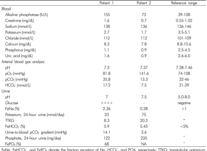
Table 1. Laboratory findings of our patients
Patient 1 Patient 2 Reference range
Blood
Alkaline phosphatase (IU/L) 155 73 39-108
Creatinine (mg/dL) 1.6 0.7 0.55-1.02
Sodium (mmol/L) 138 136 136-146
Potassium (mmol/L) 2.7 1.7 3.5-5.1
Chloride (mmol/L) 112 112 101-109
Calcium (mg/dL) 8.3 7.8 8.8-10.6
Phosphorus (mg/dL) 1.1 0.9 2.5-4.5
Uric acid (mg/dL) 1.6 0.9 2.6-6.0
Arterial blood gas analysis
pH 7.3 7.37 7.38-7.46
pO2(mmHg) 81.8 141.6 74-108
pCO2(mmHg) 35.8 13.3 32-46
HCO3-(mmol/L) 17.2 7.5 21-29
Urine
pH 7 7.5 5.0-8.0
Glucose ++++ - negative
FeNa (%) 2.36 0.28 <1
Potassium, 24-hour urine (mmol/day) 33 75
TTKG 8.3 20.3 *
FeHCO3-(%) 5.9 5.45 <5%
Urine-to-blood pCO2 gradient (mmHg) 14.1 3.6
Phosphate, 24-hour urine (mg/day) 122 235 ‡
FePO4(%) 68 NA ‡
FeNa, FeHCO3-, and FePO4 denote the fraction excretion of Na, HCO3-, and PO4, respectively; TTKG: transtubular potassium gradient; N/A: not available.
*In the setting of hypokalemia, the 24-hour urine potassium excretion level and TTKG should be less than 15-20 mmol/day and 3-4, respectively.
†If the plasma HCO3 level is higher than 23-25 mEq/L, the urine-to-blood pCO2 gradient should be higher than 20 mmHg.
‡In the setting of hypophosphatemia, the 24-hour urine phosphate excretion level and FePO4 level should be <100 mg/day and
<5%, respectively.
fractures were noted on dual-energy X-ray absorptiom- etry and bone scan, respectively.
Oddly, the urine dipstick test revealed a strong posi- tivity for glucose at the serum glucose level of 126 mg/dL.
The blood chemistry and arterial blood gas analysis sho- wed hypokalemia, hyperchloremic metabolic acidosis, hy- pophosphatemia, hypouricemia, and elevated alkaline pho- sphatase (ALP) level with a modestly decreased kidney function (Table 1). The urine anion gap, fractional excre- tion or daily excretion of several electrolytes, and transtu- bular potassium gradient are shown in Table 1: positive urine anion gap in the setting of normal anion gap meta-
bolic acidosis, normoglycemic glucosuria, enhanced pota- ssium secretion leading to hypokalemia, and renal wasting of phosphate. These findings were consistent with Fan- coni syndrome with renal tubular acidosis (RTA). More- over, her serum sodium level increased up to 151 mmol/L on the sixth day of admission, although 0.9% normal saline or hypotonic fluid was administered. The urine os- molality was inappropriately low (190 mOsm/kg), despite adequate plasma antidiuretic hormone secretion to incre- ased serum osmolality (304 mOsm/kg); such a finding re- flected a urine concentration defect. Potassium citrate powder was administered daily with intravenous potas-
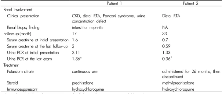
Table 2. Treatment and its outcomes in our patients
Patient 1 Patient 2
Renal involvement
Clinical presentation CKD, distal RTA, Fanconi syndrome, urine
concentration defect Distal RTA
Renal biopsy finding interstitial nephritis NA
Follow-up (month) 17 33
Serum creatinine at initial presentation 1.6 0.7
Serum creatinine at the last follow-up 2 0.59
Urine PCR at initial presentation 2.11 1.33
Urine PCR at the last exam 1.36* 0.36†
Treatment
Potassium citrate continuous use administered for 26 months, then
discontinued
Steroid prednisolone methylprednisolone
Immunosuppressant hydroxychloroquine hydroxychloroquine
CKD: chronic kidney disease; RTA: renal tubular acidosis; NA: not available; PCR: protein-to-creatinine ratio.
*The last exam was performed 13 months after the initial presentation.
†The last exam was performed 3 months after the initial presentation.
Fig. 1. Mild interstitial in- flammation with tubular injury (PAS stain, original magnification ×200) sium phosphate replacement.
A renal biopsy was performed to determine the cause of chronic kidney disease and associated renal tubule dys- function. The light microscopy showed a mild degree of interstitial nephritis and focal acute tubular injury with normal glomerulus (Fig. 1). However, the immunofluores- cence and electron microscopy did not reveal any abnor- mal finding.
Further investigations revealed a speckled pattern of antinuclear antibodies (ANA) (1:160) with positivity for both anti-SS-A and anti-SS-B antibodies. She had a history of gritty sensation in the eyes and decreased tear flow on the Schirmer’s test. Based on the clinical history and laboratory findings, interstitial nephritis associated with
Fanconi syndrome, RTA, and nephrogenic diabetes insi- pidus secondary to Sjögren’s syndrome were diagnosed.
It was likely that RTA and hypophosphatemia were res- ponsible for the multiple fractures, suggesting osteomala- cia overlapping with osteoporosis.
When the total carbon dioxide (CO2) level increased to 24.7 mmol/L via the administration of potassium citrate, the fractional excretion of bicarbonate and urine-to-blood CO2 tension gradient (U-B pCO2) were calculated as <15%
and <20 mmHg, respectively, indicating distal RTA. Fur- thermore, her urine pH level persistently ranged from 6.5 to 8.0, which supported this type of RTA (Table 1).
Thereafter, she has been on prednisolone and hydroxy- chloroquine with potassium citrate powder and maintai- ned normal serum potassium, phosphate, and total CO2
level. The ALP level also decreased to the normal range.
However, the serum creatinine level increased to 2.0 mg/dL after 17 months (Table 2).
Case 2
A 19-year-old girl was admitted with fever and weak- ness of both lower extremities. She was diagnosed with acute bronchitis and treated with intravenous levofloxa-
cin. On admission, very low levels of serum potassium and phosphate were noted and thought to cause weakness on both lower extremities and rhabdomyolysis. In addi- tion, hyperchloremic metabolic acidosis was accompa- nied. An excess urinary potassium loss and increased trans- tubular potassium gradient indicated excessive renal pota- ssium excretion (Table 1). Moreover, an inappropriately high phosphate excretion in the presence of hypophos- phatemia indicated renal phosphate wasting. Since hypo- kalemia of a renal origin and positive urine anion gap in- dicated the presence of RTA, the bicarbonate loading test was conducted to identify its specific type. Its results iden- tified the diagnosis of distal RTA based on the decreased U-B pCO2 without excessive bicarbonate wasting (Table 1).
Thereafter, sodium bicarbonate and potassium citrate were administered orally. Her symptoms resolved as the serum potassium and phosphate levels increased to the normal range.
Further evaluations for the etiology of RTA identified positive ANA (1:640, speckled nucleolar pattern) with po- sitive anti-Ro and anti-La antibodies. However, she re- ported no dry eyes or mouth. In addition, the salivary scan and Schirmer’s test did not show decreased salivary and tear flow. It was concluded that her motor weakness was attributed to distal RTA secondary to Sjögren’s synd- rome. Then, she has been treated with immunosuppre- ssive agents of methylprednisolone and hydroxychloro- quine and does not show any electrolyte imbalance and acidosis even after the discontinuation of alkali and potas- sium replacement (Table 2).
Discussion
pSS can be manifested in different ways beyond dry mouth and eyes, sometimes even before sicca symptoms are developed. The renal involvement in pSS is a well- known extraglandular manifestation with a prevalence of 5-14% in most European studies2,3). In most cases, it affe- cts the renal tubules through TIN and occasionally auto- antibodies against a certain transporter. All segments of the nephron can be involved in pSS leading to distal and
proximal RTA, Fanconi syndrome, diabetes insipidus, and less commonly Gitelman’s syndrome and Bartter’s syndrome4). Of these, distal RTA is the most frequent tubular dysfunction in pSS. The hypokalemia and meta- bolic acidosis in distal RTA cause muscle weakness, peri- odic paralysis5), and osteomalacia. In case of Fanconi syn- drome, hypophosphatemia due to defective renal phos- phate reabsorption is an additional contributing factor for osteomalacia.
Most RTAs in pSS are associated with TIN, which re- sembles lymphocyte infiltration in the exocrine glands.
In these cases, the decreased expression of both H+- ATPase and anion exchanger 1 (AE1) in α-intercalated cells has been well observed without the presence of auto- antibodies against those transporters6-8). However, under- lying cellular events linking TIN and the absence of those two transport proteins have yet to be discovered.
There are previous reports of osteomalacia secondary to pSS leading to bone fractures9). The use of bisphospho- nate in patient 1 could be responsible for the atypical left femoral fracture. However, according to the 2013 revised definition of atypical femur fractures10), incom- plete fractures originating from the medial side of the sub- trochanteric region were not consistent with atypical fem- oral fractures in which the fracture line starts from the lateral cortex. The looser zone on the right femur and multiple rib fractures with biochemical abnormalities of hypophosphatemia and elevated ALP levels suggest the presence of osteomalacia. A previous investigation repor- ted that osteomalacia in pSS is a consequence of chronic metabolic acidosis and hypophosphatemia rather than pSS itself11). In a recent review of literature regarding previou- sly reported cases of osteomalacia in pSS, the most common symptoms were bone pain and muscle weakness (85.3%) followed by fracture and pseudofractures (44.1%), and the most frequent laboratory findings were elevated ALP levels (79.4%) followed by reduced calcium and phosphate levels (70.6%)12).
Patient 1 appeared to have a long duration of disease, which was enough to cause softening of the bones. In cont- rast, patient 2 was diagnosed presumably at the early course
of Sjögren’s syndrome, whose manifestation was only muscle weakness. Although our two cases did not meet the pSS classification criteria proposed by the American-Euro- pean Consensus Group13), pSS was a reasonable clinical diagnosis in the context of positive autoantibodies and its above-mentioned characteristic renal involvement.
Moreover, the pSS classification criteria were not devel- oped for use in clinical practice.
A course of glucocorticoid is the most widely used treat- ment option for patients with TIN secondary to pSS. It may prevent the development of interstitial fibrosis and tubular atrophy and resolve tubular dysfunction as well as the sicca symptoms as in patient 2. Maripuri et al. re- ported that prednisolone alone or with immunosuppre- ssants, such as hydroxychloroquine, cyclophosphamide, or rituximab, improved the renal function of patients with pSS who had TIN on biopsy14). Although our pa- tients were treated with the combination of corticosteroid and hydroxychloroquine, the efficacy of immunosuppre- ssants as steroid-sparing strategies remains unknown in this setting, since clinical trials are difficult to conduct owing to the paucity of cases.
The involvement of multiple segments of the tubules in pSS as in patient 1 is an unusual phenomenon. She had a decreased renal function and lymphocyte interstitial infiltration reflecting moderate renal disease activity ac- cording to the EULAR Sjögren’s syndrome Disease Acti- vity Index3). Unfortunately, the disease had progressed until the development of fractures. If she had been diag- nosed earlier, such an event would have been prevented and renal function preserved with appropriate treatment.
In patient 2, the immunosuppressive agent induced clin- ical remission, which was enough to discontinue alkali and potassium replacement with marked reduction of proteinuria (Table 2). It is a challenge for physicians to make a diagnosis when encountering a patient with pSS without sicca symptoms. Since a prompt diagnosis may prevent serious complications and enable achievement of remission, it is of importance that Sjögren’s syndrome should be considered in an unknown origin of hypokale- mia and hyperchloremic metabolic acidosis.
Conflict of Interest: The authors declare no relevant financial interests.
References
1. Longo DL, Fauci AS, Kasper DL, Hauser SL, Jameson JL, Loscalzo J: Sjögren’s Syndrome. In: Harrison’s princi- ples of internal medicine. 18th ed., New York, McGraw- Hill, 2012, p2770-2773
2. Goules AV, Tatouli IP, Moutsopoulos HM, Tzioufas AG:
Clinically significant renal involvement in primary Sjogren’s syndrome: clinical presentation and outcome. Arthritis Rheum 65:2945-2953, 2013
3. Seror R, Ravaud P, Bowman SJ, et al.: EULAR Sjogren’s syndrome disease activity index: development of a con- sensus systemic disease activity index for primary Sjogren’s syndrome. Ann Rheum Dis 69:1103-1109, 2010 4. Francois H, Mariette X: Renal involvement in primary
Sjogren syndrome. Nat Rev Nephrol 12:82-93, 2016 5. Yilmaz H, Kaya M, Ozbek M, K UU, Safa Yildirim I:
Hypokalemic periodic paralysis in Sjogren’s syndrome se- condary to distal renal tubular acidosis. Rheumatol Int 33:1879-1882, 2013
6. Han JS, Kim GH, Kim J, et al.: Secretory-defect distal renal tubular acidosis is associated with transporter de- fect in H(+)-ATPase and anion exchanger-1. J Am Soc Nephrol 13:1425-1432, 2002
7. Walsh S, Turner CM, Toye A, et al.: Immunohistochemi- cal comparison of a case of inherited distal renal tubular acidosis (with a unique AE1 mutation) with an acquired case secondary to autoimmune disease. Nephrol Dial Transplant 22:807-812, 2007
8. Kim HY, Kim SS, Bae EH, Ma SK, Kim SW: Decreased Renal Expression of H(+)-ATPase and Pendrin in a Pa- tient with Distal Renal Tubular Acidosis Associated with Sjogren’s Syndrome. Intern Med 54:2899-2904, 2015 9. Khandelwal D, Bhattacharya S, Gadodia A, Khadgawat
R, Tandon N, Ammini AC: Metabolic bone disease as a presenting manifestation of primary Sjogren’s syndrome:
Three cases and review of literature. Indian J Endocrinol Metab 15:341-345, 2011
10. Shane E, Burr D, Abrahamsen B, et al.: Atypical subtro- chanteric and diaphyseal femoral fractures: second report of a task force of the American Society for Bone and Mineral Research. J Bone Miner Res 29:1-23, 2014 11. Fulop M, Mackay M: Renal tubular acidosis, Sjogren syn-
drome, and bone disease. Arch Intern Med 164:905-909, 2004
12. Geng Y, Zhao Y, Zhang Z: Tubulointerstitial nephritis- induced hypophosphatemic osteomalacia in Sjogren’s syn- drome: a case report and review of the literature. Clin Rheumatol 2017
13. Vitali C, Bombardieri S, Jonsson R, et al.: Classification
criteria for Sjogren’s syndrome: a revised version of the European criteria proposed by the American-European Consensus Group. Ann Rheum Dis 61:554-558, 2002 14. Maripuri S, Grande JP, Osborn TG, et al.: Renal involve- ment in primary Sjogren’s syndrome: a clinicopathologic study. Clin J Am Soc Nephrol 4:1423-1431, 2009