Operative Treatment with Intramedullary Fibular Strut Allograft for Osteoporotic Proximal Humerus Fracture
Yong-Min Chun, Wonyong Lee
Department of Orthopaedic Surgery, Arthroscopy and Joint Research Institute, Severance Hospital, Yonsei University College of Medicine, Seoul, Korea
Background: The purpose of this study was to investigate the clinical and radiological outcomes of locking plate fixation with fibular strut allograft to manage unstable osteoporotic proximal humerus fractures.
Methods: We retrospectively reviewed 15 patients who underwent open reduction and locking plate fixation with fibular strut allograft for osteoporotic proximal humerus fracture between July 2011 and June 2015. For functional evaluation, we evaluated visual analogue scale (VAS) pain score, American Shoulder and Elbow Surgeons (ASES) score, University of California Los Angeles (UCLA) shoulder score, and active range of motion. For radiological evaluation, shoulder true anteroposterior (AP) and AP in 20° external rotation, as well as the axillary view were taken at two weeks, six weeks, three months, six months, and one year. And the neck-shaft angle was measured on the AP view in 20° external rotation view.
Results: At the one-year follow-up, mean VAS pain score and all shoulder scores, including ASES score and UCLA shoulder score, ex- hibited satisfactory clinical outcomes. All patients obtained bone union between three and six months post-procedure. Moreover, the mean immediate postoperative neck-shaft angle was 138° ± 4°, and at one-year follow-up, the neck shaft angle was 137° ± 5°. There was no significant difference between the preoperative and postoperative values (p=0.105).
Conclusions: For the unstable two-part and three-part osteoporotic proximal humerus fractures with medial calcar comminution, the use of fibular strut allograft with locking plate fixation was effective in maintaining the initial status of reduction and exhibiting the satis- factory functional and radiological outcomes.
(Clin Shoulder Elbow 2017;20(2):95-99)
Key Words: Humerus; Aged; Osteoporosis; Fibula; Fracture fixation Clinics in Shoulder and Elbow Vol. 20, No. 2, June, 2017
https://doi.org/10.5397/cise.2017.20.2.95
Received July 27, 2016. Revised March 1, 2017. Accepted March 20, 2017.
Correspondence to: Wonyong Lee
Department of Orthopaedic Surgery, Arthroscopy and Joint Research Institute, Severance Hospital, Yonsei University College of Medicine, 50-1 Yonsei-ro, Seodaemun-gu, Seoul 03722, Korea
Tel: +82-2-2228-5679, Fax: +82-2-363-6248, E-mail: [email protected] IRB approval (No. 4-2016-0584).
Financial support: None. Conflict of interests: None.
Introduction
Proximal humerus fractures in aged patients occur in relative- ly low-energy trauma, such as fall, and are usually related with osteoporosis.1-4) Although most cases are minimally displaced fractures that can be treated conservatively with satisfactory re- sults. Nonetheless about 20% of proximal humerus fractures still require surgical intervention and poor bone quality has been an issue.3,5,6)
This poor bone quality at the proximal humerus precluded secure fixation, particularly in the setting of medial calcar com-
minution, despite the development of locking plate that has biological and mechanical advantages over the conventional plate or techniques.7) Previous investigators described the impor- tance of anatomic reduction and mechanical support of medial calcar in proximal humerus fractures, reporting high failure rate or varus malunion without medial column support in cases of concomitant comminuted fracture at the medial calcar.7-12)
Since Walch et al.13) used the intramedullary bone peg technique in treating nonunion at the humeral surgical neck, Gardner et al.14) used a fibular strut allograft as an endosteal im- plant and support for proximal humerus fractures in small series
reporting satisfactory outcomes: All fractures healed without varus collapse or loosening.7) However, there have been a few studies using this strut allograft in treating proximal humerus frac- tures.14-18)
The purpose of this study was to investigate the clinical and radiological outcomes of locking plate fixation with a fibular strut allograft for unstable osteoporotic proximal humerus fractures.
We hypothesized that the use of fibular strut allograft as an in- ternal pillar may be a good option for preventing varus collapse and maintaining the initial reduction status.
Methods
Between July 2011 and June 2015, 18 consecutive patients underwent open reduction and locking plate fixation with fibu- lar strut allograft for osteoporotic proximal humerus fracture by a single surgeon. As most patients underwent surgery on the day of or the day after admission, there was no time to examine bone mineral density. We determined the use of fibular strut allograft during the surgery in accordance with the bone quality and frac- ture characteristics at the surgical field. The indication for using a fibular strut allograft was an osteoporotic comminuted fracture at the surgical neck, including medial calcar and severe cancellous bone loss within the humeral head. The inclusion criteria were two-part (surgical neck) or three-part (surgical neck and greater tuberosity) fracture according to the Neer classification and the availability of follow-up data for a minimum one year after sur- gery. The exclusion criteria were as follows: (1) Four-part proxi- mal humerus fracture, (2) comminution at the greater tuberosity, (3) poly-trauma patient, and (4) concomintant rotator cuff tear.
Finally, 15 patients met the above inclusion and exclusion cri- teria and were included in this study. Our institutional review board approved this study with a waiver of informed consent.
Functional and Radiological Assessments
For functional evaluation, visual analogue scale (VAS) pain score, American Shoulder and Elbow Surgeons (ASES) score, and University of California Los Angeles (UCLA) shoulder score, as well as the active range of motion (ROM) were reviewed. Three movements were included to measure the active ROM: Forward flexion in the scapular plane, external rotation with the arm at the side, and internal rotation. The internal rotation was estimat- ed by determining how far the patients could reach their thumb along the spinal segments. For the purpose of statistical analysis, the spinal segment was converted into numbers: segment at T1 through T12 were designated as 1 through 12, segments at L1 through L5 were designated as 13 through 17, and the sacrum was designated as 18. In this study, we defined postoperative stiffness as 120° or less in forward flexion and abduction.19,20)
For radiological evaluation and determination of the Neer classification,21) the anteroposterior (AP) view and 3-dimentional
computed tomography of the shoulder were taken prior to sur- gery. After surgery, the shoulder true AP and AP in 20° external rotation, as well as the axillary view were taken at two weeks, six weeks, three months, six months, and one year. On the AP view in 20° external rotation view, the neck-shaft angle was measured. The neck-shaft angle was evaluated and measured by an independent examiner who was blinded to patient data preoperatively and postoperative follow-up.
Surgical Procedure
In the 20° beach chair position and under general anesthesia, all patients underwent surgery using a standard deltopectoral approach. The biceps was tenotomized and subsequent biceps tenodesis was performed at the upper border of humeral inser- tion of the pectoralis major. The cross-section of fibular strut al- lograft was triangular shaped. Thus, the exact length and width were difficult to measure in advance. Considering the inner di- ameter of humeral metaphysis and bone loss within the humeral head, the approximate length and width were determined.
Moreover, the appropriate length and diameter of fibular strut allograft was determined by considering the inner diameter of humeral meta-diaphysis and bone loss within the humeral head.
Then, after several trial insertions into both the metaphysis and humeral head, we made sure the graft was appropriate in size.
After placing the strut bone into the meta-diaphysis, the humeral head was placed on the strut bone. Satisfactory bone contact should be obtained between the meta-diaphysis and humeral head. Under fluoroscopic guidance, the neck-shaft angle was adjusted appropriately in the setting of satisfactory reduction, particularly at the medial calcar. Generally, a temporary fixation using Kirschner wires through the plate was performed prior to
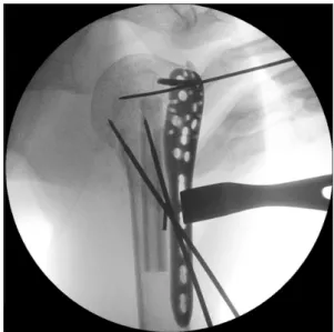
Fig. 1. After temporary fixation with two terminal threaded pins, the locking plate placement was performed, right shoulder.
a definite fixation of locking screws. If the fracture site or reduc- tion was unstable, we performed a temporary fixation using 2.5 mm terminal threaded pins prior to plate placement (Fig. 1).
Then, a locking compression plate (PHILOS; Synthes, Paoli, PA, USA) was applied along the lateral border of the bicipital groove, and its proximal tip was placed approximately 5 mm below the greater tuberosity. The plate was secured temporarily with Kirschner wires and a definite locking screw fixation was per- formed.
Postoperative Rehabilitation
The affected arm was kept in a sling for 6 weeks after surgery.
The pendulum exercise and self-assisted circumduction exercise were started on the first day after surgery. As tolerated, gradual passive ROM exercises was encouraged. After six weeks, active ROM exercise was started. After three months, isotonic exercises with an elastic band were started, and patients were gradually allowed to return to daily activities.
Results
Our consecutive 15 series were composed of 9 two-part fractures and 6 three-part fractures. There were 3 men and 12 women. Their mean age at the time of surgery was 71.3 years (ranging from 65–79 years). The dominant arm was involved in seven patients. The interval between injury and surgery was 0.6 day. The mean follow-up period after surgery was 19.3 ± 9.9 months (range, 12–49 months) (Table 1).
At the first year follow-up, the mean VAS pain score was 1.4 ± 1.3, mean ASES score was 87.3 ± 9.4, and mean UCLA shoulder score was 27.9 ± 3.2. The active ROM in forward flexion, external rotation, and internal rotation was 130° ± 15°, 36° ± 12°, and 15 ± 1, respectively. Postoperative stiffness was identified in five patients (33.3%) despite our rehabilitation pro- tocol and additional intra-articular steroid injection after three months postoperatively. There were no other complications,
such as infection or nerve injury.
Bone union was obtained in all patients post-surgically be- tween three months and six months. On the AP view in 20°
external rotation, the mean immediate postoperative neck- shaft angle was 138° ± 4°, and at one-year follow-up, the neck shaft angle was 137° ± 5°. There was no significant difference between the preoperative and postoperative values (p=0.105).
During the follow-up period, no avascular necrosis, screw pen- etration or loosening, or varus collapse was identified.
Discussion
In the two-part and three-part osteoporotic proximal humer- us fractures with osteoporosis, as we hypothesized, the use of fibular strut allograft was effective in maintaining the initial status of reduction and preventing varus collapse until bone union. In our series, all patients achieved bone union until the follow-up between three and six months after surgery, and at final follow- up, the neck-shaft angle at the immediate postoperative time was maintained without any significant difference. Although we were unable to compare the postoperative value with the preoperative value in this study, as we evaluated the functional scores only after surgery, the postoperative functional scores in this study was comparable to them in other previous stud- ies.15-17,22,23)
In surgical intervention for the unstable proximal humerus fractures with osteoporosis, the anatomical reduction at the medial calcar has been emphasized;7,11,12) in particular, if me- taphyseal comminution is combined, and therefore anatomical reduction and its maintenance were not feasible, other options, such as inferomedial calcar screw or intramedullary strut bone graft, should be considered. Gardner et al.7) emphasized the importance of medial support (either by anatomical reduction of medial cortex or oblique locking screw placement for medial calcar support) in locking the plate fixation of proximal humerus fractures. In their study, even with the use of locking plates, 29%
of patients had screw penetration of the articular surface. There was a significant loss of humeral height indicating varus collapse in patients without medial support compared with patients with the medial support. Zhang et al.23) also reported that patients without medial support screw exhibited a significantly higher failure rate and loss of neck-shaft angle loss at final follow-up, as well as better functional outcomes compared with patients with medial support screw.
On the other hand, another option for creating medial sup- port, intramedullary strut bone graft, was introduced. This option was first suggested by Walch et al.13) for nonunion of the hu- merus surgical neck; and for acute proximal humerus fracture, Gardner et al.14) employed this graft as an endosteal implant.
Recently, there have been several studies that demonstrated su- perior biomechanical properties of a fibular strut bone augmen- Table 1. Patient Demographics
Variable Value
Sex (male/female) 3/12
Age (yr) 71.3 ± 4.7 (65–79)
Neer classification
Two-part 9
Three-part 6
Dominant arm involvement 7
Time to surgery (day) 0.6 ± 0.7 (0–3) Follow-up period (mo) 19.3 ± 9.9 (12–49) Values are presented as number only or mean ± standard deviation (range).
tation in addition to locking plate fixation for proximal humerus fracture models.24-26) Bae et al.24) reported that strut bone aug- mentation significantly increased the maximum failure load and stiffness of construct with locking plate fixation in proximal hu- merus fracture compared with the construct with locking plate fixation alone. Saltzman et al.27) reported in their systemic review that fibular strut allograft was a viable option in treating proximal humerus fractures, despite great heterogeneity in the literature regarding the use of fibular strut allografts as an adjunct to open reduction internal fixation of proximal humerus fractures.
In the current study, our indications of using the fibular strut graft were osteoporotic comminuted fracture at the metaphysis of proximal humerus, including medial calcar comminution and severe cancellous bone loss within the humeral head. Our study used a similar indication for using the strut bone as other previ- ous studies.14,23)
Our study has several limitations. First, our study was a retro- spective case-series that did not have a control group to use as a comparison. If we compared the locking plate fixation without the fibular strut graft in our series, our study would be weighted.
However, although the setting of medial calcar comminution and situation of subsequent reduction loss were expected, we were unable to omit the use of fibular strut allograft. Second, the decision to use the fibular strut allograft was made subjectively at the surgical field. Third, we were unable to assess long-term complications as this study did not include a long-term follow- up.
Conclusion
For the unstable two-part and three-part osteoporotic proxi- mal humerus fractures with medial calcar comminution, the use of fibular strut allograft with locking plate fixation was effective in maintaining the initial status of reduction and exhibiting satisfac- tory functional and radiological outcomes. Fibular strut allograft, as one of the methods to support medial calcar, may be one of the most satisfactory solutions in osteoporotic unstable proximal humerus fractures.
References
1. Court-Brown CM, Caesar B. Epidemiology of adult fractures: a review. Injury. 2006;37(8):691-7.
2. DeFranco MJ, Brems JJ, Williams GR Jr, Iannotti JP. Evaluation and management of valgus impacted four-part proximal hu- merus fractures. Clin Orthop Relat Res. 2006;442:109-14.
3. Maier D, Jaeger M, Izadpanah K, Strohm PC, Suedkamp NP.
Proximal humeral fracture treatment in adults. J Bone Joint Surg Am. 2014;96(3):251-61.
4. Namdari S, Voleti PB, Mehta S. Evaluation of the osteoporotic proximal humeral fracture and strategies for structural aug-
mentation during surgical treatment. J Shoulder Elbow Surg.
2012;21(12):1787-95.
5. Nho SJ, Brophy RH, Barker JU, Cornell CN, MacGillivray JD.
Management of proximal humeral fractures based on current literature. J Bone Joint Surg Am. 2007;89 Suppl 3:44-58.
6. Edwards SL, Wilson NA, Zhang LQ, Flores S, Merk BR. Two- part surgical neck fractures of the proximal part of the hu- merus. A biomechanical evaluation of two fixation techniques.
J Bone Joint Surg Am. 2006;88:2258-64.
7. Gardner MJ, Weil Y, Barker JU, Kelly BT, Helfet DL, Lorich DG.
The importance of medial support in locked plating of proxi- mal humerus fractures. J Orthop Trauma. 2007;21(3):185-91.
8. Krappinger D, Bizzotto N, Riedmann S, Kammerlander C, Hengg C, Kralinger FS. Predicting failure after surgical fixation of proximal humerus fractures. Injury. 2011;42(11):1283-8.
9. Lee CW, Shin SJ. Prognostic factors for unstable proximal hu- meral fractures treated with locking-plate fixation. J Shoulder Elbow Surg. 2009;18(1):83-8.
10. Lescheid J, Zdero R, Shah S, Kuzyk PR, Schemitsch EH. The biomechanics of locked plating for repairing proximal humerus fractures with or without medial cortical support. J Trauma.
2010;69(5):1235-42.
11. Gerber C, Werner CM, Vienne P. Internal fixation of complex fractures of the proximal humerus. J Bone Joint Surg Br. 2004;
86(6):848-55.
12. Hertel R. Fractures of the proximal humerus in osteoporotic bone. Osteoporos Int. 2005;16 Suppl 2:S65-72.
13. Walch G, Badet R, Nové-Josserand L, Levigne C. Nonunions of the surgical neck of the humerus: surgical treatment with an intramedullary bone peg, internal fixation, and cancellous bone grafting. J Shoulder Elbow Surg. 1996;5(3):161-8.
14. Gardner MJ, Boraiah S, Helfet DL, Lorich DG. Indirect medial reduction and strut support of proximal humerus fractures us- ing an endosteal implant. J Orthop Trauma. 2008;22(3):195- 200.
15. Neviaser AS, Hettrich CM, Beamer BS, Dines JS, Lorich DG.
Endosteal strut augment reduces complications associated with proximal humeral locking plates. Clin Orthop Relat Res.
2011;469(12):3300-6.
16. Matassi F, Angeloni R, Carulli C, et al. Locking plate and fibular allograft augmentation in unstable fractures of proximal hu- merus. Injury. 2012;43(11):1939-42.
17. Panchal K, Jeong JJ, Park SE, et al. Clinical and radiological outcomes of unstable proximal humeral fractures treated with a locking plate and fibular strut allograft. Int Orthop.
2016;40(3):569-77.
18. Hinds RM, Garner MR, Tran WH, Lazaro LE, Dines JS, Lorich DG. Geriatric proximal humeral fracture patients show similar clinical outcomes to non-geriatric patients after osteosynthesis with endosteal fibular strut allograft augmentation. J Shoulder Elbow Surg. 2015;24(6):889-96.
19. Oh JH, Kim SH, Kwak SH, Oh CH, Gong HS. Results of con- comitant rotator cuff and SLAP repair are not affected by un- healed SLAP lesion. J Shoulder Elbow Surg. 2011;20(1):138- 45.
20. Kim SJ, Jung M, Lee JH, Kim C, Chun YM. Arthroscopic repair of anterosuperior rotator cuff tears: in-continuity technique vs.
disruption of subscapularis-supraspinatus tear margin: com- parison of clinical outcomes and structural integrity between the two techniques. J Bone Joint Surg Am. 2014;96(24):2056- 61.
21. Neer CS 2nd. Displaced proximal humeral fractures. I. Classifi- cation and evaluation. J Bone Joint Surg Am. 1970;52(6):1077- 89.
22. Benegas E, Ferreira Neto AA, Gracitelli ME, et al. Shoulder function after surgical treatment of displaced fractures of the humeral shaft: a randomized trial comparing antegrade intra- medullary nailing with minimally invasive plate osteosynthesis.
J Shoulder Elbow Surg. 2014;23(6):767-74.
23. Zhang L, Zheng J, Wang W, et al. The clinical benefit of me- dial support screws in locking plating of proximal humerus
fractures: a prospective randomized study. Int Orthop.
2011;35(11):1655-61.
24. Bae JH, Oh JK, Chon CS, Oh CW, Hwang JH, Yoon YC. The biomechanical performance of locking plate fixation with in- tramedullary fibular strut graft augmentation in the treatment of unstable fractures of the proximal humerus. J Bone Joint Surg Br. 2011;93(7):937-41.
25. Chow RM, Begum F, Beaupre LA, Carey JP, Adeeb S, Bouliane MJ. Proximal humeral fracture fixation: locking plate construct
± intramedullary fibular allograft. J Shoulder Elbow Surg.
2012;21(7):894-901.
26. Mathison C, Chaudhary R, Beaupre L, Reynolds M, Adeeb S, Bouliane M. Biomechanical analysis of proximal humeral fixa- tion using locking plate fixation with an intramedullary fibular allograft. Clin Biomech (Bristol, Avon). 2010;25(7):642-6.
27. Saltzman BM, Erickson BJ, Harris JD, Gupta AK, Mighell M, Romeo AA. Fibular strut graft augmentation for open reduc- tion and internal fixation of proximal humerus fractures: a sys- tematic review and the authors’ preferred surgical technique.
Orthop J Sports Med. 2016;4(7):2325967116656829.