Longitudinal Supraspinatus Tear Associated with Antegrade Humeral Intramedullary Nailing: A Case Report and Literature Review with Focus Placed on Nail Entry Point
Min Soo Shon, Tae Jung Bang, Jae Chul Yoo1
Department of Orthopedic Surgery, Center for Joint Surgery, National Medical Center, 1Department of Orthopaedic Surgery, Samsung Medical Center, Sungkyunkwan University School of Medicine, Seoul, Korea
Iatrogenic damage of the rotator cuff followed by postoperative shoulder function loss is a potential complication after antegrade intra- medullary nailing (AIN) for a humeral fracture. The authors present a case of arthroscopic rotator cuff repair and subacromial decom- pression of a non-healed rotator cuff tendon (mainly supraspinatus) and secondary impingement syndrome caused either by the tear or a proud nail after AIN for a mid-shaft humeral fracture. At presentation, the patient complained of right shoulder pain and ‘snapping’, especially during forward elevation and abduction of the shoulder, of 4 years duration. Right shoulder pain started sometime after pain due to the humeral shaft fracture, operation had subsided, and persisted after nail removal. Arthroscopic findings showed a longitudi- nal rotator cuff tear at the nail entry point that had not healed and severe fibrous hypertrophy on the acromion underspace, which is a unique finding since most longitudinal splits of tendon fibers are more likely to heal than conventional rotator cuff tears detached from bone. The torn rotator cuff was repaired after debridement and placing side-to-side sutures. At his 34-month follow-up after rotator cuff repair, the patient showed complete recovery and had excellent clinical scores.
(Clin Shoulder Elbow 2015;18(1):47-51)
Key Words: Rotator cuff; Complications; Intramedullary nailing; Arthroscopy
Copyright © 2015 Korean Shoulder and Elbow Society. All Rights Reserved. pISSN 2383-8337
Clinics in Shoulder and Elbow Vol. 18, No. 1, March, 2015 http://dx.doi.org/10.5397/cise.2015.18.1.47
Received February 2, 2015. Revised February 10, 2015. Accepted February 11, 2015.
Correspondence to: Jae Chul Yoo
Department of Orthopaedic Surgery, Samsung Medical Center, 81 Irwon-ro, Gangnam-gu, Seoul 135-710, Korea Tel: +82-2-3410-3501, Fax: +82-2-3410-0061, E-mail: [email protected]
Financial support: None. Conflict of interests: None.
Antegrade intramedullary nailing (AIN) has been recently recognized as a valid option and a reliable alternative to lock- ing plate fixation for humeral fractures, and is more in accord with the principles of minimal invasive surgery.1) In fact, AIN has several advantages, that is, better rotational stability, a good lever arm effect, less soft tissue damage at the fracture site, shorter op- erative time, lower infection rates, and earlier functional recov- ery. However, it is also associated with persistent shoulder pain and an unsatisfactory outcome rate of 20% to 40%, in some cases shoulder function does not return to normal even after nail removal.1) Complications associated with poor shoulder function after antegrade intramedullary nail insertion in the humerus in- clude, rotator cuff damage near the point of insertion, impinge- ment or superior protrusion of the nail in the subacromial space, nail loosening, screw back-out, and joint stiffness.1)
Here, we present a case of a non-healed longitudinal tear of the supraspinatus tendon at the insertion point of an intramedul- lary humeral nail with severe secondary impingement, its clinical results after cuff repair, and subacromial decompression. The pa- tient provided informed consent for the publication of his case information in the form of a case report.
Case Report
A 29-year-old man presented complaining of a 4-year his- tory of right shoulder pain and snapping, especially during forward elevation and abduction of the right shoulder. The pa- tient sustained a mid-shaft fracture of the right humerus during a fall from a height of 8 m while working in July 2004. At that time, he was treated at a local hospital immediately, although
preoperative radiographs were not available to us, and his brief operative and radiographic reports showed that the fracture had been treated using an antegrade humeral intramedullary nail of the bent type with proximal curvature (Fig. 1). He could not remember precisely the time of shoulder pain onset, but it
gradually worsened and shoulder function decreased even after achieving clinical union at the fracture site. He underwent nail removal at the local hospital after bone union in August 2006, but even after nail removal, the shoulder pain and loss of func- tion persistent. Several months of conservative treatment with
A B C
Fig. 1. (A) Preoperative anteroposterior radiographs showing complete union of the fracture site on the mid-shaft of the right humerus with no sign of humeral malrotation or malalignment. True anteroposterior (B) and axillary views showing no specific abnormality, excepting a bony irregularity at the nail entry point on the greater tuberosity (C).
Fig. 2. Preoperative fat suppressed T1-weight- ed and T2-weighted images showing poor integrity of the supraspinatus and abnormal tissues filling up the hole at the entry point of the antegrade intramedullary nailing (arrows).
non-steroidal anti-inflammatory drugs and physical therapy did not relieve his symptoms.
The patient visited our clinic in August 2008, that is, 2 years after nail removal. Inspection of the involved shoulder revealed an incisional scar corresponding to the anterolateral approach.
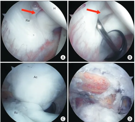
On physical examination, range of shoulder motion was not restricted in any direction, but he experienced aggravated pain, especially during forward elevation and abduction. Greater tuberosity tenderness was moderate to severe. Functional out- come scores were poor; visual analogue scale for pain 6 points, American Shoulder and Elbow Surgeons (ASES) score 60 points, and Constant score 75 points. Preoperative radiographs showed complete union of the fracture site on the mid-shaft of the hu- merus with no signs of an acromial spur, osteoarthritis of the glenohumeral joint, or malrotation or malalignment of the hu- merus (Fig. 1). However, magnetic resonance imaging revealed the presence of rotator cuff pathology at the sulcus medial to the greater tuberosity, posterior to bicipital groove as the insertion point of the nail (bent type) (Fig. 2), and subsequent diagnostic arthroscopy confirmed the presence of a rotator cuff tear. Rou- tine anterior and posterior portals were made for glenohumeral joint evaluation, the articular surface of the rotator cuff was found to be torn slightly above (or lateral to) the rotator cable near the insertion site of the greater tuberosity (Fig. 3A, B). The
scope was then moved to evaluate the bursal surface, acromio- plasty was performed because of evidence of impingement with severe fibrous hypertrophy on the acromion underspace caused either by the tear or by the proud nail (Fig. 3C, D). The supra- spinatus tendon was found to have a >25 mm longitudinal tear (Fig. 4A, B), and this was repaired using three side-to-side stitches with non-absorbable sutures (No. 2/0 Ethibond) (Fig. 4C, D).
Postoperatively, the patient underwent a rotator cuff repair re- habilitation protocol, involving, 3 weeks of immobilization in an abduction brace, followed by gentle range of motion exercises, and then (after achieving full range of motion) resistive exercises.
At his 34-month follow-up, the patient was asymptomatic with a painless full range of motion, improved clinical outcome scores (ASES score 100 points and a Constant score of 94 points).
Discussion
Antegrade intramedullary humeral nailing is mainly criticized for its potentially deleterious effects on shoulder function caused by iatrogenic damage to the rotator cuff at the nail entry point.1) To the best of our knowledge, this is the first arthroscopic report to describe a non-healing tear of the rotator cuff at the nail inser- tion point arising as a complication of AIN.
A B
C D
H
Ra
b
Ac
Rc
Fig. 3. (A) Arthroscopic image of the right shoulder showing the intra-articular site of the rotator cuff tear (arrow) from the pos- terior viewing portal with the patient in the lateral decubitus position. (B) Arthroscopic image of the full-thickness rotator cuff tear (arrow) as shown by the probe. Bursoscopy of the right shoulder in the lateral decubitus position. (C) Arthroscopic image of the right shoulder from the posterior portal showing fibrous hypertrophy on the acromion un- derspace and rotator cuff impingement. (D) Arthroscopic image from the anterolateral portal showing complete subacromial de- compression with acromioplasty. Ra: articu- lar surface of the rotator cuff, b: long head of the biceps tendon, H: humeral head, Ac:
underspace of the acromion, Rc: bursal side of the rotator cuff.
The correct identification and creation of the nail insertion point is crucial for anatomic reduction and primary stability.
However, the entry point chosen for antegrade intramedullary nail insertion may contribute to damage of the supraspinatus tendon.2) Gierer et al.3) described the direct visualization and quantification of microcirculatory dysfunction of the supraspi- natus tendon using the orthogonal polarization spectral imaging technique after stabilizing a humerus fracture by AIN, concluded that implantation of an antegrade humeral intramedullary nail, which requires splitting the rotator cuff, nearly halves the func- tional capillary density of the supraspinatus tendon.
When conventional methods are adopted through the an- terolateral deltoid split approach, which is the most commonly used for the AIN of a humeral fracture, the standard entry point for a bent type nail is at the medial sulcus of the greater tuberos- ity.4) However, this approach requires incising the supraspinatus tendon in its midsubstance (a critical hypovascular region), this procedure is not uncommonly followed by poor shoulder func- tion. García-Bógalo et al.5) performed sonographic assessments on rotator cuffs during nail insertion in 23 patients that under- went AIN for a humeral diaphyseal fracture, and postoperatively, partial tears of between 26 mm and 30 mm were found in 4 patients. Interestingly, they also found that Constant scores did not correlate with sonographic findings, that is, that cuff damage did not correspond to functional deficit.
Technological developments aimed at decreasing trauma to rotator cuffs at nail insertion sites, may lead to better results for the intramedullary nailing of humeral fractures. Recently, some authors suggested a more medial articular starting point for the straight nail at the apex (highest point) of the humeral head and splitting the rotator cuff through more muscle and well-vascular- ized tissue. Furthermore, in a recent report by Hatzidakis et al.,6) patients managed by antegrade locking intramedullary nailing of surgical neck fracture of the humerus via an articular entry point demonstrated reliable fracture-healing, favorable clinical outcomes, and little residual shoulder pain. In addition, several approaches, such as, the rotator interval splitting approach,7) approach via Neviaser ioportal,8) and an all-arthroscopic tech- nique,9) have been developed that involve nail insertion through a more medial entry point to minimize postoperative shoulder pain and preserve the critical hypovascular region of the rota- tor cuff. However, these techniques result in a tradeoff at the cost of articular cartilage damage, and its long-term effects are unknown. Other authors have described an extra-articular, extra-rotator cuff entry point located more lateral to the greater tuberosity using an angulated nail that preserves the articular surface and rotator cuff integrity.10) Nonetheless, each of these techniques has its unique advantages and disadvantages, and therefore, the most appropriate technique should be selected af- ter considering factors known to influence functional outcomes,
Ac
Sp
A B
C D
Fig. 4. (A) Arthroscopic image of the right shoulder showing a rotator cuff tear (arrow) from the anterolateral portal in the lateral de- cubitus position. (B) Arthroscopic image of a full-thickness rotator cuff tear as shown by the probe. (C) Arthroscopic image of the intra- articular portion from the posterior portal in the lateral decubitus position. The initial suture loaded with polydioxanone (PDSTM) suture (arrow) was used for full-layer repair of the torn rotator cuff. During this procedure, care is needed not to damage the long head of the biceps tendon. (D) Arthroscopic rotator cuff repair using three side-to-side stitches.
Ac: underspace of the acromion, Sp: supraspi- natus tendon.
such as, patient characteristics, bone quality, fracture type, and surgeon’s experience.
Although it is not clear when it occurred, our experience indi- cates that iatrogenic damage of the rotator cuff in the described patient could have happened at any step of the conventional AIN technique, such as, during awl insertion, reaming, or nail implantation or removal. However, we wholly agree with oth- ers that it is crucial to meticulously avoid violating the tendinous insertion of the rotator cuff on the greater tuberosity to prevent rotator cuff-related complications.
This case report cautions that the identification of rotator cuff damage after AIN is of considerable clinical importance, and unique as the longitudinal split of the supraspinatus tendon did not healed after a considerable time. To achieve painless, good functional results after AIN, the procedure should be performed carefully, and in particular, meticulous dissection should be con- ducted to minimize soft tissue damage at the nail entry point and rotator cuff splits should be repaired as completely as pos- sible. Furthermore, the possibility of rotator cuff damage should be considered after inserting or removing an AIN nail. Finally, we suggest modified techniques that spare the rotator cuff should be considered under selected conditions.
References
1. Zhu Y, Lu Y, Shen J, Zhang J, Jiang C. Locking intramedullary nails and locking plates in the treatment of two-part proximal humeral surgical neck fractures: a prospective randomized trial with a minimum of three years of follow-up. J Bone Joint Surg Am. 2011;93(2):159-68.
2. Stedtfeld HW, Mittlmeier T. Fixation of proximal humeral fractures with an intramedullary nail: Tipps and Tricks. Eur J
Trauma Emerg Surg. 2007;33(4):367-74.
3. Gierer P, Scholz M, Beck M, et al. Microcirculatory sequelae of the rotator cuff after antegrade nailing in proximal humerus fracture. Arch Orthop Trauma Surg. 2010;130(5):687-91.
4. Robinson CM, Bell KM, Court-Brown CM, McQueen MM.
Locked nailing of humeral shaft fractures. Experience in Edinburgh over a two-year period. J Bone Joint Surg Br.
1992;74(4):558-62.
5. García-Bógalo R, Larrainzar-Garijo R, Díez-Nicolás E, Llanos- Alcázar LF. Clinical and sonographic assessment of rotator cuff damage during antegrade humeral nailing. Rev Chir Orthop.
2008;52(1):2-8.
6. Hatzidakis AM, Shevlin MJ, Fenton DL, Curran-Everett D, Nowinski RJ, Fehringer EV. Angular-stable locked intramedul- lary nailing of two-part surgical neck fractures of the proximal part of the humerus. A multicenter retrospective observational study. J Bone Joint Surg Am. 2011;93(23):2172-9.
7. Park JY, Pandher DS, Chun JY, Md ST. Antegrade humeral nail- ing through the rotator cuff interval: a new entry portal. J Or- thop Trauma. 2008;22(6):419-25.
8. Knierim AE, Bollinger AJ, Wirth MA, Fehringer EV. Short, locked humeral nailing via Neviaser portal: an anatomic study.
J Orthop Trauma. 2013;27(2):63-7.
9. Lill H, Katthagen C, Hertel A, Gille J, Voigt C. All-arthroscopic intramedullary nailing of 2- and 3-part proximal humeral frac- tures: a new arthroscopic technique and preliminary results.
Arch Orthop Trauma Surg. 2012;132(5):641-7.
10. Mihara K, Tsutsui H, Suzuki K, Makiuchi D, Nishinaka N, Ya- maguchi K. New intramedullary nail for the surgical neck frac- ture of the proximal humerus in elderly patients. J Orthop Sci.
2008;13(1):56-61.