New Fixation Method Using Two Crossing Screws and Locking Plate for Cubitus Varus Deformity in Young Adult Elbow: Case Report
Byoung Jin Kim, Jong Hwan Seol, Myung Sun Kim
Department of Orthopaedic Surgery, Chonnam National University Hospital, Gwangju, Korea
Many types of osteotomy have been proposed for the treatment of cubitus varus deformity of the elbow, and various methods for fixa- tion of the osteotomy site have also been described. However, no method has been perfect. We treated two cases of cubitus varus elbow deformity with step-cut osteotomy using a new fixation method with two crossing screws and an anatomically designed locking plate. Active assisted elbow range of motion (ROM) exercise was permitted at postoperative 3 days, after removal of the drainage. Pre- operative and postoperative humerus-elbow-wrist angles and ranges of motion of the two patients were compared. At 3 months follow- up, each patient had recovered the preoperative elbow ROM, and achieved the complete bony union of the osteotomy site and proper correction of the cubitus varus deformity. In addition, the appropriate remodeling of the lateral bony protrusion was observed. Therefore, we introduce a new fixation method for achievement of stable fixation allowing immediate postoperative elbow motion after corrective osteotomy for cubitus varus deformity in young adults.
(Clin Shoulder Elbow 2016;19(1):43-47)
Key Words: Cubitus varus deformity; Corrective osteotomy; Step cut osteotomy; Crossing screw
Copyright © 2016 Korean Shoulder and Elbow Society. All Rights Reserved. pISSN 2383-8337
Clinics in Shoulder and Elbow Vol. 19, No. 1, March, 2016 http://dx.doi.org/10.5397/cise.2016.19.1.43
Received December 26, 2015. Revised January 9, 2016. Accepted January 10, 2016.
Correspondence to: Myung Sun Kim
Department of Orthopaedic Surgery, Chonnam National University Hospital, 42 Jebong-ro, Dong-gu, Gwangju 61469, Korea Tel: +82-62-220-6336, Fax: +82-62-225-7794, E-mail: [email protected]
Financial support: None. Conflict of interests: None.
Cubitus varus deformity is a common complication of pe- diatric supracondylar fracture of the humerus. This deformity is caused by angular and rotational malunion, due to physeal growth arrest or malunion. The deformity is thought to consist of extension, internal rotation, and varus angulation. Although cubitus varus does not cause functional disability, surgery is often required for cosmetic reasons. Corrective osteotomy is usually performed in children or young adults. In the past, because of the high rate of complications of valgus osteotomy, including stiffness, loss of fixation, infection, myositis ossificans, and neu- rovascular injury, corrective osteotomy was rarely performed.1) Thus, major concerns for surgical correction of cubitus varus de- formity are how to achieve sufficient correction and also restore preoperative arc of motion, without loss of fixation after surgery.
To achieve satisfactory results, accurate correction through oste- otomy should be followed by rigid fixation and early motion of the elbow.
Recently, various types of osteotomy and fixation have been proposed to correct this deformity, including lateral closing wedge osteotomy,2) dome osteotomy,3) three-dimensional os- toetomy,4) and step cut translational osteotomy.5) Of these oste- otomy techniques, step cut osteotomy is a popular technique.
Step cut osteotomy increases the contract area at the osteotomy site and the lateral spike remains on the distal part, which is fur- ther fixed to the proximal segment with a screw.
Placement of a lag screw using a screw increases fixation stability on the osteotomy site, resulting in better fracture heal- ing,6) and, fixation using 2 crossing screws can provide additional stability on rotation and displacement at the osteotomy site.7) We designed a new fixation technique, which not only creates a large contact area for rigid fixation but also achieves rigid fixation using a cross screw and locking plate for rigid fixation and early motion and early union.
Case Report
Two patients were treated for cubitus varus deformity using 2 crossing screws and locking plate fixation after step cut oste- otomy. Both were male, ages 22 and 37 years (Table 1). The deformity resulted from a malunited supracondylar fracture that occurred in the childhood period, respectively (Fig. 1). The two patients complained of the cubitus varus deformity due to cos- metic problem, not limitation of range of motion (ROM) of the elbow joint. Corrective osteotomy was scheduled for correction of the cubitus varus deformity.
Anteroposterior radiographs of both elbows with the elbows in full extension and supination were obtained before surgery.
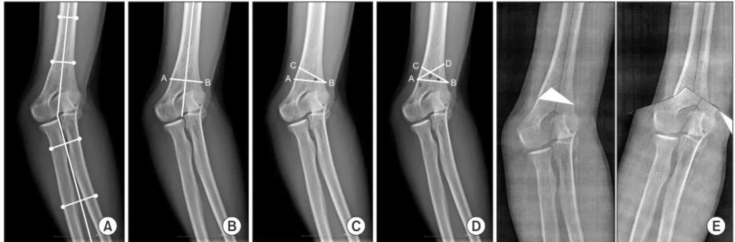
The humerus-elbow-wrist (HEW) angle was measured on the anteroposterior radiographs (Fig. 2A), and the correction angle was determined by comparing the difference of HEW angle be-
tween the affected and the normal elbow. Preoperative planned correction degree and postoperative achieved correction degree of both patients are described in Table 1.
Patients underwent surgery under tourniquet control in the lateral decubitus position. A posterior longitudinal skin incision was made in the posterior midline of the distal humerus, while the ulnar nerve was decompressed and protected, and the tri- ceps muscle was dissected using a paratricipital approach. The initial transverse osteotomy line was made 1.0 cm superior to the olecranon fossa and perpendicular to the long axis of the hu- merus (Line AB) (Fig. 2B). Then, from the medial end of this first line (Point B), the second line was drawn (Line BC), which made an angle between the first and second lines equal to the desired correction angle (Angle *) (Fig. 2C). Next, from the lateral end of the first line, the third line was drawn (Line AD), which made an angle between the second and third lines equal to the angle
Table 1. Clinical Data of Two Patients
Variable Case 1 Case 2
Age (yr)/Aff ected side 37/Right 22/Left
Follow-up (mo) 5 5
Humerus-elbow-wrist angle (°)
Preoperative (aff ected side/normal side) 18 varus/12 valgus 9 varus/13 valgus
Postoperative (aff ected side/normal side) 13 valgus/12 valgus 15 valgus/13 valgus
Correction degree
Preoperative planned correction degree 30 24
Postoperative achieved correction degree 31 26
Range of motion (°)
Preoperative 0–140 0–140
Postoperative 0–140 0–140
Fig. 1. (A) Pictures show patient with cubitus varus of right elbow. (B) Radiographs of both elbows.
A B
between the first line and the lateral supracondylar ridge line (Angle #) (Fig. 2D). Finally, our desired triangle was outlined and removed (Fig. 2E).
The osteotomy site was fixed with K-wire temporarily (Fig.
3A, B). Then, we checked the HEW angle and elbow ROM in- traoperatively. To obtain rigid fixation of the osteotomy site, one 4.5 mm cannulated screw and one 3.5 mm cortical screw were
used in cross configuration. After step cut osteotomy, a lateral spike was too small for the 4.5 mm cannulated screw, therefore we used the 3.5 mm cortical screw for lateral fixation. A 4.5mm cannulated screw was placed from anteromedial to postero- lateral direction, and a 3.5 mm cortical screw was placed from posterolateral to anteromedial direction (Fig. 3C, D). Two screws were placed in crossing configuration on the coronal and saggital Fig. 2. (A) Th e humerus-elbow-wrist (HEW) angle was measured, and determined the correction angle by comparing the HEW angle of the aff ected and the normal elbow. (B) AB is perpendicular to the long axis of humerus and 1 cm above from the olecranon fossa. (C) Line BC was drawn to make an angle equal to the desired correction angle (angle *). (D) Line AD was drawn to make an angle equal to the angle between line AB and lateral supracondylar ridge line (angle #).
(E) Triangular portion is removed and distal part of the osteotomy was repositioned.
A B C D E
A B
C
A B B
C A
D
* #
#*
Fig. 3. (A) Desired triangle was removed after osteotomy. (B) Reduction of the oste- otomized fragement and temporary fi xation with K-wire. (C) Intraoperative photo after fi xation with two crossing screw. (D) Fluoro- scopic anteroposterior (AP) view after fixa- tion with two cross screw. (E) Fluoroscopic AP view aft er fi nal fi xation. (F) Fluoroscopic lateral view aft er fi nal fi xation.
A B C
D E F
planes. Final fixation was performed to the posterolateral surface of the humerus by application of an anatomically designed, con- gruent locking plate (Synthes, Oberdorf, Switzerland) (Fig. 3E, F).
There was no impingement between screws of the plate and the two crossing screws. However, if impingement occurred, it could be resolved with change of the plate position to more proximal or distal. Active assisted ROM exercises including flexion, exten- sion, supination, and pronation began at postoperative 3 days, after removal of drainage. Active elbow motion was allowed at postoperative 6 weeks and strengthening exercise was started at postoperative 12 weeks. Patients returned to daily activity when radiographs showed union of the fracture site. Preoperative HEW angles were 18o varus and 9o varus, respectively. Postop- erative HEW angles were corrected to 13o valgus and 15o valgus.
Each patient had recovered the preoperative elbow ROM at 6 weeks and 8 weeks (Fig. 4A). Postoperative ROM of the elbow joint was 0o to 140o. Union was defined as the absence of pain and the presence of a bridging callus in 3 of the 4 cortices seen on the anteroposterior and lateral radiographic views. They achieved the complete bony union of the osteotomy site and proper correction of the cubitus varus deformity at postoperative 3 months. In addition, the appropriate remodeling of the lateral
bony protrusion was observed at the last follow-up (Fig. 4B).
There was no complication in both patients, and both patients were satisfied with correction of the cubitus varus deformity.
Discussion
Various surgical techniques for cubitus varus deformity were introduced. These techniques have been categorized as oste- otomy methods and fixation methods. Three major types are the simple lateral closing wedge, the step cut lateral closing wedge, and the dome rotational osteotomy. Lateral closing wedge oste- otomy is easy and safe, but achievement of strong internal fixa- tion is difficult. A dome osteotomy can reorient the distal frag- ment in both the coronal and the horizontal plane. However, it is often difficult to rotate the distal portion in the coronal plane due to the contracture of the surrounding soft tissue. Step cut osteotomy provides relatively more contact area. Some authors have reported on lateral condyle protrusion after step-cut oste- otomy. However, in these cases, the appropriate remodeling of the lateral bony protrusion was observed during the follow-up period (Fig. 3B).
The fixation methods after osteotomy include internal fixation and external fixation. Internal fixation is a one-stage operation that requires accurate planning. K-wire fixation is simple, but external immobilization is usually recommended with simple K-wire fixation.2,3) Plate and screw fixation offer the best stabil- ity, and allow early movement of the elbow.8,9) However, some authors reported insufficient length for fixation of the distal frag- ment.2) But, we achieved adequate fixation of the distal fragment using 2 screws in addition to an anatomically designed locking plate. External fixation including a simple uniplanar fixator and Ilizarov ring fixator may provide some advantages, but can be in- convenient and lead to pin track infection or elbow stiffness, etc.
In addition, those are not tolerated as well as internal fixation.10) Recently, several authors reported on an internal fixation method using a plate. Gong et al.8) described fixation with a lag screw and lateral plating. And Lim et al.9) described a fixa- tion with double plating through a lateral approach. Excellent results were obtained using each technique. We agree that both techniques have advantages of stable fixation and allowing early recovery of elbow motion. However, our technique can obtain not only fixation of both medial and lateral cortex, but also increase the contact area of bone surface. We think that it can lead to better fracture healing.
Therefore, we designed a new fixation method for step cut osteotomy for achievement of rigid fixation permitting immedi- ate postoperative elbow ROM exercise after corrective osteoto- my for cubitus varus deformity in young adults. There are several advantages of this fixation technique. We used 2 crossing screws (1 cannulated screw and 1 cortical screw). Fixation using two crossing screws through the osteotomy site can provide stronger Fig. 4. (A) Photos show good correction of cubitus varus deformity of right
elbow and range of motion at postoperative 3 months. (B) Radiographs at postoperative 3 months.
A
B
fixation at the osteotomy site. In addition, the placed lag screw using a cannulated screw and locking plate increases fixation sta- bility, leading to better fracture healing.
Therefore, our new fixation method for correction of cubitus varus deformity could achieve stable fixation permitting immedi- ate postoperative elbow motion after corrective osteotomy in young adults. We achieved the preoperative elbow ROM and complete bony union of the osteotomy site and proper correc- tion of cubitus varus deformity. Our new fixation method using 2 crossing screws and a locking plate after corrective osteotomy can be a reasonable alternative for correction of a cubitus varus deformity.
Consent
Written informed consent was obtained from the patient for publication of this case report and any accompanying images.
References
1. Davids JR, Lamoreaux DC, Brooker RC, Tanner SL, Westberry DE. Translation step-cut osteotomy for the treatment of post- traumatic cubitus varus. J Pediatr Orthop. 2011;31(4):353-65.
2. Bellemore MC, Barrett IR, Middleton RW, Scougall JS, White- way DW. Supracondylar osteotomy of the humerus for correc-
tion of cubitus varus. J Bone Joint Surg Br. 1984;66(4):566-72.
3. Tien YC, Chih HW, Lin GT, Lin SY. Dome corrective osteotomy for cubitus varus deformity. Clin Orthop Relat Res. 2000;(380):
158-66.
4. Chung MS, Baek GH. Three-dimensional corrective osteoto- my for cubitus varus in adults. Journal of shoulder and elbow surgery. J Shoulder Elbow Surg. 2003;12(5):472-5.
5. Kim HT, Lee JS, Yoo CI. Management of cubitus varus and val- gus. J Bone Joint Surg Am. 2005;87(4):771-80.
6. Uhl RL. The biomechanics of screws. Orthop Rev. 1989;18(12):
1302-7.
7. Kirchwehm WW. Cross screw compression fixation technique in proximal osteotomies of the first metatarsal for correction of hallux abducto valgus. J Foot Surg. 1988;27(5):412-7.
8. Gong HS, Chung MS, Oh JH, Cho HE, Baek GH. Oblique closing wedge osteotomy and lateral plating for cubitus varus in adults. Clin Orthop Relat Res. 2008;466(4):899-906.
9. Lim TK, Koh KH, Lee do K, Park MJ. Corrective osteotomy for cubitus varus in middle-aged patients. J Shoulder Elbow Surg.
2011;20(6):866-72.
10. Handelsman JE, Weinberg J, Hersch JC. Corrective supracon- dylar humeral osteotomies using the small AO external fixator.
J Pediatr Orthop B. 2006;15(3):194-7.