Increased Biceps Translation: A Clinical Sign of Complete Distal Biceps Tendon Rupture
Karan Malhotra , Abdul Waheed
Department of Orthopaedic Surgery, Basildon University Hospital, Basildon, UK
Various tests to help in the clinical diagnosis of distal biceps tendon ruptures have been described. In our experience these tests are painful in the acute setting. We suggest a simple alternative test wherein the biceps muscle belly is held by the examiner and translated medially and laterally. This is done with the forearm flexed to 90 degrees. It is first performed with the biceps relaxed and subsequently performed with the forearm flexed against resistance. In the relaxed forearm the biceps easily translates over 50% of its width. When placed under tension (by flexing against resistance) this translation is significantly reduced. In cases of complete distal biceps tendon rup- ture, the biceps still translates, even under resisted flexion of the forearm. This simple test is less painful than other described tests, is easy to perform, and aids in clinical diagnosis of distal biceps tendon ruptures.
(Clin Shoulder Elbow 2016;19(1):48-50)
Key Words: Distal biceps; Tendon injuries; Physical examination
Clinics in Shoulder and Elbow
CiSE
Copyright © 2016 Korean Shoulder and Elbow Society. All Rights Reserved.
This is an Open Access article distributed under the terms of the Creative Commons Attribution Non-Commercial License (http://creativecommons.org/licenses/by-nc/4.0) which permits unrestricted non-commercial use, distribution, and reproduction in any medium, provided the original work is properly cited.
pISSN 2383-8337 eISSN 2288-8721
TECHNICAL NOTE
Clinics in Shoulder and Elbow Vol. 19, No. 1, March, 2016 http://dx.doi.org/10.5397/cise.2016.19.1.48
Received July 22, 2015. Revised October 22, 2015. Accepted November 23, 2015.
Correspondence to: Karan Malhotra
Department of Orthopaedic Surgery, Basildon University Hospital, Basildon, Essex, SS16, 5NL, UK Tel: +44-1268-524900, Fax: +44-1268-394654, E-mail: [email protected]
Financial support: None. Conflict of interests: None.
Introduction
Distal biceps tendon ruptures result in loss of strength in forearm flexion of up to 30% and in forearm supination of up to 40%.1) As these injuries often occur in active patients there is an increasing trend towards repair. Expedient diagnosis and repair is important to improve outcome. A number of clinical tests have been described to confirm the diagnosis. Perhaps the most commonly used test is the hook test.2) We find this test difficult in the acute setting as it is painful for the patient and palpation of the tendon is difficult in the presence of swelling. We suggest a simple alternative clinical test for diagnosis of distal biceps ten- don rupture.
Technique
To begin the patient gently flexes the forearm to 90 degrees and fully supinates. The patient’s arm may be allowed to hang by their side, if more comfortable, and the forearm can rest either against their lap or be held gently by the examiner. The
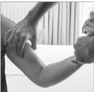
examiner first gently grasps the patient’s forearm with one hand and the patient’s biceps muscle belly with the other (Fig. 1). The patient is instructed to keep the forearm and biceps relaxed. The examiner then attempts to gently translate the patient’s biceps muscle belly medially and laterally. In the relaxed arm the biceps muscle belly will easily translate over 50% of the biceps width (Fig. 2). The amount of translation is noted. With no change in position, the patient is then asked to flex their forearm against resistance provided by the examiner holding down their fore- arm. The translation test is then repeated. If the tendon is intact the translation observed will be significantly reduced to less than 50% of the biceps’ width. If the tendon is ruptured the transla- tion will not be significantly reduced. This may be compared with the contralateral (uninjured) side. This is illustrated for clar- ity in Fig. 3.
Discussion
The relaxed biceps can be easily translated as the distal ten- don is relatively slack, allowing movement of the muscle. How-
Increased Biceps Translation in Distal Biceps Tendon Rupture Karan Malhotra and Abdul Waheed
www.cisejournal.org
49
ever, on resisted contraction the biceps pulls the tendon taut, which causes a relative tethering of the muscle belly, preventing translation. With a complete tendon rupture this tethering effect is lost and translation can therefore still occur. We have found that in most patients, in the relaxed state, the biceps muscle belly can be translated to over 50% of its width. In patients with intact tendons/aponeurosis this translation is significantly reduced but the exact amount of translation that remains is vari-
able. It is therefore important to perform the test without resis- tance first and compare the change in translation. One limitation of this test is that it may be difficult in patients with fatty tissue or atrophic muscles. In such cases the examiner may have difficulty in grasping the bicep muscle, and normal translation may be increased. Therefore, in these patients comparison of translation with the contralateral side is particularly important in order to better assess the expected change in translation.
Fig. 1. With the patient’s forearm supinated and fl exed to 90 degrees the ex- aminer gently grabs the biceps muscle belly and attempts to translate it medi- ally and laterally.
Fig. 2. Th is demonstrates the translation (medially in this case) which will occur with a relaxed biceps. Th is degree of translation will not be present in resisted fl exion with an intact distal biceps tendon.
Fig. 3. Th is is an illustration of the mechanism of this test. Th e biceps is shown from the anterior and lateral views. (A) Th is fi gure shows the normal resting posi- tion of the biceps. (B) Th is fi gure shows the translating force applied by the examiner, against active resistance. When the distal tendon is intact it is pulled taut on testing and prevents lateral translation. In (C), the distal tendon is ruptured and the translating force can therefore produce movement of the muscle as it is unop- posed.
50
www.cisejournal.orgClinics in Shoulder and Elbow Vol. 19, No. 1, March, 2016
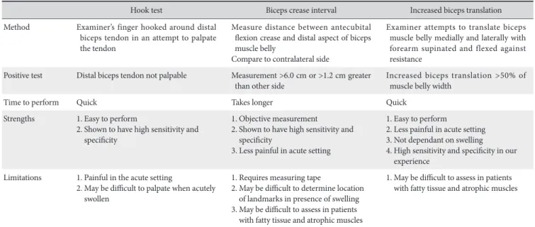
As the site of pressure applied on the biceps during this test is proximal to the injury, this test causes less pain and is less af- fected by swelling / bruising than the hook test. It is also simple to perform requiring little specialist experience to learn. It may therefore be easily taught to doctors and nurses in orthopaedic and emergency departments, and to general practitioners. A comparison between our test and the other commonly used tests in clinical practice is shown in Table 1.2,3)
We have used this technique successfully to help in diagnosis in 10 consecutive cases of patients presenting with suspected distal biceps rupture. Patients were all male, with acute injuries, presenting on the day of injury. The mean age of the patients ex- amined was 49.1 years (31 to 64 years). All patients had a posi- tive translation sign, and all patients underwent subsequent mag- netic resonance imaging which also showed distal biceps tendon tears. All patients proceeded to surgical repair and the tears were confirmed in all 10 cases (no false negatives, 100% sensitivity).
We also examined 20 normal, uninjured volunteers and found no cases of increased biceps translation (no false positives, 100%
specificity). We plan to validate this test in the future by assessing inter-observer reliability between health care professionals from different specialities (orthopaedic surgeons, emergency doctors, general practitioners, and emergency nurse practitioners) and comparing this with the traditional hook test.
This technique is simple, less painful than previously de- scribed tests and reproducible. We have found this a reliable test in the cohort of patients assessed, and we feel this may be help- ful in earlier clinical diagnosis of distal biceps tendon ruptures.
References
1. Spencer EE Jr, Tisdale A, Kostka K, Ivy RE. Is therapy necessary after distal biceps tendon repair? Hand (N Y). 2008;3(4):316-9.
2. O’Driscoll SW, Goncalves LB, Dietz P. The hook test for distal biceps tendon avulsion. Am J Sports Med. 2007;35(11):1865-9.
3. ElMaraghy A, Devereaux M, Tsoi K. The biceps crease interval for diagnosing complete distal biceps tendon ruptures. Clin Orthop Relat Res. 2008;466(9):2255-62.
Table 1. Comparing Our Technique (Increased Biceps Translation) for Diagnosis of Distal Biceps Tendon Rupture with Previously Described Techniques
Hook test Biceps crease interval Increased biceps translation
Method Examiner’s finger hooked around distal biceps tendon in an attempt to palpate the tendon
Measure distance between antecubital fl exion crease and distal aspect of biceps muscle belly
Compare to contralateral side
Examiner attempts to translate biceps muscle belly medially and laterally with forearm supinated and flexed against resistance
Positive test Distal biceps tendon not palpable Measurement >6.0 cm or >1.2 cm greater than other side
Increased biceps translation >50% of muscle belly width
Time to perform Quick Takes longer Quick
Strengths 1. Easy to perform
2. Shown to have high sensitivity and specifi city
1. Objective measurement
2. Shown to have high sensitivity and specifi city
3. Less painful in acute setting
1. Easy to perform
2. Less painful in acute setting 3. Not dependant on swelling
4. High sensitivity and specifi city in our experience
Limitations 1. Painful in the acute setting
2. May be diffi cult to palpate when acutely swollen
1. Requires measuring tape
2. May be diffi cult to determine location of landmarks in presence of swelling 3. May be diffi cult to assess in patients with fatty tissue and atrophic muscles
1. May be diffi cult to assess in patients with fatty tissue and atrophic muscles
Strengths and limitations are listed.