ISSN 0378-6471 (Print)⋅ISSN 2092-9374 (Online)
http://dx.doi.org/10.3341/jkos.2016.57.5.786
Original Article
스펙트럼영역 빛간섭단층촬영기를 이용한 정상인의 망막층별 두께 측정치의 반복성
The Repeatability of Retinal Layer Thickness Measurements with Spectral-Domain Optical Coherence Tomography in Normal Eyes
강민석⋅유승영⋅곽형우
Min Seok Kang, MD, Seung-Young Yu, MD, PhD, Hyung Woo Kwak, MD, PhD
경희대학교 의학전문대학원 경희대학교병원 안과학교실
Department of Ophthalmology, Kyung Hee University Hospital, Kyung Hee University School of Medicine, Seoul, Korea
Purpose: To evaluate the repeatability of retinal layer thickness measurements in normal eyes imaged using spectral domain op- tical coherence tomography (SD-OCT).
Methods: Sixty-eight eyes of 34 healthy subjects were enrolled in this study. Imaging was performed 4 times using 9 × 9 mm macular scans with SD-OCT (RS 3000 Advance HD OCT, NIDEK, Gamagori, Japan) at the same visit by an experienced examiner. After automatic retinal segmentation (layering) in 5 layers, the thickness of each layer was calculated. Macular thick- ness of 9 Early Treatment of Diabetic Retinopathy Study (ETDRS)–like regions was obtained. Repeatability for each of the 9 subfield areas was calculated by their repeatability coefficients and intraclass correlation coefficients (ICCs).
Results: There was no significant difference in average retinal thickness and each retinal layer thickness between all measure- ments acquired by the experienced examiner. The ICCs of retinal layer thickness ranged from 0.826 to 0.847 for the ganglion cell layer + inner plexiform layer, inner nuclear layer + outer plexiform layer and outer nuclear layer + external limiting membrane in the fovea. The ICCs were greater than 0.909 for the other intra-retinal layers in all 9 ETDRS subfield thickness between all meas- urement pairs.
Conclusions: Excellent repeatability was observed for SD-OCT retinal segmented layer thickness measurements in healthy subjects.
J Korean Ophthalmol Soc 2016;57(5):786-793
Keywords: Automated segmentation, Intraclass correlation coefficients, Repeatability, Retinal layer thickness, Spectral domain optical coherence tomography
■Received: 2015. 11. 5. ■ Revised: 2016. 4. 21.
■Accepted: 2016. 4. 22.
■Address reprint requests to Hyung Woo Kwak, MD, PhD Department of Ophthalmology, Kyung Hee University Hospital,
#23 Kyungheedae-ro, Dongdaemun-gu, Seoul 02447, Korea Tel: 82-2-958-8451, Fax: 82-2-966-7340
E-mail: [email protected]
ⓒ2016 The Korean Ophthalmological Society
This is an Open Access article distributed under the terms of the Creative Commons Attribution Non-Commercial License (http://creativecommons.org/licenses/by-nc/3.0/) which permits unrestricted non-commercial use, distribution, and reproduction in any medium, provided the original work is properly cited.
빛간섭단층촬영기(optical coherence tomography, OCT) 의 도입은 망막 분야를 필두로 전안부 및 녹내장 영역을 비
롯한 안과 전 분야에 걸쳐 질환의 이해와 치료에 있어 획기 적인 발전을 가져왔다. 빛간섭단층촬영기가 처음 도입되어 사용된지 약 20여 년이 지난 지금, 다양한 빛간섭단층촬영 기의 개발과 발전이 이뤄져 기기의 보편화에 힘입어 여러 빛간섭단층촬영기가 임상적으로 사용되고 있다. 빛간섭단 층촬영기는 비침습적 방법으로 망막 내 구조를 이미지화시 켜 보여주고, 황반부의 두께 및 망막의 신경섬유다발 두께 까지도 정량화하여 나타냄으로써 질환의 경과와 치료 방법 의 결정 등에 객관적인 임상 근거로 활용되고, 치료 후 효
Figure 1. Early Treatment Diabetic Retinopathy Study
(ETDRS) subfield (left eye). F = fovea; S1 = superior inner;T1 = temporal inner; I1 = inferior inner; N1 = nasal inner;
S2 = superior outer; T2 = temporal outer; I2 = inferior out- er; N2 = nasal outer.
과를 판정하는 데에도 매우 중요한 역할을 하고 있다.1-4 최 근에는 빛간섭단층촬영기의 급속한 발전에 힘입어 많은 새 로운 기능이 추가됨에 따라 망막층별 두께의 측정이 가능 해졌고, 질환의 진단 및 경과 관찰에 핵심적인 역할을 하고 있는 실정이다. 망막층별 두께에 관해서는 이미 녹내장 및 근시 환자에서 망막신경섬유층(retinal nerve fiber layer, RNFL)의 두께가 감소한다는 연구5,6를 비롯하여, 녹내장이 진행함에 따라 신경절세포층(ganglion cell complex, GCC) 두께의 감소에 관한 보고7 등 다양한 연구가 발표되었다.
또한 Matsumoto et al8은 중심성장액성맥락망막병증 환자 에서 중심와의 외핵층(outer nuclear layer) 두께가 시력과 연관이 있음을 발표하였고, Acton et al9은 나이관련황반변 성 환자에서 초기에 광수용체 외분절(photoreceptor outer segment)이 얇아지고 망막색소상피층(retinal pigment epi- thelium, RPE)이 두꺼워짐에 따라 시력 변화가 나타난다고 하였다.
기존의 시간영역 빛간섭단층촬영기(time-domain OCT, TD-OCT)부터, 현재 임상적으로 가장 많이 사용하고 있는 푸리에 변환을 통해 더 빠르고 해상도가 좋은 영상 획득이 가능한 스펙트럼영역 빛간섭단층촬영기(spectral-domain OCT, SD-OCT)를 비롯, 가장 최근에 소개되고 있는 파장 가변 빛간섭단층촬영기(swept-source OCT, SS-OCT)에 이 르기까지 점차 해상도가 뛰어난 영상을 통해 더욱 정확한 진단이 가능하게 되었다.1,10,11 이러한 측정 장비가 실제로 임상적으로 이용되려면 검사의 신뢰성이 보장되어야 하는 데, 실제 검사 간의 오차 정도를 나타내는 반복성이 뒷받침 되어야 한다. 특히 측정 장비의 반복성은 그 결과에 대한 해석에 많은 영향을 미칠 수 있기 때문에, 일치도와 신뢰성 평가에 중요한 지표가 된다.2,12,13 이미 많은 연구에서 빛간 섭단층촬영기의 반복성과 재현성에 관한 연구가 발표되었 고, 실제로 그 결과는 믿을 만한 수준의 높은 신뢰성을 보 이고 있음이 증명되었다. 하지만 망막층별 두께를 측정할 수 있는 자동화 알고리즘 소프트웨어가 개발되어 빛간섭단 층촬영기의 기능에 추가된 것은 불과 얼마 되지 않은 일이 고, 망막층별 두께 측정의 임상적 중요성과 활용에 비해 그 신뢰성을 뒷받침할 수 있는 연구는 아직까지 충분히 보고 되지 않았다.
따라서 이번 연구에서는 정상 한국인에서 스펙트럼영역 빛 간섭단층촬영기를 이용하여 황반부를 9개의 Early Treatment Diabetic Retinopathy Study (ETDRS) 구획(subfield)으로 나 누어 구역별 평균 망막 두께와 층별 두께를 측정하여 반복성 및 일치도 여부를 파악하고, 측정자 내 반복성(repeatability) 을 통해 스펙트럼영역 빛간섭단층촬영기의 정확성을 알아 보고자 하였다.
대상과 방법
본원 안과 외래를 방문한 환자 가운데 안과적 질환이나 당뇨, 고혈압 등의 다른 전신 질환이 없고 굴절교정 수술을 포함한 다른 안과 수술의 기왕력이 없는 성인 34명, 총 68 안을 대상으로 하였다. 각각의 대상안에 대하여 최대교정 시력(best corrected visual acuity [BCVA], snellen), 비접촉 성 안압계(TX-10 Puff Non-Contact Tonometer, Canon, Tokyo, Japan)를 이용한 안압 및 자동굴절계(Humphrey HARK-599 Autorefractor Keratometer, Carl Zeiss Meditec AG, Jena, Germany)를 이용한 구면수차(spherical equiv- alent)를 측정하였고, 한 명의 검사자가 세극등 검사로 전안 부 및 후안부의 이상 유무를 검사하였다. 모든 빛간섭단층촬 영은 스펙트럼영역 빛간섭단층촬영기인 RS-3000 Advance HD OCT (NIDEK, Gamagori, Japan)를 이용하여 무산동 상태로 macular map scan (9 × 9 mm, 512 × 128)으로 측정 하였다. 한 명의 검사자가 2회 연속으로 황반부 두께를 측 정하고, 1시간 간격을 두고 동일한 검사자가 다시 2회씩 추 가 시행하여 한 대상안에 대하여 모두 4회의 검사를 시행 하였다.
황반부를 중심으로 ETDRS subfield로 나누어, 즉 중심와를 기준으로 1 mm, 3 mm, 6 mm 직경의 3개의 동심원인 중심원 (central circle), 안쪽 원(inner ring), 바깥쪽 원(outer ring) 구 역을 중심와 영역(fovea), 상내측(superior inner, SI), 비내측 (nasal inner, NI), 하내측(inferior inner, II), 이내측(temporal inner, TI), 상외측(superior outer, SO), 비외측(nasal outer, NO), 하외측(inferior outer, IO), 이외측(temporal outer, TO)의 총 9개 구역의 평균 망막 두께와 망막층별 두께를
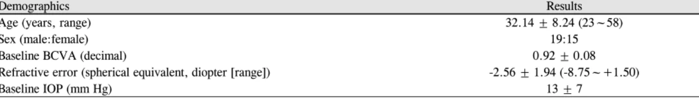
Table 1. Demographics
Demographics Results
Age (years, range) 32.14 ± 8.24 (23~58)
Sex (male:female) 19:15
Baseline BCVA (decimal) 0.92 ± 0.08
Refractive error (spherical equivalent, diopter [range]) -2.56 ± 1.94 (-8.75~+1.50)
Baseline IOP (mm Hg) 13 ± 7
Values are presented as mean ± SD unless otherwise indicated.
BCVA = best corrected visual acuity; IOP = intraocular pressure.
Table 2. Repeatability of thickness measurements for 5 intra-retinal layers in the fovea
Subfield Layer segmentation Inter-session
Mean ± SD (μm) ICC COV (%)
Fovea TRT 262.78 ± 15.53 0.959 5.91
ILM + RNFL 10.65 ± 1.67 0.834 15.68
GCL + IPL 34.28 ± 6.22 0.847 18.14
INL + OPL 39.34 ± 6.31 0.826 16.04
ONL + ELM 103.17 ± 10.07 0.962 9.76
IS/OS + RPE 77.26 ± 6.02 0.941 7.79
Values are presented as mean ± SD unless otherwise indicated.
ICC = intraclass correlation coefficient; COV = coefficient of variance; TRT = total retinal thickness; ILM = inner limiting membrane;
RNFL = retinal nerve fiber layer; GCL = ganglion cell layer; IPL = inner plexiform layer; INL = inner nuclear layer; OPL = outer plexi- form layer; ONL = outer limiting layer; ELM = external limiting membrane; IS/OS = photoreceptor inner segment/outer segment; RPE = retinal pigment epithelium.
Figure 2. Spectral domain optical coherence tomography
(SD-OCT) automatic segmentation of the retinal layers in the macula. ILM = inner limiting membrane; RNFL = retinal nerve fiber layer; GCL = ganglion cell layer; IPL = inner plexiform layer; INL = inner nuclear layer; OPL = outer plexiform layer; ONL = outer limiting layer; ELM = ex- ternal limiting membrane; IS/OS = inner segment/outer seg- ment photoreceptor layer; RPE = retinal pigment epithelium.측정하였다(Fig. 1).14 층별 두께는 선형 스캔(linear scan) 방 식으로 기기의 자동화 알고리즘(automated segmentation al- gorithm) 소프트웨어를 이용, 각각의 망막층의 반사도(reflectivity
index)에 대응하는 서로 다른 명암(shades of gray)에 따라 망막의 안쪽에서부터 바깥쪽 방향으로 (1) 내경계막(inner limiting membrane, ILM) + 신경세포층(retinal nerve fiber layer, RNFL), (2) 신경절세포층(ganglion cell layer, GCL) + 내망상층(inner plexiform layer, IPL), (3) 내핵층(inner nuclear layer, INL) + 외망상층(outer plexiform layer, OPL), (4) 외핵층(outer nuclear layer, ONL) + 외경계막 (external limiting membrane, ELM), (5) 광수용체 내/외분 절 (photoreceptor inner segment/outer segment, IS/OS) + 망막색소상피(retinal pigment epithelium, RPE)의 5개의 층 으로 나누어 측정하였다(Fig. 2).
통계적 분석은 SPSS version 18.0 (SPSS Inc., Chicago, IL, USA)을 사용하였고, 각 측정치마다 검사된 두 값 간에 유의한 차이가 있는지를 알아보기 위해 쌍표본 t-검정 (paired t-test)을 실시하여 반복성을 검정하였다. 모든 통계 적 유의성은 p값 0.05 미만으로 하였고, 급내상관계수(intraclass correlation coefficients, ICCs)와 변동계수(coefficient of varia- tion, COV)를 분석하여 반복된 측정치 간의 관계 및 일치 정도를 정량적으로 평가하였다.
결 과
대상자 34명의 평균 나이는 32.14 ± 8.24세(23-58세)였으 며, 이 중 남자가 19명, 여자가 15명이었다. 최대교정시력
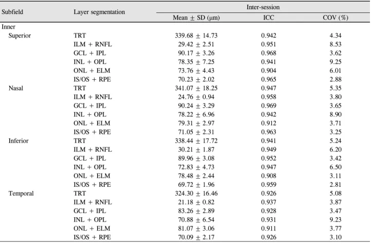
Table 3. Repeatability of thickness measurements for 5 intra-retinal layers in the inner circle
Subfield Layer segmentation Inter-session
Mean ± SD (μm) ICC COV (%)
Inner
Superior TRT 339.68 ± 14.73 0.942 4.34
ILM + RNFL 29.42 ± 2.51 0.951 8.53
GCL + IPL 90.17 ± 3.26 0.968 3.62
INL + OPL 78.35 ± 7.25 0.941 9.25
ONL + ELM 73.76 ± 4.43 0.904 6.01
IS/OS + RPE 70.23 ± 2.02 0.965 2.88
Nasal TRT 341.07 ± 18.25 0.947 5.35
ILM + RNFL 24.76 ± 0.94 0.958 3.80
GCL + IPL 90.24 ± 3.29 0.969 3.65
INL + OPL 78.22 ± 6.96 0.942 8.90
ONL + ELM 79.31 ± 2.97 0.912 3.71
IS/OS + RPE 71.05 ± 2.31 0.963 3.25
Inferior TRT 338.44 ± 17.72 0.941 5.24
ILM + RNFL 30.21 ± 1.87 0.949 6.20
GCL + IPL 89.96 ± 3.08 0.952 3.42
INL + OPL 72.83 ± 4.73 0.947 6.50
ONL + ELM 78.48 ± 2.44 0.908 3.11
IS/OS + RPE 69.72 ± 1.96 0.959 2.81
Temporal TRT 324.30 ± 16.46 0.926 5.08
ILM + RNFL 21.18 ± 0.82 0.937 3.87
GCL + IPL 83.26 ± 2.89 0.928 3.47
INL + OPL 70.88 ± 6.54 0.931 9.23
ONL + ELM 81.07 ± 3.06 0.911 3.77
IS/OS + RPE 70.09 ± 2.17 0.926 3.10
Values are presented as mean ± SD unless otherwise indicated.
ICC = intraclass correlation coefficient; COV = coefficient of variance; TRT = total retinal thickness; ILM = inner limiting membrane;
RNFL = retinal nerve fiber layer; GCL = ganglion cell layer; IPL = inner plexiform layer; INL = inner nuclear layer; OPL = outer plexi- form layer; ONL = outer limiting layer; ELM = external limiting membrane; IS/OS = photoreceptor inner segment/outer segment; RPE = retinal pigment epithelium.
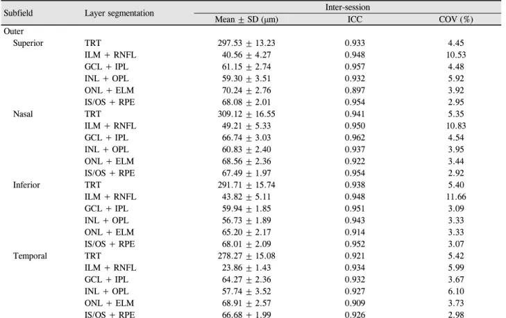
(snellen)은 0.92 ± 0.08이었으며, 안압은 13 ± 7 mmHg로 모두 정상범위 내였다. 평균 구면렌즈 대응치는 -2.56 ± 1.94D였다(Table 1). 스펙트럼영역 빛간섭단층촬영기 RS-3000 Advance HD OCT (NIDEK)로 측정한 평균 망막 두께는 중심와 262.78 ± 15.53 μm였고, 구역별 평균 망막두께는 직경 3 mm의 안쪽 원의 경우 비측(nasal)-상측(superior)-하 측(inferior)-이측(temporal)이 각각 341.07 ± 18.25 μm, 339.68 ± 14.73 μm, 338.44 ± 17.72 μm, 324.30 ± 16.46 μm 로 측정되었다. 직경 6 mm의 바깥 원 역시 비측(nasal)-상 측(superior)-하측(inferior)-이측(temporal)의 순서로 각각 309.12 ± 16.55 μm, 297.53 ± 13.23 μm, 291.71 ± 15.74 μm, 278.27 ± 15.08 μm로 얇아지는 양상을 보였다. 평균 망막 두께의 급내상관계수는 9개 구획 모두에서 0.921-0.947 범 위의 아주 높은 반복성을 보였다(Table 2-4).
망막층별 두께의 경우, 앞서 언급한 대로 ETDRS sub- field에 따라 9개의 구획으로 나누어 5개의 층으로 나누어 측정한 결과, 각각 층별 두께의 범위는 10.65 μm에서
103.17 μm 사이로 나타났다. 급내상관계수의 경우, 중심와 의 내경계막(ILM)과 신경섬유층(RNFL), 신경절세포층(GCL) 과 내망상층(IPL), 내핵층(INL)과 외망상층(OPL)에서 제외 한 모든 구획, 모든 층에서 0.909 이상의 매우 높은 일치도 를 보였으며, 위의 중심와 3개층의 급내상관계수도 각각 0.834, 0.847, 0.826으로 0.8 이상의 비교적 높은 반복성을 보였다(Table 2-4). 측정값을 각각 쌍표본 t-검정(paired t-test)에 적용해 두 측정치의 차이를 검정한 결과, 모든 구 역에서 평균 망막 두께 및 층별 두께에서 측정치 간 통계학 적으로 의미 있는 차이는 없었다.
고 찰
여러 망막질환에서 황반두께의 변화를 측정하여 정량적 으로 평가하는 것은 치료시기의 판정이나 치료효과 유무, 예후 등의 평가에 매우 중요한 객관적 지표로써 빛간섭단 층촬영기를 이용한 황반두께 측정은 임상적으로 중요한 의
Table 4. Repeatability of thickness measurements for 5 intra-retinal layers in the outer circle
Subfield Layer segmentation Inter-session
Mean ± SD (μm) ICC COV (%)
Outer
Superior TRT 297.53 ± 13.23 0.933 4.45
ILM + RNFL 40.56 ± 4.27 0.948 10.53
GCL + IPL 61.15 ± 2.74 0.957 4.48
INL + OPL 59.30 ± 3.51 0.932 5.92
ONL + ELM 70.24 ± 2.76 0.897 3.92
IS/OS + RPE 68.08 ± 2.01 0.954 2.95
Nasal TRT 309.12 ± 16.55 0.941 5.35
ILM + RNFL 49.21 ± 5.33 0.950 10.83
GCL + IPL 66.74 ± 3.03 0.962 4.54
INL + OPL 60.83 ± 2.40 0.937 3.95
ONL + ELM 68.56 ± 2.36 0.922 3.44
IS/OS + RPE 67.49 ± 1.97 0.954 2.92
Inferior TRT 291.71 ± 15.74 0.938 5.40
ILM + RNFL 43.82 ± 5.11 0.948 11.66
GCL + IPL 59.94 ± 1.85 0.951 3.09
INL + OPL 56.73 ± 1.89 0.943 3.33
ONL + ELM 65.20 ± 2.17 0.914 3.33
IS/OS + RPE 68.01 ± 2.09 0.952 3.07
Temporal TRT 278.27 ± 15.08 0.921 5.42
ILM + RNFL 23.86 ± 1.43 0.934 5.99
GCL + IPL 64.27 ± 2.36 0.932 3.67
INL + OPL 57.74 ± 3.52 0.927 6.10
ONL + ELM 68.91 ± 2.57 0.909 3.73
IS/OS + RPE 66.68 ± 1.99 0.926 2.98
Values are presented as mean ± SD unless otherwise indicated.
ICC = intraclass correlation coefficient; COV = coefficient of variance; TRT = total retinal thickness; ILM = inner limiting membrane;
RNFL = retinal nerve fiber layer; GCL = ganglion cell layer; IPL = inner plexiform layer; INL = inner nuclear layer; OPL = outer plexi- form layer; ONL = outer limiting layer; ELM = external limiting membrane; IS/OS = photoreceptor inner segment/outer segment; RPE = retinal pigment epithelium.
미를 갖는다. 최근에는 파장 가변 빛간섭단층촬영기(swept- source OCT, SS-OCT)도 개발되면서 망막 구조뿐만 아니 라 맥락막(choroid)과 같은 깊은 구조층까지 이미지화하는 것이 가능해졌고 스펙트럼 영역 빛간섭단층촬영기 역시 많 은 기능들이 추가 개발되어 망막 각각의 개별적인 층(layer) 까지도 구분하여 구현화할 수 있게 되었다.11,15
망막층별 두께의 임상적 이용은 앞서 언급한 것 외에도 특히, 최근에는 당뇨황반부종(diabetic retinal edema, DME) 환자나 나이관련황반변성(age-related macular degeneration, AMD) 환자에서 스펙트럼영역 빛간섭단층촬영기를 이용한 en face 이미지를 통해 환자의 망막의 내분절/외분절 (IS/OS)까지 포함한 망막 전 층(all layer) 및 특정 층의 길 이나 넓이까지도 시각화하는 것이 가능하여 시력이나 치료 전후 분석에 많이 이용되고 있다.16-18 또한 Watanabe et al19 의 연구에 따르면 변시증(metamorphopsia)을 호소하는 망 막전막(epiretinal membrane, ERM) 환자에서 내핵층(inner nuclear layer)의 두께가 증상과 관련이 있음을 보고하고, 이는 수술적 치료의 적응증이 될 수 있음을 발표하였다. 또
다른 연구에서, Koo et al20은 망막전막 환자에서 시수용체 층(photoreceptor layer)의 손상이 없을 경우 중심와 및 중심 와 주변의 망막내층(inner retina), 특히 내핵층(inner nu- clear layer)의 두께가 시력변화와 중요한 관련이 있음을 밝 히기도 하였다. 이렇듯 단순히 황반부 망막 두께 측정을 넘 어서 망막의 각 층별 두께 측정의 중요성이 점차 대두되고 있는 실정이고, 실제로 망막층별 두께 측정이 가능한 빛간 섭단층촬영기가 개발됨에 따라 임상적으로도 활발히 이용 되고 있다.
이와 같이 다양한 빛간섭단층촬영기가 개발되고, 지속적 으로 새로운 기능과 편의성이 추가된 빛간섭단층촬영기가 활발하게 사용됨에 따라 이를 이용한 검사에서 검사의 신 뢰성이 무엇보다 중요하다. 이러한 신뢰성을 뒷받침할 수 있는 지표로서 주로 반복성과 재현성을 평가하는데, 반복 성은 동일한 검사자가 똑같은 장비와 동일한 대상을 여러 번 검사하였을 때 결과가 얼마나 일치하는지를 나타내는 지표이고, 재현성은 검사자가 다를 때 검사자 간의 측정치 가 얼마나 일치하는가를 보여주는 지표이다.12,13,21 이미 많
은 기존의 연구에서 시간영역 빛간섭단층촬영기와 스펙트 럼 영역 빛간섭단층촬영기 측정에 관한 반복성과 재현성 연구가 이뤄졌으며 그 신뢰성이 증명되었다. 특히 황반부 두께나 신경절 세포층(ganglion cell layer) 측정의 반복성에 대해서는 다양한 빛간섭단층촬영기를 이용한 연구가 보고 되고 있는데 비해, 아직까지 망막층별 두께 측정의 신뢰성 을 뒷받침할 수 있는 반복성에 관한 연구는 충분히 보고되 어 있지 않다.22,23 따라서 본 연구에서는 다양한 스펙트럼 영역 빛간섭단층촬영기 가운데 최근에 개발되어 임상적으로 사용 중인 RS-3000 Advance HD OCT (NIDEK)를 이용하여 망막층별 두께를 측정하여 측정자 내 반복성(repeatability)을 통해 측정의 정확성을 알아본 연구라는 점에 그 의의가 있다.
반복성의 차이를 알아보기 위해 이번 연구에서는 급내상 관계수(intraclass correlation coefficient, ICC)를 이용하여 비교하였다. 급내상관계수는 검사 시점이 서로 다른 숫자 변수 간의 상관 계수를 계산하는 것으로 반복성의 정도를 평가할 수 있는 척도인데, 0에서 1 사이의 값을 가지며 보 통 1에 가까울수록 두 수치의 상관성이 높아 측정 방법의 반복성이 뛰어나다는 것을 뜻한다. 일반적으로 급내상관계 수의 값이 0.8-1.0이면 매우 신뢰할 수 있고, 0.60-0.79이면 중등도의 신뢰, 0.6 미만일 경우 낮은 신뢰도로 반복성이 없음을 의미한다.24,25 또한 측정값의 평균이나 단위가 다를 경우 산포도 비교가 어렵기 때문에 이러한 단위에 영향을 받지 않도록 객관적으로 수치화시킨 변동계수(coefficient of variation, COV)를 사용하여 데이터의 퍼짐 정도를 비교 하였는데, 일반적으로 변동계수는 값이 작을수록 산포가 적어 측정치 간의 차이가 없는 균일한 것을 의미하여, 보통 10% 미만일 경우 높은 반복성이 있음을 의미한다.26
본 연구에서 사용한 RS-3000 Advance HD OCT (NIDEK) 의 장점은 기존의 스펙트럼영역 빛간섭단층촬영기와 비교 하여 좀 더 높은 영상 스캔 속도를 가진다는 점이다. 일반적 으로 스펙트럼 영역 빛간섭단층촬영기의 스캔속도가 초당 25,000 A-scan의 속도를 보이는 반면, 최근 개발된 RS-3000 Advance HD OCT (NIDEK)는 초당 53,000 A-scan의 속도 를 가져 기존의 스펙트럼 영역 OCT보다 2배가 넘는 속도 로 높은 해상도를 보여 훨씬 더 자세한 망막 내 구조를 보 여줄 수 있다. 또한 기계 내에 eye tracking system을 갖춰 고해상도의 이미지와 더불어 환자들 가운데 나이가 많거나 협조가 잘 되지 않아 정확한 측정이 어려운 환자들에게서 도 기존의 스펙트럼 영역 OCT보다 조금 더 정확한 측정이 가능하게 되었다. 게다가 9 × 9 mm의 wide area scan 범위 를 가져 기존의 6 × 6 mm보다 전반적이고 한 눈에 주변부 까지 볼 수 있는 장점을 가진다. 무엇보다 망막층별로 두께 측정이 가능하여 망막의 각각의 층별(layer segmentation)
변화를 보는 연구에서도 유용하게 이용할 수 있을 것으로 보인다.
이번 연구에서 RS-3000 Advance HD OCT (NIDEK)의 기능을 이용하여 황반부의 구역별 평균 망막 두께와 망막 층별 두께를 측정한 결과, 내경계막(ILM)에서부터 신경세 포층(RNFL), 신경절세포층(GCL), 내망상층(IPL), 내핵층 (INL), 외망상층(OPL), 외핵층(ONL), 외경계막(ELM), 광 수용체 내/외분절(IS/OS), 망막색소상피(RPE)에 이르기까 지 급내상관계수 0.826 이상의 신뢰할 수 있는 결과를 나타내 었다. 그러나 중심와의 내경계막(ILM)과 신경세포층(RNFL), 신경절세포층(GCL), 내망상층(IPL), 내핵층(INL), 외망상 층(OPL)을 제외하면 급내상관계수는 0.909 이상으로 조금 더 커지고, 중심와의 이 3개층의 변동계수는 15.68-18.14로 다른 층이 평균 6-7%의 값을 나타낸 것에 비해 높은 수치 를 보였다. 이는 중심와 부근에서 위 3개층의 두께가 안쪽 원 및 바깥 원에 비해 1/3 정도로 급격하게 얇아지고, 이에 따른 segmentation error에 의한 차이라고 볼 수 있다. 하지 만 이 역시 높은 급내상관계수를 나타내었으며 중심와 평 균 망막 두께의 경우 급내상관계수 0.959의 매우 높은 일치 도를 보인 것으로 미루어 보아 변동계수의 차이가 중요한 의미를 갖지는 않음을 알 수 있다.
본 연구는 단일 검사자가 2회 연속으로 측정 후, 1시간 간격을 두고 2회를 재측정하여 총 4회의 검사를 통하여 반 복성을 평가한 연구이다. 따라서 반복성(repeatability)과 함 께 검사의 신뢰성을 더 높일 수 있는 재현성(reproducibility) 에 대한 고찰이 이뤄지지 않은 점은 본 연구의 제한점이라 할 수 있을 것이다. 그러므로 검사자 간에 발생할 수 있는 측정값의 차이에 의한 bias를 보여줄 수 있는 재현성에 대 한 연구도 추후 뒷받침되어야 할 것이다. 또한 본 연구는 망막 질환이 없는 정상안만을 대상으로 실시한 결과이므로, 실제로 황반 변성이나 황반 부종 등의 망막병증이 있을 경 우에는 결과를 신뢰할 수 있는 측정의 정확성을 보장할 수 없다는 데에 이 연구의 또 다른 한계점이 있다. 따라서 황 반부 질환이나 이상이 있을 경우에도 측정의 신뢰성을 뒷 받침할 수 있도록 병변이 있는 환자에 대한 추가적인 연구 도 필요할 것이다. 실제로 망막 질환이 있을 경우 정상인에 비해 환자의 시력 저하나 상의 왜곡에 따른 주시도 감소가 있을 것이고, 나이에 따라 환자 순응도에도 차이가 있어 해 상도 역시 감소할 것으로 예상되는 바, 이러한 오류 분석에 대한 연구도 같이 병행되어야 할 것이다.26
결론적으로 스펙트럼 영역 RS-3000 Advance HD OCT (NIDEK)로 측정한 정상안의 평균 망막 두께와 망막층별 두께 측정치는 모두 신뢰할 만한 반복성을 보였다. 따라서 본 연구는 망막층별 두께 측정의 임상적 활용도가 높아지
고 있는 최근 안과 질환의 진단과 치료 경향으로 미루어 보 건데, 중요한 의의를 가진다고 할 수 있다. 앞으로 더 나아 가, 여러 안과 질환별 측정치의 반복성 및 재현성에 관한 추가적인 연구가 필요할 것이다.
REFERENCES
1) Leung CK. Cheung CY, Weinreb RN, et al. Comparison of macular thickness measurements between time domain and spectral domain optical coherence tomography. Invest Ophthalmol Vis Sci 2008;
49:4893-7.
2) Menke MN, Dabov S, Knecht P, Sturm V. Reproducibility of reti- nal thickness measurements in healthy subjects using spectralis op- tical coherence tomography. Am J Ophthalmol 2009;147:467-72.
3) Han IC, Jaffe GJ. Comparison of spectral- and time-domain optical coherence tomography for retinal thickness measurements in healthy and diseased eyes. Am J Ophthalmol 2009;147:847-58, 858.e1.
4) Vizzeri G, Weinreb RN, Gonzalez-Garcia AO, et al. Agreement be- tween spectral-domain and time-domain OCT for measuring RNFL thickness. Br J Ophthalmol 2009;93:775-81.
5) Hess DB, Asrani SG, Bhide MG, et al. Macular and retinal nerve fi- ber layer analysis of normal and glaucomatous eyes in children us- ing optical coherence tomography. Am J Ophthalmol 2005;139:
509-17.
6) Leung CK, Mohamed S, Leung KS, et al. Retinal nerve fiber layer measurements in myopia: an optical coherence tomography study.
Invest Ophthalmol Vis Sci 2006;47:5171-6.
7) Kotowski J, Folio LS, Wollstein G, et al. Glaucoma discrimination of segmented cirrus spectral domain optical coherence tomography (SD-OCT) macular scans. Br J Ophthalmol 2012;96:1420-5.
8) Matsumoto H, Sato T, Kishi S. Outer nuclear layer thickness at the fovea determines visual outcomes in resolved central serous chorioretinopathy. Am J Ophthalmol 2009;148:105-10.e1.
9) Acton JH, Smith RT, Hood DC, Greenstein VC. Relationship be- tween retinal layer thickness and the visual field in early age-re- lated macular degeneration. Invest Ophthalmol Vis Sci 2012;53:
7618-24.
10) Huang J, Liu X, Wu Z, et al. Macular thickness measurements in normal eyes with time-domain and Fourier-domain optical coher- ence tomography. Retina 2009;29:980-7.
11) Mansouri K, Medeiros FA, Tatham AJ, et al. Evaluation of retinal and choroidal thickness by swept-source optical coherence tomog- raphy: repeatability and assessment of artifacts. Am J Ophthalmol 2014;157:1022-32.
12) Muscat S, Parks S, Kemp E, Keating D. Repeatability and reprodu- cibility of macular thickness measurements with the Humphrey
OCT system. Invest Ophthalmol Vis Sci 2002;43:490-5.
13) Oh SB, Cho WB, Moon JW, Kim HC. Repeatability and agreement of macular thickness measurement using time domain OCT and spectral domain OCT in normal subjects. J Korean Ophthalmol Soc 2009;50:710-16.
14) Photocoagulation for diabetic macular edema. Early Treatment Diabetic Retinopathy Study report number 1. Early Treatment Diabetic Retinopathy Study research group. Arch Ophthalmol 1985;103:1796-806.
15) Pierro L, Gagliardi M, Iuliano L, et al. Retinal nerve fiber layer thickness reproducibility using seven different OCT instruments.
Invest Ophthalmol Vis Sci 2012;53:5912-20.
16) Kiernan DF, Zelkha R, Hariprasad SM, et al. En face spectral-do- main optical coherence tomography outer retinal analysis and rela- tion to visual acuity. Retina 2012;32:1077-86.
17) Mohammad F, Wanek J, Zelkha R, et al. A method for En Face OCT Imaging of subretinal fluid in age-related macular degeneration. J Ophthalmol 2014;2014:720243.
18) Geitzenauer W, Hitzenberger CK, Schmidt-Erfurth UM. Retinal optical coherence tomography: past, present and future perspectives.
Br J Ophthalmol 2011;95:171-7.
19) Watanabe A, Arimoto S, Nishi O. Correlation between meta- morphopsia and epiretinal membrane optical coherence tomog- raphy findings. Ophthalmology 2009;116:1788-93.
20) Koo HC, Rhim WI, Lee EK. Morphologic and functional associa- tion of retinal layers beneath the epiretinal membrane with spec- tral-domain optical coherence tomography in eyes without photo- receptor abnormality. Graefes Arch Clin Exp Ophthalmol 2012;
250:491-8.
21) Moon SW, Kim ES, Kim YG, et al. The comparison of macular thickness measurements and repeatabilities between time domain and spectral domain OCT. J Korean Ophthalmol Soc 2009;50:
1050-9.
22) Carpineto P, Aharrh-Gnama A, Ciciarelli V, et al. Reproducibility and repeatability of ganglion cell-inner plexiform layer thickness measurements in healthy subjects. Ophthalmologica 2014;232:
163-9.
23) Ng DS, Gupta P, Tham YC, et al. Repeatability of perimacular gan- glion cell complex analysis with spectral-domain optical coher- ence tomography. J Ophthalmol 2015;2015:605940.
24) Müller R, Büttner P. A critical discussion of intraclass correlation coefficients. Stat Med 1994;13:2465-76.
25) Kang NH, Kim HJ, Lee JH. The measurements of macular thick- ness and volume with SD-OCT in normal eyes. J Korean Ophthalmol Soc 2011;52:1182-8.
26) Polito A, Shah SM, Haller JA, et al. Comparison between retinal thickness analyzer and optical coherence tomography for assess- ment of foveal thickness in eyes with macular disease. Am J Ophthalmol 2002;134:240-51.
= 국문초록 =
스펙트럼영역 빛간섭단층촬영기를 이용한 정상인의 망막층별 두께 측정치의 반복성
목적: 정상인에서 스펙트럼영역 빛간섭단층촬영기(Spectral-domain optical coherence tomography, SD-OCT)를 이용하여 황반부 를 구역별로 나누어 망막층별 두께를 측정하고 그 반복성을 알아보고자 하였다.
대상과 방법: 정상인 34명의 총 68안을 대상으로 스펙트럼영역 빛간섭단층촬영기를 이용하여 한 명의 검사자가 2회 측정한 후, 1시간 간격을 두고 2회 추가 시행하여 각각의 측정치를 비교 분석하였다. 황반부를 9개의 Early Treatment Diabetic Retinopathy Study (ETDRS) 구획(subfield)으로 나누어 구역별 평균 망막 두께와 층별 두께를 측정하여 측정치 간의 급내상관계수(intraclass correlation coefficients, ICCs)를 분석하여 반복성을 평가하였다.
결과: 단일 검사자에 의한 평균 망막 두께는 측정치 간 유의한 차이를 보이지 않았고(p>0.05), 급내상관계수는 모든 구획에서 0.918 이상의 높은 반복성을 보였다. 또한 층별 두께 역시 측정치 간 유의한 차이를 보이지 않았으며, 중심와의 내경계막과 신경섬유층, 신경절세포층과 내망상층, 내핵층과 외망상층에서 급내상관계수 0.826-0.847의 비교적 높은 반복성을 보였고, 이 중심와 3개층을 제외한 모든 구획의 나머지 전 층(all layer)에서 급내상관계수 0.909 이상의 매우 높은 반복성을 보였다.
결론: 스펙트럼영역 빛간섭단층촬영기는 황반부 구역별 평균 망막 두께와 층별 두께 측정에서 반복성이 뛰어난 결과를 나타내어 높은 신뢰성을 보였다.
<대한안과학회지 2016;57(5):786-793>