ISSN 0378-6471 (Print)⋅ISSN 2092-9374 (Online)
https://doi.org/10.3341/jkos.2017.58.2.165
Original Article
단안 망막분지정맥폐쇄 환자에서 반대눈과 대조군 눈의 망막내층의 두께 비교
Comparison of Inner Retinal Thickness between the Fellow Eyes of Unilateral Branch Retinal Vein Occlusion and Normal Control
정규철1,2⋅이동은1,2⋅윤창기1,2⋅김현웅1⋅김정림1,2
Gyu Chul Chung, MD1,2, Dong Eun Lee, MD1,2, Chang Ki Yoon, MD1,2, Hyun Woong Kim, MD, PhD1, Jung Lim Kim, MD, PhD1,2
인제대학교 의과대학 부산백병원 안과학교실1, 인제대학교 의과대학 부산백병원 안신생혈관질환 치료기술개발센터2 Department of Ophthalmology, Busan Paik Hospital, Inje University College of Medicine1, Busan, Korea
Therapeutics Center for Ocular Neovascular Disease, Busan Paik Hospital, Inje University College of Medicine2, Busan, Korea
Purpose: The purpose of this study was to compare inner retinal thickness and retinal nerve fiber layer (RNFL) thickness be- tween fellow eyes with unilateral branch retinal vein occlusion (BRVO) and normal control eyes.
Methods: Retrospective cross-sectional study including 59 patients diagnosed with unilateral BRVO and 51 control subjects.
Using spectral domain optical coherence tomography, we investigated the average, 4 quadrant, and 12 clock-hour RNFL thick- nesses and the average, minimum, superior, superonasal, superotemporal, inferior, inferonasal, and inferotemporal thicknesses of the ganglion cell-inner plexiform layer (GCIPL) layer.
Results: Patients with unilateral BRVO had a higher incidence of hypertension. In the fellow eyes of the unilateral BRVO pa- tients, 7 and 11 o’clock RNFL thicknesses were significantly thinner than for the control eyes. There was no significant difference in the GCIPL thickness between the two groups
Conclusions: The RNFL thickness of the fellow eyes of the unilateral BRVO patients showed significant decreases in the 7 and 11 o’clock sectors (p=0.005, 0.017, respectively), whereas there was no significant difference in the GCIPL thickness between the two groups. In the RNFL thickness analysis, the 7 and 11 o’clock sectors were found to be dominant locations of decreased RNFL thickness for the open angle glaucoma. Further, glaucomatous change should be carefully monitored in the fellow eyes of unilateral BRVO patients.
J Korean Ophthalmol Soc 2017;58(2):165-170
Keywords: Fellow eye, Ganglion cell layer thickness, Glaucoma, Retinal nerve fiber layer thickness, Unilateral retinal vein occlusion
■Received: 2016. 8. 25. ■ Revised: 2016. 11. 3.
■Accepted: 2017. 1. 18.
■Address reprint requests to Jung Lim Kim, MD, PhD Department of Ophthalmology, Inje University Busan Paik Hospital, #75 Bokji-ro, Busanjin-gu, Busan 47392, Korea Tel: 82-51-890-6016, Fax: 82-51-890-6329
E-mail: [email protected]
* This study was supported by a grant from the Korea Healthcare Technology R&D Project, Ministry of Health and Welfare Affairs, Republic of Korea (grant #: HI15C1142).
ⓒ2017 The Korean Ophthalmological Society
This is an Open Access article distributed under the terms of the Creative Commons Attribution Non-Commercial License (http://creativecommons.org/licenses/by-nc/3.0/) which permits unrestricted non-commercial use, distribution, and reproduction in any medium, provided the original work is properly cited.
망막정맥폐쇄와 개방각녹내장의 연관성은 여러 연구에 서 보고되었다. 망막폐쇄안에서 개방각녹내장의 유병률과 평균 안압이 높다고 보고하며 안압상승이 혈관을 압박하고 혈관내막의 증식을 유도하여 망막혈관벽을 붕괴시켜서 망 막정맥폐쇄를 유발한다고 보고한 연구가 있고,1 개방각녹내 장 환자에서 망막정맥폐쇄의 발생률이 더 높으며 유두출혈 이 발생한 증례를 보고하며 망막정맥폐쇄는 혈관내피세포 가 증식하면서 혈류저항을 증가시키고 시신경유두출혈은
A B
C D
Figure 1. Photographs of the retinal nerve fiber layer (RNFL) of a 65 year old man with branch retinal vein occlusion (BRVO) (A, B).
This patient had BRVO in the left eye (B) and red-free fundus photography showed a wedge-shaped defect (between arrows) in the right eye (A). Optical coherence tomography (C) of the right eye showed that RNFL thickness was identified as abnormal by the
< 1% criterion from 10-o’clock to 11-o’clock area. Ganglion cell analysis map of the right eye showed thinning of the ganglion cell-inner plexiform layer (red color area) on deviation map of right eye. A glaucomatous visual field change was noted in the corre- sponding hemifield location (D). TEM P = temporal; SUP = superior; NAS = nasal; INF = inferior; S = superior; N = nasal;
I = inferior; T = temporal; MD = mean deviation; PSD = pattern standard deviation.
이와 유사한 기전으로 혈류장애를 일으켜 녹내장발생을 유 발할 것이라고 주장한 연구가 있다.2-4 또 다른 연구들에서 는 개방각녹내장과 망막정맥폐쇄 두 질환 모두에서 고혈압, 당뇨 등의 공통적인 혈역학적인위험인자를 가지며 인슐린 저항성과 혈관자동조절기능이상이 두 질환의 중요한 원인 일 수 있다고 주장하였다.5-8
본 연구에서는 망막정맥폐쇄와 녹내장이 발병과 관련된 전신적 위험인자를 공통적으로 가진다고 가정하고 이런 위 험인자들이 망막정맥폐쇄가 있는 눈뿐만 아니라 반대눈에도 영향을 미칠 것이라 판단하여 망막분지정맥폐쇄 환자의 반 대안과 대조군에서 스펙트럼 빛간섭단층촬영을 이용하여 초 기녹내장성변화를 반영할 수 있는 망막신경섬유층의 두께와 함께 신경절세포복합체의 두께를 비교해 보고자 하였다.
대상과 방법
의무기록을 후향적으로 조사하여 2011년 1월부터 2015년 12월까지 인제대학교 부산백병원 안과에 내원하여 단안 망 막정맥폐쇄로 진단 받은 환자를 대상으로 연구기준에 적합 한 59명과, 동일 기간 내에 동일병원에서 검진목적으로 녹 내장검사를 시행 받은 적이 있는 대상자들 중에서 망막정맥 폐쇄로 진단 받은 환자와 성별과 연령을 맞춘 48명을 대조 군으로 하였다.
본원에서 처음으로 단안망막정맥폐쇄로 진단 받은 환자 를 환자군으로 하였고 환자군의 반대안에 망막정맥폐쇄가 발생하였거나 망막출혈이나 황반부종을 유발할 수 있는 망 막질환이나 포도막염 등의 질환이 있거나 백내장수술 이외
RVO Fellow eye (n = 59) Control (n = 48) p-value
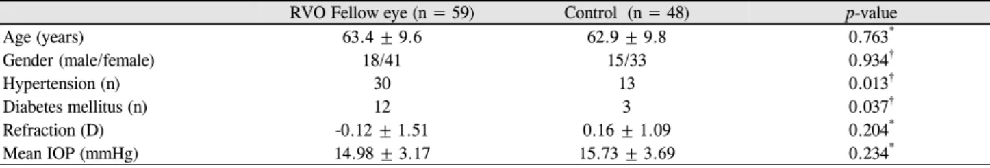
Age (years) 63.4 ± 9.6 62.9 ± 9.8 0.763*
Gender (male/female) 18/41 15/33 0.934†
Hypertension (n) 30 13 0.013†
Diabetes mellitus (n) 12 3 0.037†
Refraction (D) -0.12 ± 1.51 0.16 ± 1.09 0.204*
Mean IOP (mmHg) 14.98 ± 3.17 15.73 ± 3.69 0.234*
Values are presented as mean ± SD unless otherwise indicated.
RVO = retinal vein occlusion; D = diopter; IOP = intraocular pressure.
*By student t-test; †By chi-square test.
Table 1. Characteristics of RVO fellow eyes and control groups
의 다른 안구내수술 기왕력이 있거나 이전에 녹내장으로 진단 받고 치료를 받고 있는 경우, 시신경이나 망막에 영향 을 줄 만한 신경과적 질환이 있는 경우를 제외하였다. 대조 군은 안압이 21 mmHg 이상인 경우, 수직유두함몰비가 0.7 이상인 경우, 시신경유두테의 얇아짐이나 결손이 있는 경 우, 국소적인 망막신경섬유층결손이 있는 경우를 제외하였 고 이전 녹내장진단을 받았거나 치료를 받은 적이 있는 경 우, 망막정맥폐쇄가 발생하였거나 시신경염, 시신경병증, 당뇨병성망막병증, 망막출혈을 유발할 수 있는 기타 망막질 환이 동반되어 있는 경우, 시야에 영향을 줄 수 있는 다른 안과적, 신경학적 이상이 있는 경우도 제외하였다. 과거 병 력, 양안 시력, 굴절력, 골드만 안압검사, 세극등 검사, 전방 각 검사, 산동 후 안저검사, 빛간섭 단층촬영을 시행 받았던 경우를 포함시켰고 교정시력이 20/40 미만, 굴절률이 ±5D 이상인 경우, 전방각이 좁은 경우는 배제하였으며 대조군 중에서 양안 모두 조건에 맞을 경우에는 2안 중 1안만을 임 의적으로 택했다. 분석에 사용한 자료는 환자군에서 망막분 지정맥폐쇄 진단 당시에 측정한 값을 사용하였다. 본 연구 는 헬싱키 선언에 입각한 부산백병원 의학연구 윤리심의위 원회의 승인 아래 진행되었다(승인번호: 16-0126).스펙트럼영역 빛간섭단층촬영기는 Cirrus HD-OCT (Carl Zeiss Meditec, Dublin, CA, USA)를 이용하여 산동 후 촬영 하였다. 신호강도가 6 미만이거나 눈운동 및 눈깜박임으로 촬영된 이미지의 질이 떨어지는 경우, 분할(segmentation)에 오류가 있는 경우는 제외하였다. Optic disc cube 200 × 200 scan mode를 이용하여 평균 시신경유두함몰비, 수직 시신 경유두함몰비를 측정하였고 시신경유두주위 망막신경섬유 층은 평균 망막신경섬유층 두께, 12시각 망막신경섬유층 두 께, 4사분면 망막신경섬유층 두께를 측정하였다. 검사는 한 검사자가 각각 2차례씩 측정한 값의 평균을 사용하였다. 12 시각 망막신경섬유층두께의 경우 우안을 기준으로 하여 9 시 방향을 이측으로 3시 방향을 비측으로 정의하였다. 그리 하여 양안 모두에서 9시 방향을 이측으로 하여 12시각 방향 의 망막신경섬유층두께를 분석하였다. 망막내층의 두께는
ganglion cell analysis algorithm으로 분석한, 황반신경절세포 층과 속얼기층의 두께를 합한 신경절세포-속얼기층(gan- glion cell-inner plexiform layer, GCIPL)으로 평가하였고, 평 균 신경절세포복합체의 두께, 최소 신경절세포복합체의 두 께, 상측, 상비측, 상이측, 하측, 하비측, 하이측 총 6방향의 신경절세포복합체의 두께를 측정하여 비교하였다(Fig. 1).
통계적 분석은 statistic package for social science version 22.0 program (SPSS Inc., Chicago, IL, USA)을 이용하였고 연속형 자료는 Student t-test를 사용하였고 범주형 자료는 chi-square test or Fisher’s exact test를 사용하여 비교하였다.
p값이 0.05 미만인 경우를 통계학적 의의가 있는 것으로 판 정하였다.
결 과
대상 환자 59명 중 남성이 18명, 여성이 41명이며 평균 연령은 63.4 ± 9.6세, 대조군 51명 중 남성이 16명, 여성이 35명이며, 평균연령은 63.0 ± 9.8세였다. 단안 망막분지정맥 폐쇄가 있는 환자와 대조군을 비교하였을 때 고혈압과 당 뇨유병률은 단안망막분지정맥폐쇄가 있는 환자에서 대조군 보다 유의하게 많았지만(p=0.013, p=0.037) 나이, 성별, 굴절 률, 평균안압은 양 군 간의 차이를 보이지 않았다(Table 1).
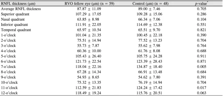
단안 망막정맥폐쇄가 있는 환자의 반대눈과 대조군의 망 막신경섬유층의 두께를 비교하였을 때 12시각 망막신경섬 유층 두께 분석에서 7시 방향 망막신경섬유층의 두께는 118.04 ± 22.16 μm로 대조군 134.87 ± 18.40 μm와 비교하여 의미 있는 차이를 보였고(p=0.005), 단안망막정맥폐쇄가 있 는 환자의 반대눈의 11시 방향 망막신경섬유층의 두께는 112.59 ± 21.83 μm로 대조군 124.24 ± 17.42 μm (p=0.017)와 비교하여 의미 있는 차이를 보였다. 나머지 부분은 양 군 사 이에 의미 있는 차이를 보이지 않았다(Table 2).
신경절세포복합체의 두께를 비교하였을 때는 평균 신경 절세포복합체의 두께와 최소 신경절세포복합체의 두께, 상 비측, 상측, 상이측, 하이측, 하측, 하비측 신경절세포복합
RNFL thickness (μm) RVO fellow eye (μm) (n = 59) Control (μm) (n = 48) p-value*
Average RNFL thickness 87.87 ± 11.09 89.00 ± 7.46 0.705
Superior quadrant 107.29 ± 17.05 109.28 ± 15.06 0.286
Nasal quadrant 63.85 ± 8.98 66.34 ± 7.06 0.104
Inferior quadrant 111.91 ± 22.05 114.69 ± 12.38 0.551
Temporal quadrant 65.97 ± 10.54 65.51 ± 9.70 0.821
1-o’clock 101.04 ± 21.35 100.45 ± 22.18 0.390
2-o’clock 75.51 ± 14.94 77.52 ± 13.23 0.704
3-o’clock 55.73 ± 7.87 55.62 ± 7.98 0.764
4-o’clock 60.36 ± 10.00 61.76 ± 8.08 0.688
5-o’clock 105.43 ± 26.40 105.75 ± 24.28 0.911
6-o’clock 121.73 ± 22.54 123.39 ± 28.43 0.871
7-o’clock 118.04 ± 22.16 134.87 ± 18.40 0.005
8-o’clock 67.28 ± 14.34 66.91 ± 13.48 0.684
9-o’clock 54.93 ± 8.65 54.62 ± 7.80 0.391
10-o’clock 75.32 ± 13.35 76.19 ± 14.94 0.704
11-o’clock 112.59 ± 21.83 124.24 ± 17.42 0.017
12-o’clock 118.49 ± 19.24 115.76 ± 20.51 0.063
Values are presented as mean ± SD unless otherwise indicated.
RNFL = retinal nerve fiber layer; RVO = retinal vein occlusion.
*By student t-test.
Table 2. Comparison between RNFL thickness in the fellow eyes of RVO patients and controls
GCIPL thickness (μm) RVO Fellow eye (μm) (n = 59) Control (μm) (n = 48) p-value*
Average 79.05 ± 11.22 80.08 ± 6.64 0.640
Minimum 73.52 ± 13.97 76.77 ± 8.91 0.223
Superornasal 81.46 ± 12.33 81.33 ± 7.36 0.995
Superior 79.32 ± 12.07 80.23 ± 6.23 0.375
Superotemporal 78.43 ± 11.63 79.33 ± 5.99 0.223
Inferotemporal 79.51 ± 10.46 80.54 ± 7.47 0.600
Inferior 76.68 ± 12.64 79.29 ± 7.06 0.395
Inferonasal 78.13 ± 13.04 79.37 ± 8.94 0.986
Values are presented as mean ± SD unless otherwise indicated.
GCIPL = ganglion cell-inner plexiform layer; RVO = retinal vein occlusion.
*By student t-test.
Table 3. Comparison between GCIPL thickness in the fellow eyes of RVO patients and controls
체의 두께 모두에서 양 군의 의미 있는 차이를 보이지 않았다 (Table 3).
고 찰
망막정맥폐쇄 환자들에서 녹내장 혹은 고안압 환자의 유 병률이 높으며 안압 상승이 혈관을 압박하고 혈관내막의 증식을 유도하여 망막혈관벽을 붕괴시켜서 망막정맥폐쇄를 유발한다는 연구가 있다.1 본 연구에서는 망막분지정맥폐쇄 반대안과 대조군에서 평균 안압은 통계적인 차이는 보이지 않았고 망막분지정맥폐쇄 반대안에서 안압이 가장 높았던 경우는 22 mmHg였다. 이는 망막정맥폐쇄안의 안압을 측정 한 것이 아니므로 이전 연구와는 차이를 보일 수 있을 것으 로 생각된다. 또한 본 연구에서는 안압의 일중 변동을 측정 하지 않아 일중 최고 안압을 반영하지 못하였으므로 이전
연구와는 차이가 있을 수 있겠다. 하지만 유사한 조건에서 측정한 양 군 간의 평균 안압이 통계적으로 유의한 차이가 없었다는 결과는 망막정맥폐쇄가 있는 환자들의 반대쪽 안 에 상이측, 하이측 망막신경섬유층의 두께가 차이를 보이는 것이 안압 이외의 다른 요소가 작용할 가능성이 있음을 나 타낸다고 볼 수 있을 것이다.
망막정맥폐쇄와 개방각 녹내장은 고혈압, 당뇨 등의 전신 적인 혈역학적 위험인자를 공통적으로가진다는 연구들이 있다.5-13 이번 연구에서 단안망막분지정맥폐쇄를 일으킨 환 자에서 대조군보다 더 높은 고혈압 비율과 당뇨병의 비율 을 보였다(p=0.013, p=0.037). 고혈압, 당뇨 같은 전신적인 혈역학적 위험인자가 망막분지정맥폐쇄 환자의 반대눈의 망막신경섬유층의 두께에 영향을 주었을 가능성도 있다고 생각된다.
본 연구에서 시신경유두함몰비를 비교하였을 때 단안망
막정맥폐쇄가 있는 환자의 반대눈은 평균 시신경유두함몰 비는 0.63 ± 0.14, 수직 시신경유두함몰비는 0.58 ± 0.15였고 대조군의 경우 평균 시신경유두함몰비는 0.51 ± 0.08, 수직 시신경유두함몰비는 0.49 ± 0.09로 양 군 간 의미 있는 차이 를 보였다(indepent t-test, p=0.002). 또한 단안 망막정맥폐쇄 가 있는 환자의 반대눈의 7시, 11시 방향 망막신경섬유층의 두께는 대조군과 비교하여 의미 있는 차이를 보였다. 단안 망막정맥폐쇄가 있는 환자의 반대눈에서 7시, 10시, 11시 방향의 망막신경섬유층의 두께가 대조군에 비해 유의하게 차이가 있었다는 기존 연구결과와 일치하는 소견을 보였 다.14 7시, 11시 방향은 시신경의 상이측과 하이측에 해당하 는 부분으로 구조적으로 녹내장성 변화가 빈번히 발생하는 곳이다. 단안 망막정맥폐쇄가 있는 환자의 반대눈에 이런 변화가 있었다는 것은 녹내장성 변화와 관련이 있을 가능 성을 시사하는 소견으로 볼 수 있다.15,16
망막내층, 특히 망막신경절세포는 허헐성 손상에 영향을 많이 받는다고 알려져 있다.17,18 망막신경섬유층은 망막신 경절세포의 축삭을 포함하고 있고 신경절세포층과 속얼기 층은 망막신경절세포의 핵과 수상돌기로 이루어지는데 황 반의 망막신경절 세포의 핵과 수상돌기의 손상이 망막신경 섬유층의 손상과 비교하였을 때 더 빠르고 더 일관되게 나 타난다고 알려져 있다.19-21 신경절 세포복합체는 신경절 세 포층과 속얼기층으로 정의되며 스펙트럼 빛간섭단층촬영을 통해 신경절 세포복합체의 두께를 측정할 수 있다.22 본 연 구에서는 망막분지정맥폐쇄 반대안에서 신경절세포복합체 의 두께가 전신적인 위험인자에 민감하게 영향을 받을 것 이라고 예상하였지만 대조군과 유의한 차이를 보이지 않았 다. 이는 신경절세포복합체의 두께는 중심오목에 중심을 맞 춘 타원모양의 영역이 상측, 상비측, 상이측, 하측, 하비측, 하이측 6개로 구분되어, 12시각으로 나누어 분석하는 망막 신경섬유층 두께검사보다 더 넓은 범위의 평균 두께를 측 정하므로 국소적인 망막내층의 변화를 평가하는 데 제한이 있을 것으로 생각된다. 또한 망막신경섬유층의 두께 차이가 난 부분이 시신경의 7시, 11시 방향으로 황반부에서 상대적 으로 주변부에 해당하므로 황반부의 망막내층두께가 이 부 분의 차이를 잘 반영하지 못하였을 것으로 생각된다. 추후 에 신경절세포복합체의 두께를 좀 더 광범위하게 측정하고 세분화하여 분석될 수 있다면 이번 결과와 차이를 보일 수 도 있을 것으로 생각된다. 마지막으로 망막내층 특히 망막 신경절세포는 급성 저산소성손상에 민감하다고 알려져 있 으므로 만성적인 혈역학적 변화를 알아보는 것과는 차이가 있었을 것으로 생각된다.17,18
망막정맥폐쇄와 녹내장의 관계를 비교하려면 망막정맥 폐쇄가 있는 눈의 변화를 관찰하는 것이 좋지만, 일단 망막
정맥폐쇄가 일어나면, 초기에는 망막부종에 의해, 후기에는 망막정맥폐쇄가 있던 부분에서 나타나는 망막의 두께감소 에 의해, 녹내장과 관련된 망막신경섬유층 및 신경절세포복 합체의 두께변화를 정확하게 측정할 수 없게 된다.14 전신적 인 위험인자에 의한 혈관의 병리학적 변화에 의해 망막정 맥폐쇄가 발생하였다면 반대눈에도 역시 유사한 변화가 있 을 가능성이 높다고 판단하여 이번 연구에서는 망막정맥폐 쇄가 있는 환자의 반대눈과 대조군을 비교하였다.
본 연구에서 3차 의료기간에 내원하여 망막정맥폐쇄를 진단 받은 환자와 검진 목적으로 녹내장검사를 시행 받은 경우를 대상으로 하였고, 개체수가 상대적으로 적어 선택 비뚤림의 가능성이 있다. 또한 대상자 중 시야검사를 시행 하지 않은 경우와 시야검사의 신뢰도가 떨어지는 경우가 있어 시야에 대한 분석은 시행하지 못하였다. 망막정맥폐쇄 진단 당시 시행한 빛간섭단층촬영으로 측정한 망막신경섬 유층과 망막신경절복합체 두께를 단면적으로 분석하였으므 로 앞으로 망막신경섬유층과 망막신경절복합체의 두께 변 화를 확인해 보는 것이 필요할 것으로 사료된다.
결론적으로 본 연구에서 망막분지정맥폐쇄 환자들의 반 대안에서 대조군에 비해 시신경의 상이측, 하이측에 해당하 는 7시, 11시 방향의 망막신경섬유층의 두께가 얇았으며 이 것은 간접적으로 개방각녹내장과 망막분지정맥폐쇄가 공통 된 병인을 공유한다는 것을 시사할 수 있는 근거 중 하나가 될 수 있을 것으로 생각된다. 따라서 단안에 국한된 망막정 맥폐쇄 환자에서도 반대안의 녹내장성 변화가 있는지 주의 깊은 관찰이 필요할 것으로 생각된다.
REFERENCES
1) Luntz MH, Schenker HI. Retinal vascular accidents in glaucoma and ocular hypertension. Surv Ophthalmol 1980;25:163-7.
2) Krakau CE. Disk hemorrhages and retinal vein occlusions in glaucoma. Surv Ophthalmol 1994;38 Suppl:S18-21; discussion S22.
3) Sonnsjö B, Krakau CE. Arguments for a vascular glaucoma etiology. Acta Ophthalmol (Copenh) 1993;71:433-44.
4) Yoo YC, Park KH. Disc hemorrhages in patients with both normal tension glaucoma and branch retinal vein occlusion in different eyes. Korean J Ophthalmol 2007;21:222-7.
5) Rath EZ, Frank RN, Shin DH, Kim C. Risk factors for retinal vein occlusions. A case-control study. Ophthalmology 1992;99:509-14.
6) Risk factors for branch retinal vein occlusion. The Eye Disease Case-control Study Group. Am J Ophthalmol 1993;116:286-96.
7) Risk factors for central retinal vein occlusion. The Eye Disease Case-Control Study Group. Arch Ophthalmol 1996;114:545-54.
8) Stewart RM, Clearkin LG. Insulin resistance and autoregulatory dysfunction in glaucoma and retinal vein occlusion. Am J Ophthalmol 2008;145:394-6.
9) Bonovas S, Peponis V, Filioussi K. Diabetes mellitus as a risk fac-
= 국문초록 =
단안 망막분지정맥폐쇄 환자에서 반대눈과 대조군 눈의 망막내층의 두께 비교
목적: 단안 망막분지정맥폐쇄환자에서 망막분지정맥폐쇄가 발생한 눈의 반대눈의 망막신경섬유층의 두께와 망막내층의 두께가 감소 되어 있는지 알아보고자 하였다.
대상과 방법: 단안 망막분지정맥폐쇄로 진단 받은 환자(59명, 59안)와 동일 기간 내에 동일 병원에서 검진 목적으로 녹내장검사를 시행 받았던 환자(48명, 48안)를 대조군으로 하여 후향적으로 비교하였다. 스펙트럼영역 빛간섭단층촬영기를 이용하여 각 군의 평균 망막신경섬유층 두께, 12시각 망막신경섬유층 두께, 4사분면 망막신경섬유층 두께 및 평균 신경절세포복합체의 두께, 최소 신경절세 포복합체의 두께, 상측, 상비측, 상이측, 하측, 하비측, 하이측 신경절세포복합체의 두께를 각각 측정하여 비교분석하였다.
결과: 단안 망막분지정맥폐쇄가 있는 환자와 대조군과 비교하였을 때 고혈압은 단안 망막분지정맥폐쇄가 있는 환자에서 대조군보다 유의하게 많았고, 망막신경섬유층의 두께는 단안 망막분지정맥폐쇄가 있는 환자의 반대눈의 7시와 11시 방향에서 대조군과 비교하여 의미 있는 차이를 보였지만(p-value=0.005, 0.017) 나머지 부분은 의미 있는 차이를 보이지 않았고 신경절세포복합체의 두께는 통계 적으로 두 군 간의 차이를 보이지 않았다.
결론: 망막분지정맥폐쇄 환자들의 반대안에서 대조군에 비해 시신경의 상이측, 하이측에 해당하는 7시, 11시 방향의 망막신경섬유층의 두께가 얇았다. 따라서 단안에 국한된 망막정맥폐쇄 환자에서도 반대안의 녹내장성 변화가 있는지 주의 깊은 관찰이 필요할 것으로 생각된다.
<대한안과학회지 2017;58(2):165-170>
tor for primary open-angle glaucoma: a meta-analysis. Diabet Med 2004;21:609-14.
10) Ellis JD, Evans JM, Ruta DA, et al. Glaucoma incidence in an un- selected cohort of diabetic patients: is diabetes mellitus a risk fac- tor for glaucoma? DARTS/MEMO collaboration. Diabetes Audit and Research in Tayside Study. Medicines Monitoring Unit. Br J Ophthalmol 2000;84:1218-24.
11) Pasquale LR, Kang JH, Manson JE, et al. Prospective study of type 2 diabetes mellitus and risk of primary open-angle glaucoma in women. Ophthalmology 2006;113:1081-6.
12) Gordon MO, Beiser JA, Brandt JD, et al. The Ocular Hypertension Treatment Study: baseline factors that predict the onset of primary open-angle glaucoma. Arch Ophthalmol 2002;120:714-20; dis- cussion 829-30.
13) Hulsman CA, Vingerling JR, Hofman A, et al. Blood pressure, ar- terial stiffness, and open-angle glaucoma: the Rotterdam study.
Arch Ophthalmol 2007;125:805-12.
14) Kim MJ, Woo SJ, Park KH, Kim TW. Retinal nerve fiber layer thickness is decreased in the fellow eyes of patients with unilateral retinal vein occlusion. Ophthalmology 2011;118:706-10.
15) Quigley HA, Addicks EM. Regional differences in the structure of the lamina cribrosa and their relation to glaucomatous optic nerve damage. Arch Ophthalmol 1981;99:137-43.
16) Jonas JB, Gusek GC, Naumann GO. Optic disc morphometry in
chronic primary open-angle glaucoma. I. Morphometric intra- papillary characteristics. Graefes Arch Clin Exp Ophthalmol 1988;226:522-30.
17) Janáky M, Grósz A, Tóth E, et al. Hypobaric hypoxia reduces the amplitude of oscillatory potentials in the human ERG. Doc Ophthalmol 2007;114:45-51.
18) Tinjust D, Kergoat H, Lovasik JV. Neuroretinal function during mild systemic hypoxia. Aviat Space Environ Med 2002;73:1189-94.
19) Chhablani J, Rao HB, Begum VU, et al. Retinal ganglion cells thin- ning in eyes with nonproliferative idiopathic macular telangiectasia type 2A. Invest Ophthalmol Vis Sci 2015;56:1416-22.
20) Jeoung JW, Choi YJ, Park KH, Kim DM. Macular ganglion cell imaging study: glaucoma diagnostic accuracy of spectral-domain optical coherence tomography. Invest Ophthalmol Vis Sci 2013;54:4422-9.
21) Nouri-Mahdavi K, Nowroozizadeh S, Nassiri N, et al. Macular ganglion cell/inner plexiform layer measurements by spectral do- main optical coherence tomography for detection of early glauco- ma and comparison to retinal nerve fiber layer measurements. Am J Ophthalmol 2013;156:1297-307.e2.
22) Seong M, Sung KR, Choi EH, et al. Macular and peripapillary retinal nerve fiber layer measurements by spectral domain optical coher- ence tomography in normal-tension glaucoma. Invest Ophthalmol Vis Sci 2010;51:1446-52.