CASE REPORT
간세포암종 환자에서 경동맥화학색전술 후 발생한 담낭 천공 1예
손민영, 한병훈, 이상욱, 윤병철, 서광일, 허진도
1고신대학교 의과대학 내과학교실, 영상의학교실1
Gallbladder Perforation after Transarterial Chemoembolization in a Patient with a Huge Hepatocellular Carcinoma
Min Young Son, Byung Hoon Han, Sang Uk Lee, Byung Cheol Yun, Kwang Il Seo and Jin Do Huh1 Departments of Internal Medicine and Radiology1, Kosin University College of Medicine, Busan, Korea
Transarterial chemoembolization (TACE) is a common treatment for unresectable hepatocellular carcinoma (HCC). The most com- mon complications after TACE are non-specific symptoms called post-embolization syndrome, such as abdominal pain or fever.
Rare complications, such as liver failure, liver abscess, sepsis, pulmonary embolism, cholecystitis, can also occur. On the other hand, gallbladder perforation is quite rare. This paper reports a case of gallbladder perforation following TACE. A 76-year-old male with a single 9-cm-sized HCC underwent TACE. Five days after TACE, he developed persistent right upper quadrant pain and ileus.
An abdomen CT scan confirmed gallbladder perforation with bile in the right paracolic gutter and pelvic cavity. Percutaneous transhepatic gallbladder drainage was performed with the intravenous administration of antibiotics. After 1 month, the patient underwent right hemihepatectomy and cholecystectomy. Physicians should consider the possibility of gallbladder perforation, which is a rare complication after TACE, when unexplained abdominal pain persists. (Korean J Gastroenterol 2020;76:351-355) Key Words: Gallbladder; Rupture; Chemoembolization, therapeutic; Carcinoma, hepatocellular
Received April 6, 2020. Revised April 23, 2020. Accepted April 23, 2020.
CC This is an open access article distributed under the terms of the Creative Commons Attribution Non-Commercial License (http://creativecommons.org/licenses/
by-nc/4.0) which permits unrestricted non-commercial use, distribution, and reproduction in any medium, provided the original work is properly cited.
Copyright © 2020. Korean Society of Gastroenterology.
교신저자: 한병훈, 49267, 부산시 서구 감천로 262, 고신대학교 의과대학 내과학교실
Correspondence to: Byung Hoon Han, Department of Internal Medicine, Kosin University College of Medicine, 262 Gamcheon-ro, Seo-gu, Busan 49267, Korea. Tel:
+82-51-990-5205, Fax: +82-51-990-5055, E-mail: [email protected], ORCID: https://orcid.org/0000-0001-7645-3477 Financial support: None. Conflict of interest: None.
서 론
경동맥화학색전술(transarterial chemoembolization)은 간 절제, 간 이식 그리고 국소 치료가 어려운 간세포암종에서 환자의 수행 상태가 양호하고 주 혈관 침범이나 간 외 전이 가 없을 때 적용되는 주된 치료법이며, 환자의 생존 기간을 향상시킨다고 알려져 있다.1,2하지만 시술과 관련된 합병증 도 문헌을 통하여 보고되고 있다. 경동맥화학색전술 이후 복 통, 발열, 오심, 구토 등의 증상으로 나타나는 색전후증후군 (post-embolization syndrome)과 일시적인 간기능의 변화가 가장 흔하게 발생하는데, 대부분 특별한 치료 없이 회복된다.
그러나 일부에서는 간부전, 간농양, 췌장염, 패혈증, 폐색전,
신부전, 위장관 출혈 등의 심각한 합병증을 동반하기도 한 다.1,3경동맥화학색전술 이후 발생하는 담낭과 관련된 합병증 에는 담낭염, 담낭 경색, 담낭 천공 등이 있으나 드물게 발생 한다고 알려져 있다.4경동맥화학색전술 이후 발생한 담낭 천 공은 해외에서도 증례보고가 드물고,5,6 국내에서는 보고된 적 이 없다. 본 저자들은 원발성 간세포암종 환자에서 경동맥화 학색전술 이후 발생한 담낭 천공 증례를 경험하였기에 이를 보고하는 바이다.
증 례
76세 남자가 18년 전 만성 B형간염을 진단받고 정기적으
A B C
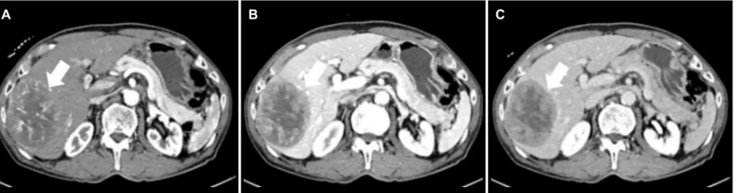
Fig. 1. Triple phase computed tomography of the liver at diagnosis. (A) A 9 cm sized heterogeneous enhancing mass of the right hepatic lobe at the arterial phase (white arrow). (B) The heterogeneous enhancing mass is washed out, showing low density at the portal and (C) delayed phase (white arrows).
A B C
Fig. 2. Transarterial chemoembolization (TACE) of the hepatocellular carcinoma. Right hepatic angiography shows (A) huge hypervascular mass in the right hepatic lobe (white arrow), (B) super selection of supplying branches of the right hepatic artery by microcatheter (white arrow), and (C) complete embolization (white arrow).
로 상복부 초음파를 시행받던 중 간 우엽에 종괴가 발견되어 전원되었다. 본원에서 시행한 간 CT에서 간의 5, 6, 7번 분획 에 9 cm 크기의 동맥기 조영 증가, 문맥기 및 지연기 조영 감소를 보이는 전형적인 간세포암종이 확인되었다(Fig. 1). 환 자는 B형간염 이외에 다른 과거력은 없었고, 하루 소주 1-2병 을 약 30년간 섭취한 음주력이 있었다. 입원 당시 활력징후는 혈압 110/70 mmHg, 맥박 53회/분, 호흡수 19회/분, 체온 36.6℃였다. 신체검진에서 공막 황달은 없었고 간과 비장은 만져지지 않았으며, 우상복부 압통이나 반동압통도 없었다. 혈 액 검사에서 백혈구 4,540/mm3 (중성구 62%), 혈색소 15.7 g/dL, 혈소판 156,000/mm3, 프로트롬빈 시간 13.4초(INR 1.17), 요 소질소 11.2 mg/dL, 크레아티닌 1.09 mg/dL, 총 단백 7.6 g/dL, 알부민 4.5 g/dL, 나트륨 137 mEq/L, 칼륨 4.3 mEq/L, 직접 빌리루빈은 0.36 mg/dL로 모두 정상이었으나 AST 73 U/L, ALT 50 U/L, 총 빌리루빈 1.33 mg/dL, AFP 11,364 ng/mL, protein induced by vitamin K absence or antagonist-ll 3,655 mAU/mL는 증가되어 있었다. Child-Turcotte-Pugh 점수는 5점으로 A등급이었고, Barcelona Clinic Liver Cancer 병기는 A, modified International Union Against Cancer
병기는 II였다. 환자 및 보호자와 치료에 대하여 상의하였으나 수술적 치료를 원하지 않아 경동맥화학색전술을 시행하였다.
우측 간혈관조영술을 통하여 우측 간동맥에서 분지된 영양동맥 을 초선택하여 독소루비신 50 mg, 리피오돌 12 mL, 젤라틴 색전 물질을 이용하여 경동맥화학색전술을 시행하였다(Fig. 2).
시술 후 1일째 환자는 우상복부 둔통을 호소하였고, 당시 체온 은 36.5℃였다. 혈액 검사에서는 백혈구 11,630/mm3 (호중구 93%)로 증가하였고, 총 빌리루빈 1.27 mg/dL, 직접 빌리루빈 0.32 mg/dL로 입원 당시와 비슷하였으나 AST 231 U/L, ALT 169 U/L 그리고 CRP는 1.6 mg/dL로 증가되어 있었다.
시술 후 5일째 환자의 우상복부 둔통이 지속되고 신체검진 에서 Murphy 징후는 없었으나 새롭게 압통이 있었다. 체온은 36.7℃였고, 혈액 검사에서 백혈구는 7,830/mm3 (호중구 86%), AST 45 U/L, ALT 44 U/L로 떨어지며 호전 추세를 보 였지만 총 빌리루빈 2.79 mg/dL, 직접 빌리루빈 1.63 mg/dL, CRP는 15 mg/dL로 증가 추세에 있었다. 또한 복부 방사선에 서 우상복부 장마비가 확인되었다(Fig. 3). 환자는 열은 없었 으나 보존적인 치료를 시행하여도 장마비가 지속되어, 시술 후 7일째 복부 CT를 시행하였다. 복부 CT에서 담낭 기저부가
A B
Fig. 4. Contrast-enhanced computed tomography (CT) at 7 days after the transarterial chemoembolization. (A) Axial and (B) coronal CT images show the perforation of the gallbladder with bile (white arrows) in the right paracolic gutter and pelvic cavity.
Fig. 5. Right hepatic angiography. The tip of the microcatheter (white arrow) is located beyond the cystic artery (black arrow) but is close between the microcatheter's tip and cystic artery.
Fig. 3. X-ray of the abdomen at 5 days after the transarterial chemoembolization. X-ray findings show the generalized distension of small and large intestine.
천공되어 담즙이 우측 주름 창자 고랑과 골반강 내에 있는 것 이 확인되었다(Fig. 4). 간담췌외과와 상의한 뒤, 정맥용 항생제 를 투여하고 경피경간담낭배액술(percutaneous transhepatic gallbladder drainage)을 시행하여 간기능과 환자의 전신 상 태가 회복된 다음에 담낭 절제술을 시행하기로 하였다. 내과 적 치료 이후 우상복부 둔통과 장마비는 호전을 보였고, CRP
수치도 안정화되었다. 또한 이후 시행한 복부 CT에서도 담낭 허탈과 복강내 담즙 소실이 확인되었으며, 새롭게 경동맥화학 색전술 이후 간세포암종의 크기가 줄어든 것이 확인되었다.
Indocyanine Green-R15 검사 결과 8.9%로 간기능이 양호하 고, MRI에서 간문맥 침범이나 림프절 비대, 전이 소견은 없어 환자 및 보호자와 치료 계획에 대하여 다시 상의한 후에 담낭 절제술과 우측 반간절제술(right hemihepatectomy)을 같 이 시행하기로 하였다. 한 달 뒤 환자는 담낭 절제술과 우측 반간 절제술을 같이 시행받았다. 수술 당시 병리 결과에서는
10.5×7.5×6.0 cm 크기의 팽창 결절형 간세포암종으로 확인되 었고, 분화도는 Edmondson-Steiner 등급 IV로 분화도가 나 빴다. 종괴 괴사 범위는 80%였고 피막 침습, 문맥 침습, 미세 혈관 침습도 있었다. 간세포암종 주변 간 조직은 만성 B형간 염, 2기 섬유화를 보였다. 환자는 수술 후 1개월 뒤 다발성 폐 전이가 확인되어 sorafenib 치료를 하였으나 5개월 뒤 간 세포암종 재발과 다발성 폐 전이 악화가 확인되어 현재 외래 에서 추적 관찰 및 대증 치료 중에 있다.
고 찰
경동맥화학색전술 이후 가장 흔하게 발생하는 합병증은 색 전후증후군과 간수치 상승이다. 색전후증후군은 환자의 약 60-80%, 최대 90%에서 나타나며, 일시적인 간수치와 백혈구 수치의 증가 또한 동반될 수 있는데, 대부분 일주일 내에 회복 된다. 이러한 증상은 경동맥화학색전술 과정에서 발생하는 간 세포 손상과 종양 괴사, 담낭 동맥의 색전으로 인한 것으로 생각된다.7,8
경동맥화학색전술 이후 발생하는 간부전, 간농양, 폐색전, 패혈증, 위장관 출혈 등과 같은 심각한 합병증은 문헌보고마 다 다르지만 환자의 약 5% 이내에서 발생하며, 사망 위험도 는 약 1% 정도이다.5,7경동맥화학색전술 이후 발생하는 담낭 염의 확률은 드물지만 0.3-10%로 다양하게 보고되고 있으 며,4본 증례와 같이 담낭 천공의 경우는 매우 드물게 발생한 다.5,6
간 주변 혈관 분지 구조의 인지 부족이나 시술 테크닉의 문제로 인한 화학 색전 물질의 역류로 표적 혈관이 아닌 담낭 동맥이나 췌장 동맥, 위십이지장 동맥 등 비표적 혈관에 색전 이 일어나는 것을 비표적 색전(nontarget embolization)이라 고 한다.4,7 이중혈관 공급을 받는 간과는 달리 담낭은 담낭 동맥을 통해서만 혈관 공급을 받는다. 이러한 담낭의 단일혈 관 공급은 경동맥화학색전술 중에 담낭이 손상에 취약하도록 하게 되는 원인이 된다.4담낭 동맥은 대부분 우측 간동맥에서 분지되기 때문에 우측 간동맥을 통하여 경동맥화학색전술을 하는 경우는 담낭 동맥에 비표적 색전이 일어날 수 있기 때문 에 주의해야 한다. 우측 간동맥을 통하여 경동맥화학색전술을 하는 경우 카테터 끝이 대부분 담낭 동맥을 지나서 위치하기 때문에, 색전 물질의 역류가 발생하면 담낭 동맥의 색전을 일 으켜 담낭 벽의 궤양, 괴사로 인한 담낭염이나 경색, 드물게는 천공을 유발한다.4,7 담낭염의 경우 대부분 항생제 치료로 호 전될 수 있으나 기종성 담낭염이나 담낭 천공의 경우는 경피 적 배액술이나 담낭 절제술이 필요하다.9
이번 증례는 간의 5, 6, 7번 분획에 9 cm 크기의 매우 큰 간세포암종 환자에서 첫 번째 경동맥화학색전술 이후 발열 소
견 없이 우상복부 둔통과 장마비가 발생하여, 색전후증후군으 로 생각하고 보존적 치료를 시행하였으나 우상복부 둔통과 장 마비가 7일간 지속되었고 원인 감별을 위하여 시행한 복부 CT에서 담낭 천공을 확인한 경우이다. 환자는 우상복부 둔통 이 지속되고 장마비 소견이 있었지만 발열은 없었고, 혈액 검 사에서도 백혈구와 간수치 증가가 발생하였다가 5일 이내에 호전 소견을 보여 색전후증후군으로 오인될 가능성이 있었다.
또한 증례 환자의 경우도 우측 간동맥에서 담낭 동맥이 분지 되고 있고, 담낭 동맥을 넘어서 미세 카테터 끝이 위치하고 있으나 담낭 동맥과의 거리가 가까워 시술 과정에서 색전 물 질의 역류가 담낭 동맥의 비표적 색전을 일으켜 담낭 천공이 발생하였을 것으로 생각된다(Fig. 5). 또한, 이번 증례처럼 Child-Turcotte-Pugh class A, modified International Union Against Cancer 병기 II, Barcelona Clinic Liver Cancer 병 기 A, 간의 우측에 크기가 큰 단일 간세포암종의 경우는 경동 맥화학색전술 이후 발생할 합병증을 고려하여 경동맥화학색 전술보다는 수술적 절제를 먼저 고려해야 한다.1
비표적 색전을 피하기 위해서는 간과 간 외 주변 혈관의 구조를 명확히 인지하고 간동맥에서 분지되는 혈관을 초선택 해야 한다. 그러기 위해서는 시술 과정에서 카테터 끝을 간동 맥의 원위부 가지에 최대한 가깝게 배치하여 모니터링을 통하 여 색전 물질을 천천히 주의 깊게 주입하여 역류를 줄이는 것이 중요하다, 또한 크기가 큰 간세포암종의 경우 시술에 사 용되는 리피오돌의 양을 30 mL 이상 주입하지 않도록 해야 한다.5,10 경동맥화학색전술 이전에 예방적으로 담낭 동맥 코 일 색전술을 시행하는 것이 담낭 관련 합병증을 줄이는 데 도움이 된다는 것은 논쟁의 여지가 있으나 예방적 담낭 동맥 코일 색전술 시행은 시술자의 경험, 담낭의 측부 혈관 유무, 색전 물질의 주입 위치 등과 같은 요소에 따라 결정된다.11,12
현재 경동맥화학색전술 이후 발생한 복통에 대한 명확한 치료 지침은 없다.1 또한 경동맥화학색전술 이후 복통, 발열, 간수치와 백혈구 수치의 증가는 드문 일이 아니기 때문에 다 른 심각한 합병증과 구별은 어려울 수 있다. 경동맥화학색전 술 이후 발생하는 색전후증후군과 심각한 합병증을 감별하기 위해서는 임상증상과 혈역학적 상태 변화에 유의해야 한다.
환자에게 이유 없이 우상복부 복통이 지속되거나 신체검진에 서 압통, 반동압통, Murphy 징후의 확인, 혈액 검사에서 백혈 수 수치, 간수치, 빌리루빈의 상승, 고열이 동반된다면 다른 심각한 합병증 확인을 위하여 초음파, CT, MRI와 같은 적극 적인 영상학적 검사를 해야 할 필요가 있다. 그중에서도 조기 에 CT를 시행하는 것이 원인 파악 및 치료를 위하여 유용하 다. 이번 증례의 경우 고열이나 Murphy 징후가 없었지만 우 상복부 복통이 지속되었고 조기에 CT를 시행함으로써 담낭 천공을 확인하여 항생제 치료와 경피경간담낭배액술을 빨리
시작할 수 있었다. 본 증례를 통하여 경동맥화학색전술 이후 흔하지 않은 합병증인 담낭 천공이 발생할 수 있음을 인지하 고 적극적인 검사를 통하여 원인 치료를 하는 것이 중요하다 고 생각된다.
REFERENCES
1. Korean Liver Cancer Association (KLCA) and National Cancer Center (NCC). 2018 Korean Liver Cancer Association-National Cancer Center Korea practice guidelines for the management of hepatocellular carcinoma. Korean J Radiol 2019;20:1042-1113.
2. Llovet JM, Bruix J. Systematic review of randomized trials for un- resectable hepatocellular carcinoma: chemoembolization im- proves survival. Hepatology 2003;37:429-442.
3. Paik YH, Chon CY, Cho JY, et al. Risk factors of acute hepatic fail- ure associated with transcatheter arterial chemoembolization for hepatocellular carcinoma. Korean J Med 2005;69:622-630.
4. Wagnetz U, Jaskolka J, Yang P, Jhaveri KS. Acute ischemic chol- ecystitis after transarterial chemoembolization of hepato- cellular carcinoma: incidence and clinical outcome. J Comput Assist Tomogr 2010;34:348-353.
5. Tu J, Jia Z, Ying X, et al. The incidence and outcome of major com- plication following conventional TAE/TACE for hepatocellular carcinoma. Medicine (Baltimore) 2016;95:e5606.
6. Lim EJ, Spanger M, Lubel JS. Gallbladder perforation following transarterial chemoembolisation; a rare but serious compli- cation. Frontline Gastroenterol 2013;4:135-137.
7. Clark TW. Complications of hepatic chemoembolization. Semin Intervent Radiol 2006;23:119-125.
8. Leung DA, Goin JE, Sickles C, Raskay BJ, Soulen MC.
Determinants of postembolization syndrome after hepatic chemoembolization. J Vasc Interv Radiol 2001;12:321-326.
9. Chan AO, Yuen MF, Hui CK, Tso WK, Lai CL. A prospective study regarding the complications of transcatheter intraarterial lip- iodol chemoembolization in patients with hepatocellular carcinoma. Cancer 2002;94;1747-1752.
10. Yamaguchi T, Seki T, Komemushi A, et al. Acute necrotizing pan- creatitis as a fatal complication following DC bead transcatheter arterial chemoembolization for hepatocellular carcinoma: a case report and review of the literature. Mol Clin Oncol 2018;9:403-407.
11. McWilliams JP, Kee ST, Loh CT, Lee EW, Liu DM. Prophylactic em- bolization of the cystic artery before radioembolization: feasi- bility, safety, and outcomes. Cardiovasc Intervent Radiol 2011;34:786-792.
12. Powerski M, Busse A, Seidensticker M, et al. Prophylactic emboli- zation of the cystic artery prior to radioembolization of liver malig- nancies--an evaluation of necessity. Cardiovasc Intervent Radiol 2015;38:678-684.