단일 치아 결손시 TiUnite TM 표면 처리한 임플란트의 생존율에 대한 후향적 연구
성균관대학교 삼성서울병원 치과진료부 치주과
김혜진․양승민․계승범․신승윤
구강 내 단일 치아를 상실한 경우 기존에는 고정성 보철물을 이용하여 수복하는 방법이 일반적이었지만, 최근
에 와서는 임플란트를 이용하여 수복하는 것이 보편화되고 있다. 본 연구는 단일 치아 결손 시 TiUniteTM 표면
처리한 임플란트를 이용하여 수복한 경우를 후향적으로 조사하여 그 생존율을 분석한 것이다. 2002년 9월부터
2006년 12월까지 삼성서울병원에서 단일 치아 결손 부위에 식립된 총 269개의 TiUniteTM 표면 처리한 임플란트
중 21개는 인접한 임플란트가 있거나, 기록이 누락되거나 관찰 기간이 짧아 연구에서 제외되었다. 248개의 임플란 트 중 상악에는 129개(52.0%) 하악에는 119개(48.0%) 식립되었다. 수술 부위에 치조골 재생술을 시행한 경우는 총 100개(40.3%)였으며, 상악동 거상술이 시행된 증례는 총 36개(14.5%)였다. 수술 당일부터 관찰 기간은 평균 26.0±11.8 개월이었으며, 그 기간 동안 실패한 것으로 간주된 임플란트는 12개로 생존율은 95.2%였다. 그 중 상악 에서 실패한 경우가 10개, 하악에서는 2개로 각각의 생존율은 92.2%, 98.3% 이다. 단일 치아 결손 시 TiUniteTM 표면 처리한 임플란트를 이용하여 수복한 경우 단기간 동안 높은 생존율을 보였다.
주요어: TiUniteTM 표면 처리, 단일 치아 결손 수복, 임플란트 생존율, 후향적 연구
(대한치과턱관절기능교합학회지 2009:25(3):267~277)
서 론
구강 내 단일 치아를 상실한 경우 고전적으로 인접치를 지대치로 사용하는 고정성 보철물을 이용하여 수복하는 방법이 사용되어 왔다. 하지 만 이는 지대치로 사용되는 치아에 인위적인 손 상을 주게 되고 경우에 따라 신경치료를 필요로 할 수 있으며, 장기적으로 사용시 이차 우식이나 치주 질환에 이환될 가능성이 높아지게 된다. 이 러한 이유로 단일 치아가 상실 된 경우 임플란트
교신저자 : 신승윤
삼성서울병원 치과진료부 치주과, 서울 강남구 일원동 삼성서울병원 135-710 대한민국.
팩스:+82-2-3410-0038, E-mail: [email protected]
원고접수일 :2009년 08월 05일, 원고수정일 :2009년 08월 20일, 원고채택일 :2009년 09월 25일
를 이용하여 수복하는 것이 고정성 보철물을 대 신하며 점차 보편화 되고 있다.1) 하지만 골량이 부족하거나 골질이 나쁜 경우, 전신적인 이유로 외과적 시술이 어려운 경우, 전치부에 치은 퇴축 등이 예상되어 심미적으로 임플란트 수술이 불 리한 경우 등에서는 아직도 고정성 보철물이 훌 륭한 대안이 될 수 있다. 하지만 지금까지 단일 치아의 상실에 있어 두 술식간의 적응증에 관한 기준이나 성공률 등에 관한 직접적인 비교 연구 는 부족한 실정이다.2)
임플란트는 1970년대 후반 치아 결손부위의 수복을 위해 식립된 이후 디자인이나 표면처리 등에서 지속적인 변화가 있어 왔다. 디자인에 있 어서는 초기의 blade 형태에서 원통형을 거쳐 현 재는 나사형이 대부분을 이루게 되었다. 나사형 임플란트는 골내 초기 안정성이 증진되고, 식립 도중 임플란트의 수직적인 위치 조절이 유리하 여 결국 더 높은 성공률을 보인다고 알려져 있 다.3)
표면처리에 있어서는 초기에는 티타늄 표면을 기계절삭한 면을 이용하였는데, 이는 골질이 나 쁜 경우 성공률이 상대적으로 낮아지게 된다. 이 후 임플란트 표면의 거칠기를 증가시킴으로써 골유착을 위한 세포 부착이 유리해짐을 여러 연 구를 통해 확인하였고,4-6) 이를 바탕으로 다양한 표면처리 방법이 발전하게 되었다. TiUniteTM표 면처리 방법도 그 중 하나로 electrochemical process 방법을 이용하여 티타늄 임플란트 표면 에 자연적인 산화막을 입히는 방법이다.
TiUniteTM 표면은 고결정성의 인산이 풍부한 산 화 티타늄으로 구성되어 있으며, 고르게 분포된 다공성 표면을 가진다.7) TiUniteTM 표면 처리한 임플란트는 이전의 turned, machined 표면의 임플 란트와 비교해 더 좋은 골반응을 보임이 여러 조 직학적 실험들에 의해 밝혀 졌다.8-11) TiUniteTM 표면 처리한 임플란트는 이런 골반응에 의해 보 다 빠르고 강한 골유착을 얻을 수 있게 된다.
이에 본 연구에서는 본원에서 단일 치아의 수 복에 TiUniteTM 표면 처리한 임플란트를 이용한 경우를 후향적으로 조사하여 임플란트에 대한 자료 및 생존율을 분석해 보고자 한다.
연구 재료 및 방법
본 연구는 2002년 9월부터 2006년 12월까지 삼 성서울병원에서 TiUniteTM표면을 가진 임플란트 를 이용하여 단일 치아를 수복한 증례를 대상으 로 하였다. 총 405명의 환자에서 수술 당일 한 개 의 임플란트를 식립하였으나, 이 중 인접한 임플
란트가 있는 경우, 보철 완료되지 않은 경우, 전 원 등에 의해 관찰이 지속되지 않은 경우는 연구 에서 제외되어 실제로 221명에서 248개 증례가 최종적으로 연구에 포함되었다. 이 중 4명에서는 인접하지 않은 임플란트 3개, 19명에서는 2개의 임플란트가 중복해서 식립되었다. 이번 연구는 후향적 연구로 수술 당일과 그 후 내원시 기록된 진료 기록을 바탕으로 환자 기본 정보, 전신질환 여부, 흡연 유무, 수술 부위, 수술 방법, 보철 수 복 시기, 합병증 여부 등을 증례 기록지에 기록 후 정리하였다. 생존율 평가를 위한 임플란트 생 존의 범주는 Buser 등,12,13)Cochran 등14)의 기준에 따라 1) 골유착에 성공하여 최종 보철물 장착이 이루어져 현재 동요도 없이 기능중인 경우 2) 방 사선 사진상에서 임플란트 주위에 방사선 투과 상이 관찰되지 않는 경우 3) 지속적인 통증이나 비가역적인 동통, 감염, 이상감각 등이 없는 경 우로 하였다.
본 연구는 성균관 대학교 삼성서울병원 IRB 위원회의 승인을 받아 진행 되었다. (승인번호:
IRB 2007-05-054)
결 과
1. 환자 분포
총 221명의 환자에서 248개의 임플란트를 식 립하였으며, 환자의 평균 연령은 48.6±12.5 (17~82)세 였으며, 성별로는 남자가 124명 여자 가 97 명 이었다. 94명(42.5%)의 환자에서 한 가 지 이상의 전신 질환을 갖고 있었는데 고혈압 (37명), 당뇨 (24명)가 제일 많았으며 그 밖에 갑 상선 질환, 신장질환, 심장질환 등이 있었다. 두 가지 이상의 질환을 중복하여 갖는 경우도 많았 다. 전체 환자 중 흡연자는 51명(23.1%), 비흡연 자는 162명(73.3%)이었으며 8명은 흡연 여부에 관한 기록이 누락되어 있었다.
2. 임플란트 분포 및 종류
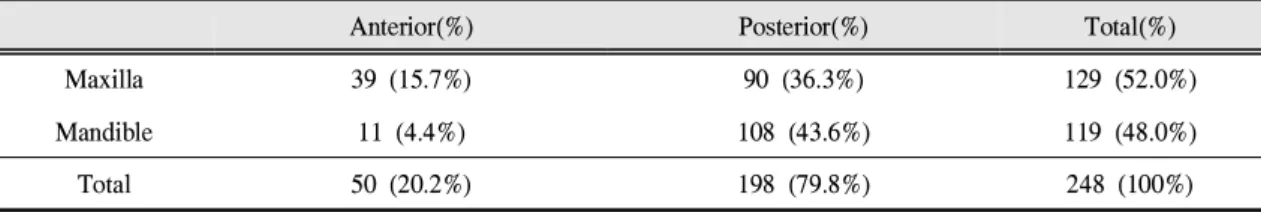
식립 위치에 따른 분포를 살펴보면 상악에 129개(52.0%), 하악에 119개 (48.0%) 식립되었으 며, 전치부에 식립된 경우가 50개 (20.2%), 구치 부에 식립된 경우는 198개 (79.8%) 였다(Table I).
식립된 임플란트의 직경을 살펴보면 18개(7.3%) 가 NP(직경 3.3mm), 144개(58.0%) 가 RP (직경 3.75 또는 4.0 mm), 86개(34.7%)가 WP(직경 5.0 mm) 였다. 18개의 NP 임플란트 중 하나만을 제 외하고는 모두 전치부에서 식립되었으며, 86개 의 WP 임플란트 중에서 전치부에는 한 경우에 만 식립되었다(Table II).
식립된 임플란트의 길이는 11.5 mm 104개 (41.9%), 13 mm 100개(40.3%)로 대부분을 차지
Anterior(%) Posterior(%) Total(%)
Maxilla 39 (15.7%) 90 (36.3%) 129 (52.0%)
Mandible 11 (4.4%) 108 (43.6%) 119 (48.0%)
Total 50 (20.2%) 198 (79.8%) 248 (100%)
Table I. Distribution of implants
Maxilla Mandible
Total
Anterior Posterior Anterior Posterior
NP (3.3mm)
7 (2.8%)
1 (0.4%)
10 (4.0%)
0 (0.0%)
18 (7.3%)
RP (3.75/4.0mm) 31 (12.5%)
61 (24.6%)
1 (0.4%)
51 (20.5%)
144 (58.0%) WP
(5.0mm)
1 (0.4%)
28 (11.3%)
0 (0.0%)
57 (23.0%)
86 (34.7%)
Total 39
(15.7%)
90 (36.3%)
11 (4.4%)
108 (43.6%)
248 (100%) Table II. Distribution of implant diameter
하였고, 그 외에는 10 mm 38개(15.3%), 15 mm 4 개(1.6%), 8.5 mm 2개(0.8%)의 순서였다(Table III).
임플란트 수술 당일 healing abutment를 연결하 는 1단계 수술법을 이용한 경우는 128개 (51.6%) 였고, 식립 후 이차 수술을 나중에 진행하는 2단 계 수술법을 이용한 경우는 120개(48.4%)였다 (Table IV).
수술 부위에 치조골 재생술(Guided bone regeneration; GBR)을 시행한 경우가 총 100개 (40.3%)였는데, 그 중 임플란트 식립 전 GBR을 시행한 경우는 9개(3.6%)였고, 식립 전과 식립 시 동시에 시행한 경우도 1개 있었다. 나머지는 모 두 임플란트 식립 시 동시에 시행하였다. GBR 시에 사용된 골 이식재는 대부분 자가골과 이종
골(BioOssⓇ, Geistlich, Swiss)이었으며 둘을 같이 섞어 사용한 경우가 가장 많았다. 또 주로 사용 된 차폐막은 흡수성 콜라겐 차폐막(Bio-GideⓇ, Geistlich, Swiss)이었으며 그 밖에 OssixⓇ (3i, Colbar R&D, Ramat Hasharon, Israel)나 비흡수성 차폐막인 TefGen (American Custom Medical Inc., Lubbock, Tex, USA)이 사용된 경우도 있었다.
상악동 거상술을 시행한 증례는 총 36개 (14.5%)였으며 그 중 대부분인 32개(12.9%)에서 osteotome technique이 이용되었고 4개(1.6%) 증 례에서만 lateral sinus augmentation technique이 시 행되었다.
Maxilla Mandible
Total
Anterior Posterior Anterior Posterior
8.5mm 0(0.0%) 0(0.0%) 0(0.0%) 2(0.8%) 2(0.8%)
10mm 1(0.4%) 20(8.1%) 0(0.0%) 17(6.9%) 38(15.3%)
11.5mm 10(4.0%) 39(15.7%) 4(1.6%) 51(20.6%) 104(41.9%)
13mm 27(10.9%) 30(12.1%) 7(2.8%) 36(14.5%) 100(40.3%)
15mm 1(0.4%) 1(0.4%) 0(0.0%) 2(0.8%) 4(1.6%)
Total 39(15.7%) 90(36.3%) 11(4.4%) 108(43.6%) 248(100%)
Table III. Distribution of implant length
Maxilla Mandible
Total
Anterior Posterior Anterior Posterior
1 stage 18
(7.2%)
35 (14.1%)
5 (2.0%)
70 (28.3%)
128 (51.6%)
2 stage 21
(8.5%)
55 (22.2%)
6 (2.4%)
38 (15.3%)
120 (48.4%)
Total 39
(15.7%)
90 (36.3%)
11 (4.4%)
108 (43.6%)
248 (100%) Table IV. Distribution of implant surgery method
3. 임플란트 생존율
임플란트를 식립한 후 부하를 가한 시기는 술 후 평균적으로 5.2±2.8개월 이였으며, 식립 당일 즉시부하를 가한 경우부터 최대 23개월 후 시행 한 경우도 있었다. 식립 위치에 따라 분류해보면 상악에서는 평균 5.7±3.1 (0~23)개월 후에 하악에 서는 4.6± 2.4 (0~18)개월 후에 부하를 가하였다.
수술 당일부터 관찰 기간은 평균 26.0±11.8 개월 이였으며, 부하를 가한 기간부터는 20.5±11.8 개 월 이였다.
임플란트 식립 후 합병증이 생긴 경우는 총 58
Months in observation
Implant at beginning of interval
Implant failures during interval
Interval failure rate(%)
Cumulative failure rate(%)
Cumulative survival rate(%)
0-6 129 8 6.2 6.2 93.8
7-12 121 1 0.8 7.0 93.0
13-18 111 0 0 7.0 93.0
19-24 75 1 1.3 7.8 92.2
25-30 52 0 0.0 7.8 92.2
31-36 33 0 0.0 7.8 92.2
37-42 25 0 0.0 7.8 92.2
43-48 15 0 0.0 7.8 92.2
49-54 9 0 0.0 7.8 92.2
55-60 3 0 0.0 7.8 92.2
61- 1 0 0.0 7.8 92.2
Table V. Cumulative survival rates for total implants placed for 0 to 62 months in observation. ( a) Maxilla , b) Mandible , c) Total )
a) Maxilla
Months in observation
Implant at beginning of interval
Implant failures during interval
Interval failure rate (%)
Cumulative failure rate(%)
Cumulative survival rate(%)
0-6 119 1 0.8 0.8 99.2
7-12 117 1 0.9 1.7 98.3
13-18 109 0 0.0 1.7 98.3
19-24 84 0 0.0 1.7 98.3
25-30 59 0 0.0 1.7 98.3
31-36 36 0 0.0 1.7 98.3
37-42 23 0 0.0 1.7 98.3
43-48 11 0 0.0 1.7 98.3
49-54 8 0 0.0 1.7 98.3
55-60 2 0 0.0 1.7 98.3
61- 0 0 0.0 1.7 98.3
b) Mandible
Months in observation
Implant at beginning of interval
Implant failures during interval
Interval failure rate (%)
Cumulative failure rate(%)
Cumulative survival rate(%)
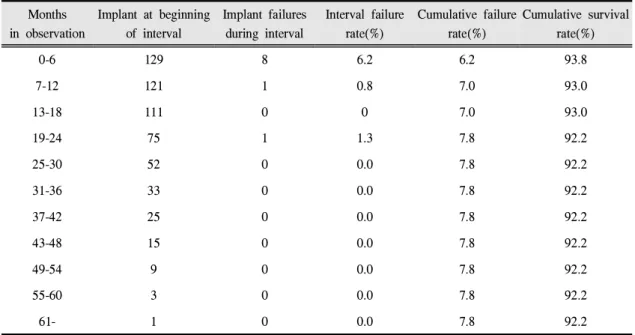
0-6 248 9 3.6 3.6 96.4
7-12 238 2 0.8 4.4 95.6
13-18 220 0 0.0 4.4 95.6
19-24 159 1 0.6 4.8 95.2
25-30 111 0 0.0 4.8 95.2
31-36 69 0 0.0 4.8 95.2
37-42 48 0 0.0 4.8 95.2
43-48 26 0 0.0 4.8 95.2
49-54 17 0 0.0 4.8 95.2
55-60 5 0 0.0 4.8 95.2
61- 1 0 0.0 4.8 95.2
c) Total (Maxilla, Mandible)
증례로 수술 후 임시 수복 전 기간 중 생긴 경우 가 34증례였으며, 임시 수복 후에 생긴 경우가 24증례였다. 수술 후 합병증으로는 1단계 수술법 으로 진행하였는데, cover screw가 노출된 경우가 가장 많았고 그 다음으로 healing abutment가 풀 린 경우가 대부분이었다. 임시 수복 후 합병증으 로는 도재 수복물의 파절이나 임플란트 주변의 염증 소견 등이 있었다.
연구에 포함된 총 248개의 임플란트 중 12개 가 실패한 것으로 간주되어 평균 26.1개월의 관 찰 기간 중 임플란트의 생존율은 95.2%였다. 그 중 상악에서 실패한 경우가 10개, 하악에서는 2 개로 각각의 생존율은 92.2%, 98.3% 이다. 또한 시기별로 보면 식립 수술 후 loading 전에 실패한 경우가 8개였으며, loading 후 실패한 경우는 4개 였다(Table V).
총괄 및 고안
이번 연구에서 평균 26.1개월의 관찰 기간 중 TiUniteTM 표면 처리한 임플란트 누적 생존율은 95.2%이다. 이는 Jung 등15)이 systemic review를 통 해 단일 임플란트로 지지되는 수복물의 5년 생존 율을 분석한 결과인 평균 96.8% (CI:95.9%-97.6%) 보다는 다소 낮은 생존율을 보여준다. 하지만 이 review에서는 ITI의 TPS (Titanium Plasma- sprayed), SLA (Sandblasted,large-grit, acid-etched), Astra의 Tioblast, Osseospeed, Bränemark의 TiUnite, 3i의 Osseotite 등 다양한 표면을 가진 임 플란트가 포함되었고, 식립 위치나 loading을 가 한 시기나 방법 등도 각기 다른 연구들을 평균 낸 것이기 때문에 이번 연구와 직접 비교하기는 한 계가 있다.
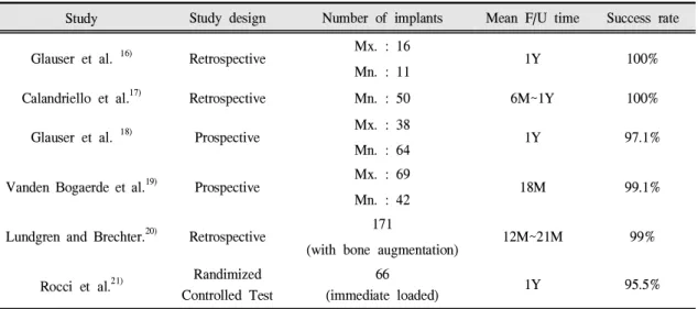
TiUniteTM 표면 처리한 임플란트에 관한 기존 연구들을 살펴보면 평균 1년 내외의 단기간 관찰 후 평균 95.5% 에서 100%의 성공률을 보인다.
Study Study design Number of implants Mean F/U time Success rate
Glauser et al. 16) Retrospective Mx. : 16
Mn. : 11 1Y 100%
Calandriello et al.17) Retrospective Mn. : 50 6M~1Y 100%
Glauser et al. 18) Prospective Mx. : 38
Mn. : 64 1Y 97.1%
Vanden Bogaerde et al.19) Prospective Mx. : 69
Mn. : 42 18M 99.1%
Lundgren and Brechter.20) Retrospective 171
(with bone augmentation) 12M~21M 99%
Rocci et al.21) Randimized Controlled Test
66
(immediate loaded) 1Y 95.5%
Table Ⅵ. Success rate of the TiUniteTMsurface implants
16-21)(Table VI) 하지만 이 연구들은 단일 임플란
트 뿐 아니라 인접하여 여러 개의 임플란트를 식 립한 경우나, 고정성은 물론 가철성 보철물로 수 복한 경우까지 모두 포함되었다. 또한 상대적으 로 높은 성공률을 보고한 경우에서는 포함 기준 을 엄격히 적용하여 충분한 골량이 확보된 경우 만 포함시키거나, 비교적 골질이 좋은 하악만 포 함시킨 경우도 있었다.
생존율을 상악과 하악을 분리해서 보면 두 군 사이에 결과가 큰 차이가 난다. 상악의 경우는 전체 식립한 129개 임플란트 중 10개가 실패하 여 누적 생존율이 92.2%인 반면 하악은 119개 중 단 2개만이 실패하여 98.3%의 누적 생존율을 보 였다. 상악에서 상대적으로 낮은 성공률을 보이 는 것은 임플란트의 실패를 야기할 수 있는 요인 중 불충분한 골질 및 골량과 관련이 있다.
Misch22)에 의하면 임플란트의 성공률을 높이기 위해서는 수직적 골 높이가 최소 10 mm 이상 되 어야 하나, 상악 구치부는 치조골 흡수와 동반되 는 상악동 함기화로 가용골이 부족한 경우가 많 고, 또한 치밀골층이 얇고 밀도가 낮은 type Ⅲ 또는 type Ⅳ 의 형태가 대부분이라고 하였다. 실
제로 이번 연구과정에서도 수술시 골질을 기록 한 경우에서 상악에서는 70개 중 54개에서 type
Ⅲ 또는 type Ⅳ의 골질을 보이고 있었다. 또한 상악의 129개 임플란트 중 36개(27.9%)의 임플란 트 식립 시 상악동 거상술을 동반하여 식립되었 다.
기존의 연구에서도 같은 종류의 임플란트를 사용했을 때 상악에서 하악보다 상대적으로 낮 은 성공률을 보인 경우가 많았다. Friberg 등23)이 TiUniteTM 표면 처리한 임플란트의 생존율에 관 한 1년 동안의 전향적 연구를 한 결과 총 478개 의 임플란트를 식립하여 그 중 5개가 실패하였 는데, 이는 모두 상악에 식립 된 임플란트였다.
누적 성공률은 상악과 하악에서 각각 98.6%, 100%로 보고하였다. Fugazzotto 등24)의 연구에서 도 TPS and SLA (sandblasted, large-grit acid etched) 표면처리한 5,526개의 ITI 임플란트를 식 립하여 최고 72개월 이상 누적성공률을 평가하 였는데, 상악에서는 94.8% 하악에서는 97.5%를 보였다.
임플란트 식립 후 합병증이 나타난 경우는 58 개로 2단계 수술 후 cover screw가 노출 된 경우
가 가장 많았는데, 이런 경우 주위 상피조직이 자라 들어오고 치태가 침착되기 쉬워 염증이 생 기기 유리한 환경이 된다. 그 결과 노출되지 않 은 경우와 비교해 치조골 소실이 많이 일어나게
된다.25,26) 그러므로 노출된 cover screw는 healing
abutment로 교체해준 후 철저한 구강 위생 관리 를 해 주어야 한다.27)본 연구에서 살펴본 증례에 서도 노출된 cover screw는 healing abutment로 교 체해 주어 해결하였는데, 추후 임플란트의 생존 율에는 영향을 미치지 않았다. 임시 보철물로 수 복된 후 나타난 합병증으로는 임시 또는 도재 수 복물이 파절되거나 임플란트 주위로 염증이 생 긴 경우 등이 있었다. 보철물 파절 시 재제작이 필요한 경우는 없었다. 임플란트 주위염이 생긴 경우에서는 항생제 복용과 소파술로 처치하였 고, 한 증례에서는 수술을 시행하였다.
Salinas와 Eckert2)는 systemic review를 통해 단 일 치아 수복에 있어 임플란트를 이용한 경우와 치아 지지 보철물을 이용한 경우의 성공률을 비 교해보고자 하였는데, 결과적으로 두 방법을 직 접 비교해 놓은 연구는 아직까지 없었고 다만 각 각의 술식에 대한 60개월의 성공률의 평균을 비 교하면 임플란트의 경우는 95.1% (CI:92.2%- 98.0%)였던데 비하여 고정성 보철물에서는 84.0% (CI:79.1%-88.9%)에 불과하였다고 하였다.
고정성 보철물의 실패는 대부분 치아우식증, 치 주질환 또는 근관내 병소 등 생물학적인 것이고
28-30) 그 밖에 구조적인 합병증으로 유지력이나
지대치의 파절 등과 관련이 있다고 하였다.31,32) 이러한 합병증들은 수복 후 시간이 지날수록 더 많은 빈도로 나타날 수 있고, 그러므로 연구 기 간이 길어질수록 보철물의 생존율은 감소하게 된다. 반면에 임플란트는 실패하는 경우 대부분 식립 초기 일년 이내에 일어나게 된다.33,34) 이번 연구 결과에서도 살펴보면 총 12개의 실패한 임 플란트 중 11개가 초기 일년 내에 발견되어 제거 되었다. 나머지 하나의 증례도 식립 후 보철 완 료 7달 후에 실패로 간주되어 제거 되었다. 물론 임플란트에서도 장기적으로 사용시 보철적 합병
증으로 인해 재제작이 필요한 경우도 있고, 임플 란트 주위염이나 과도한 교합력 등에 의한 후기 실패(late failure)가 나타나기는 하지만 실제 생존 율을 크게 감소시킬 만큼 많은 빈도가 나타나는 것은 아니다.35) 장기적인 비교 연구가 이루어질 수록 상실 치아를 임플란트로 수복한 경우와 자 연치를 지대치로 한 고정성 보철물을 사용한 경 우의 생존율 차이가 증가할 수도 있을 것이다.
결 론
본 연구에서 단일 치아를 상실한 경우 TiUniteTM 표면 처리한 임플란트를 사용하여 수 복하였을 때 평균 26.1개월의 누적 생존율은 95.2%였고, 그 중 상악에서는 92.2%, 하악에서는 98.3%의 생존율을 보여 기존의 연구들과 유사한 결과를 얻었다. 이는 고정성 보철물을 사용하여 수복했던 기존의 연구 결과에 비해 좋은 결과를 보여줌으로써 단일 치아를 상실 한 경우 임플란 트를 이용하여 수복하는 것이 인접치를 보존할 뿐 아니라 장기적인 예후 측면에서도 바람직한 술식임을 보여준다. 하지만 이번 연구는 평균 관 찰 기간이 짧고 후향적 연구라는 한계를 가지고 있기 때문에, 고정성 보철물과 임플란트의 유용 성에 관해서는 보다 높은 수준의 장기적인 비교 연구가 필요할 것으로 사료된다.
참 고 문 헌
1. Andersson B, Odman P, Lindvall AM et al.
Single-tooth restorations supported by osseointegrated implants: results and experiences from a prospective study after 2 to 3 years. Int J Oral Maxillofac Implants 1995;10:702-711.
2. Salinas TJ, Eckert SE. In patients requiring single-tooth replacement, what are the outcomes of implant- as compared to tooth-supported restorations?
Int J Oral Maxillofac Implants 2007;22 Suppl:71-95.
3. Friberg B, Grondahl K, Lekholm U. A new self-tapping Branemark implant: clinical and
radiographic evaluation. Int J Oral Maxillofac Implants 1992;7:80-85.
4. Albrektsson T, Branemark PI, Hansson HA et al.
Osseointegrated titanium implants. Requirements for ensuring a long-lasting, direct bone-to-implant anchorage in man. Acta Orthop Scand 1981;52:
155-170.
5. Jaffin RA, Berman CL. The excessive loss of Branemark fixtures in type IV bone: a 5-year analysis. J Periodontol 1991;62:2-4.
6. Jemt T. Implant treatment in resorbed edentulous upper jaws. A three-year follow-up study on 70 patients. Clin Oral Implants Res 1993;4:187-194.
7. Sul YT, Johansson CB, Kang Y et al. Bone reactions to oxidized titanium implants with electrochemical anion sulphuric acid and phosphoric acid incorporation. Clin Implant Dent Relat Res 2002;4:78-87.
8. Albrektsson T, Johansson C, Lundgren AK et al.
Experimental studies on oxidized implants. A histomorphometrical and biomechanical analysis.
Appl Osseointegration Res 2000;1:21-24.
9. Huang YH, Xiropaidis AV, Sorensen RG et al. Bone formation at titanium porous oxide (TiUnite) oral implants in type IV bone. Clin Oral Implants Res 2005;16:105-111.
10. Degidi M, Petrone G, Iezzi G et al. Histologic evaluation of a human immediately loaded titanium implant with a porous anodized surface. Clin Implant Dent Relat Res 2002;4:110-114.
11. Ivanoff CJ, Widmark G, Johansson C et al. Histologic evaluation of bone response to oxidized and turned titanium micro-implants in human jawbone. Int J Oral Maxillofac Implants 2003;18:341-348.
12. Buser D, Mericske-Stern R, Bernard JP et al.
Long-term evaluation of non-submerged ITI implants.
Part 1: 8-year life table analysis of a prospective multi-center study with 2359 implants. Clin Oral Implants Res 1997;8:161-172.
13. Buser D, Weber HP, Bragger U et al. Tissue integration of one-stage ITI implants: 3-year results of a longitudinal study with Hollow-Cylinder and Hollow-Screw implants. Int J Oral Maxillofac
Implants 1991;6:405-412.
14. Cochran DL, Buser D, ten Bruggenkate CM et al.
The use of reduced healing times on ITI implants with a sandblasted and acid-etched (SLA) surface:
early results from clinical trials on ITI SLA implants.
Clin Oral Implants Res 2002;13:144-153.
15. Jung RE, Pjetursson BE, Glauser R et al. A systematic review of the 5-year survival and complication rates of implant-supported single crowns. Clin Oral Implants Res 2008;19:119-130.
16. Glauser R, Gottlow J, Lundgren AK et al. Immediate occlusal loading of Brnemark Mk IV TiUnite implants placed in bone quality type 4. Appl Osseointegration Res 2002;3:22-24.
17. Calandriello R, Tomatis M, Vallone R et al.
Immediate occlusal loading of single lower molars using Branemark System Wide-Platform TiUnite implants: an interim report of a prospective open-ended clinical multicenter study. Clin Implant Dent Relat Res 2003;5 Suppl 1:74-80.
18. Glauser R, Lundgren AK, Gottlow J et al. Immediate occlusal loading of Branemark TiUnite implants placed predominantly in soft bone: 1-year results of a prospective clinical study. Clin Implant Dent Relat Res 2003;5 Suppl 1:47-56.
19. Vanden Bogaerde L, Pedretti G, Dellacasa P et al.
Early function of splinted implants in maxillas and posterior mandibles using Branemark system machined-surface implants: an 18-month prospective clinical multicenter study. Clin Implant Dent Relat Res 2003;5 Suppl 1:21-28.
20. Lundgren S, Brechter M. Preliminary findings of using oxidized titanium implants in reconstructive jaw surgery. Appl Osseointegr Res 2002;3:35-39.
21. Rocci A, Martignoni M, Gottlow J. Immediate loading of Branemark System TiUnite and machined-surface implants in the posterior mandible:
a randomized open-ended clinical trial. Clin Implant Dent Relat Res 2003;5 Suppl 1:57-63.
22. Misch CE. Maxillary sinus augmentation for endosteal implants: organized alternative treatment plans. Int J Oral Implantol 1987;4:49-58.
23. Friberg B, Dahlin C, Widmark G et al. One-year
results of a prospective multicenter study on Branemark System implants with a TiUnite surface.
Clin Implant Dent Relat Res 2005;7 Suppl 1:S70-75.
24. Fugazzotto PA, Vlassis J, Butler B. ITI implant use in private practice: clinical results with 5,526 implants followed up to 72+ months in function. Int J Oral Maxillofac Implants 2004;19:408-412.
25. Toljanic JA, Banakis ML, Willes LA et al. Soft tissue exposure of endosseous implants between stage I and stage II surgery as a potential indicator of early crestal bone loss. Int J Oral Maxillofac Implants 1999;14:436-441.
26. Tal H, Artzi Z, Moses O et al. Spontaneous early exposure of submerged endosseous implants resulting in crestal bone loss: a clinical evaluation between stage I and stage II surgery. Int J Oral Maxillofac Implants 2001;16:514-521.
27. Jeong SM, Choi BH, Li J et al. Influence of abutment connections and plaque control on the initial healing of prematurely exposed implants: an experimental study in dogs. J Periodontol 2008;79:1070-1074.
28. Cheung GS, Dimmer A, Mellor R et al. A clinical evaluation of conventional bridgework. J Oral Rehabil 1990;17:131-136.
29. Hochman N, Mitelman L, Hadani PE et al. A clinical and radiographic evaluation of fixed partial dentures (FPDs) prepared by dental school students: a retrospective study. J Oral Rehabil 2003;30:165-170.
30. Holm C, Tidehag P, Tillberg A et al. Longevity and quality of FPDs: a retrospective study of restorations 30, 20, and 10 years after insertion. Int J Prosthodont 2003;16:283-289.
31. Karlsson S. Failures and length of service in fixed prosthodontics after long-term function. A longitudinal clinical study. Swed Dent J 1989;13:
185-192.
32. Walton TR. An up to 15-year longitudinal study of 515 metal-ceramic FPDs: Part 2. Modes of failure and influence of various clinical characteristics. Int J Prosthodont 2003;16:177-182.
33. Avivi-Arber L, Zarb GA. Clinical effectiveness of implant-supported single-tooth replacement: the Toronto Study. Int J Oral Maxillofac Implants 1996;11:311-321.
34. Henry PJ, Laney WR, Jemt T et al. Osseointegrated implants for single-tooth replacement: a prospective 5-year multicenter study. Int J Oral Maxillofac Implants 1996;11:450-455.
35. Esposito M, Hirsch JM, Lekholm U et al. Biological factors contributing to failures of osseointegrated oral implants. (II). Etiopathogenesis. Eur J Oral Sci 1998;106:721-764.
A Retrospective Study of Survival Rate in single Brnemark TiUnite
TMImplant
Hye-Jin Kim, Seung-Min Yang, Seung-Beom Kye, Seung-Yun Shin
Department of Periodontics, The Institute of Oral Health and Science, Samsung Medical Center, Sungkyunkwan University School of Medicine.
Recently implant supported single crown is the popular treatment option to replace a single missing tooth. The purpose of this retrospective study was to analyze and evaluate the survival of implants with the TiUnite™surface for single tooth replacement. From September 2002 to December 2006, 269 TiUnite™ surfaced implants were used in single tooth replacements at the Institute of Oral Health Science, Samsung Medical Center. Twenty one cases were excluded because of neighbor implants, missing records & short follow up period. Among 248 implants, the 129 implants (52.0%) were inserted in the maxilla and 119 (48.0%) in the mandible. One hundred implants placement (40.3%) were combined with guided bone regeneration, and 36 implants placement (14.5%) were combined with sinus bone augmentation. Mean observation period was 26.0±11.8 months after implant placement. Twelve implants were recorded as failures, rendering a single implant survival rate of 95.2% over the observation period. Among failed 12 implants, 10 implants placed in the maxilla. The survival rate in the maxilla was 92.2% and in the mandible was 98.3%. The use of TiUnite™surfaced single implant placement showed high survival rate for short time period.
Key words: TiUniteTM surface, single tooth replacement, retrospective study, survival rate, implant
Correspondence to : Prof. Seung-Yun Shin
Department of Periodontics, The Institute of Oral Health Science Samsung Medical Center, 50 Irwon-dong, Gangnam-gu, Seoul, 135-710 Korea.
Fax +82-2-3410-0038, E-mail: [email protected]
Received :August 05, 2009, Last Revision :August 20, 2009, Accepted: September 25, 2009