Ⅰ. 서 론
Bra¨nemark과 Adel, Schroeder 등은 골 융합된 임플란 트가 치아 결손부위의 보철적 수복에 충분한 역할을 할 수 있음을 연구했고 그 이후 임플란트는 결손 치아의 수복을 위한 중요한 치료방법 중의 하나로 자리잡게 되었다1-3). 초 기에는 성공적인 골 융합을 위해서 임플란트 식립 수술 후 이차 수술이 이루어지기 이전 일정 기간동안 점막하에 위치 하는 시기가 필요하다고 제안되었으나 Buser와 Weber 등 은 일회법의 점막관통형 (tranmucosal, non-submerged type) 임플란트로도 기존의 2회 수술법 임플란트와 비교했
을 때 성공률의 차이가 없다고 발표하였다4,5). 1986년 Albrektsson과 Zarb 등은 임플란트의 성공 기준을 발표하 면서 이중 골흡수에 대해서는 식립 후 1년간 1-1.5 mm이 하 그 후 매년 0.2 mm이하의 골 흡수를 보이는 경우라고 주장했고6) Weber는 일회법 임플란트에 대한 연구에서 처 음 1년간은 평균 0.6 mm의 치조정 골 흡수를 보이고 그 후 매년 0.05 mm정도의 골 흡수를 보인다고 함으로써7)이회 법(submerged type)과 일회법 임플란트 모두에 대해 골 흡수량에 관한 성공의 기준이 정립되었다.
임플란트를 이용해 성공적으로 치아 결손 부위를 수복하 기 위해서 많은 치과의사들은 발치 또는 치아 상실 후 임플 박관수*∙윤규호*∙정정권*∙신재명*∙홍성철*∙전인성**
*인제대학교 부속 상계백병원 구강악안면외과학교실, **서울하버드치과의원
ITI
�implant system에 있어서 Bio-Oss
�와 Bio-Gide
�를 이용한 발치 후 즉시 임플란트의 유용성
THE USEFULNESS OF IMMEDIATE IMPLANTATION USING BIO-OSS�AND BIO-GIDE�IN ITI�IMPLANT SYSTEM
Kwan-Soo Park*, Kyu-Ho Yoon*, Jeong-Kwon Cheong*, Jae-Myung Shin*, Sung-Chul Hong*, In-Seong Jeon**
*Dept. of Oral & Maxillofacial Surgery, Sanggye Paik Hospital, Inje University
**Seoul-Harvard Dental Clinic
In the recent studies, many authors have reported that the success rate of immediate implantation has no difference compared to conventional staged implantation. Although the immediate implantation has many advantages over conventional approach, many clinicians don’t seem to practice because they think that most of the extraction sockets will have some bony gab with defects around implant and that this sit- uation makes the result of immediate implantation unpredictable.
We clinically analyzed 23 implanted sites of 18 patients treated with immediate implantation in our hos- pital from September 2003 to January 2004. The ITI�dental implant system was used and GBR procedure with Bio-Oss�and Bio-Gide� was done simultaneously. The pre & post-op. measurements were recorded such as alveolar crest-adjacent tooth CEJ distance, gingival crest-adjacent tooth CEJ distance, existence of periapical lesion, vertical defect around the extraction socket, horizontal defect around the extraction sock- et, probing depth, radiologic change of alveolar crest height.
We report a positive outcome about immediate implantation with review of literatures.
Key words : Immediate implantation, Implant, Xenograft, Bio-Oss, Bio-Gide Abstract
란트를 식립하기 위한 잘 성숙된 골이 형성되는 것을 기다 리기 위해 3개월 내지 6개월 또는 그 이상의 시간을 소모해 야 했으며 이것이 당연한 프로토콜로 여겨져 왔고 이 시기 동안 환자들은 저작과 심미에 있어 많은 불편함과 고통을 감내해야 했다. 1978년에 이르러 Schulte가 신선한 발치와 에 식립한 임플란트에 관해 발표한 이후8) 이에 관한 많은 연구가 이루어져왔다. 그러나 치료기간 단축을 포함한 즉시 식립 임플란트의 여러 가지 장점에도 불구하고 여전히 몇몇 단점이 알려져 있으며 즉시 식립 임플란트의 시술 및 이와 함께 동반될 수도 있는 골이식술 방법 등에 대해 완전히 일 치된 합의도 없는 상태이다9,10). 현재 우리나라에서도 여러 임상가와 연구자들이 발치 후 즉시 임플란트를 식립하는 치 료법에 관심을 갖고 이를 시행하고 있으나 아직도 많은 임 상가들은 발치 직후 치조골의 양이나 두께, 발치와의 크기, 치조골의 결손 등이 성숙한 골로 잘 채워진 치조골 위에 드 릴을 이용하여 구멍을 뚫고 임플란트를 식립하는 전통적인 방법을 사용하는 것에 비해 불리한 조건이 될지도 모른다는 경험적이거나 막연한 두려움을 가지고 있는 것도 사실이다.
하지만 많은 연구자들은 발치와에 즉시 식립하는 방법도 좋 은 치료 방법중의 하나라는 연구 결과를 속속 발표하고 있다11-14).
이에 저자 등은 다수의 환자에게 일회법 임플란트 중의 하 나인 ITI�시스템을 이용하여 발치 후 즉시 식립을 시행하 고 발치와의 상황에 따라 자가골 및 Bio-Oss�, Bio-Gide� 등을 이용한 골유도재생술을 동시에 시행하는 방법으로 양 호한 결과를 얻어 그 결과에 대해 임상∙방사선학적 평가 및 문헌고찰과 함께 보고하는 바이다.
Ⅱ. 환자 및 연구방법
2003년 9월부터 2004년 1월까지 인제대학교 부속 상계 백병원 치과/구강악안면외과에 내원하여 발치 후 즉시 식립 에 의해 임플란트 치료를 받은 환자 중 식립 시 발치와벽과 임플란트 몸체 간의 최대 간격이 1.5 mm 이상이면서 골유 도재생술을 동시에 시행한 경우를 선별하였다. 총 18명의 환자에게 식립된 23개의 임플란트를 대상으로 하였으며, 성별은 남성이 9명(13개의 임플란트), 여성이 9명(10개의 임플란트)이었고 연령은 16세에서 75세 사이(평균 42.7 세)였으며 발치의 원인이 된 질환으로는 만성 치주염이 8명 (13개의 임플란트), 외상이 4명(4개의 임플란트), 잔존치 근이 3명(3개의 임플란트), 유치잔존이 1명(1개의 임플란 트), 치근단 병소가 2명(2개의 임플란트)이었다. 임플란트 의 식립 부위는 상악 전치부가 7개, 하악 전치부가 4개, 상 악 구치부가 3개, 하악 구치부가 9개 였다.
임플란트는 SLA 표면처리한 solid type의 ITI�임플란트 (Institute Straumann AG, Waldenburg, Switzerland)
를 사용하였으며 직경은 4.1 mm가 20개, 4.8 mm가 3개 였고 길이는 12 mm가 10개, 10 mm가 11개, 8 mm가 2 개였다. 봉합시 연조직에 의한 healing cap 피개 정도에 따 라 submerged와 semi-submerged, non-submerged로 나누었으며 각각 2개, 9개, 12개 였다(Table 1).
치료 과정은 전층판막을 박리한 후 가능한한 최소한의 외 상이 가해지도록 조심스럽게 치아를 발거한 뒤 발치와 내부 를 충분히 소파하고 드릴링을 시행한 후 임플란트를 식립하 였다. 식립한 임플란트 주변에는 다양한 크기의 골 결손부 가 존재하였고 부위에 따라 자가골과 Bio-Oss� 및 Bio- Gide�(Geistlich AG, Wolhusen, Switzerland)를 다양한 조합으로 사용하여 골이식을 시행한 후 봉합하고 5-7일간 경구용 항생제와 진통소염제를 투여하였으며 7-10일 후 발 사를 시행하고 2-6개월 후 보철적 수복을 시행하였다 (Fig.1-1 ~ 3-7).
모든 식립부위에 대해 술전 및 술중에 다음의 항목을 측정 하였다.
1) 치조정(alveolar crest) - 인접치 CEJ까지의 수직 거리 2) 치은정(gingival crest) - 인접치 CEJ까지의 수직 거리 3) 발치와 주위의 수직적 최대 골 결손 깊이
4) 발치와 주위의 수평적 최대 골 결손 넓이 5) 치근단 부위의 병소 유무
술 후에는 각 환자마다 방사선학적 평가 항목으로 골 이식 된 치조정 최상방(grafted alveolar crest)에서 임플란트의 rough-smooth surface 경계까지의 수직 거리를 수술 완료 후 1, 3개월에 각각 측정했고, 임상적 측정 항목으로 치주 탐침을 이용해 보철 완료 후 1, 3개월에 탐침 깊이를 각각 측정하였다.
Ⅲ. 결 과
술전/술중 평가에서는 1) 치조정-인접치의 CEJ까지의 거 리가 2.0-4.0 mm, 평균 3.04 mm였고 2) 치은정-인접치 의 CEJ까지의 거리는 1.0-3.0 mm, 평균 1.85 mm였으며 3) 발치와 주위의 수직적 골 결손은 1.0-5.0 mm, 평균3.0 mm였고 4) 발치와 주위의 수평적 골결손은 1.0-3.0 mm, 평균 2.02 mm였다. 치근단 부위의 병소 유무에서는 있는 경우가 2개 부위, 없는 경우가 21개 부위였고 골이식 형태 는 Bio-Gide�만 사용한 것이 1개 부위, Bio-Oss�와 Bio- Gide�를 함께 사용한 경우가 18개 부위, 자가골과 Bio-Oss�, Bio-Gide�를 함께 사용한 경우가 3개 부위, 자가골과 Bio- Gide�를 함께 사용한 경우가 1개 부위였다(Table 2).
술후 평가에서 탐침 깊이는 평균 0.52 mm가 감소했고 방사선학적 검사에서 골이식된 치조정과 임플란트 rough- smooth surface 경계부까지의 거리가 평균 0.29 mm 감소 한 것으로 나타났다(Table 3).
Table 2.Measurements of Pre/Intra-OP. and GBR type
Case. Alveolar crest- Gingival crest- Vertical defect Horizontal defect
Periapical
No. adjacent tooth adjacent tooth around the Ext. around the Ext. lesion GBR type
CEJ(mm) CEJ(mm) socket(mm) socket(mm)
1 3.5 2.5 3.0 1.0 No G+O
2 2.5 1.5 1.0 2.0 No G+O
3 2.0 1.5 3.0 2.0 Yes G+O
4 4.0 3.0 3.0 3.0 No G+O
5 3.0 1.5 2.0 2.0 No G+O
6 2.0 1.0 3.5 2.5 No G+O
7 2.0 1.0 3.0 2.0 Yes G+O
8 2.5 1.0 3.0 2.0 No G+O
9 4.0 2.0 5.0 3.0 No G+O+A
10 4.0 2.0 2.0 1.0 No G+O
11 3.5 2.0 3.0 2.0 No G+O
12 2.0 1.0 1.0 1.0 No G
13 3.0 2.0 2.0 1.0 No G+O
14 2.5 1.0 2.0 1.0 No G+A
15 2.5 1.5 3.0 2.0 No G+O
16 4.0 2.0 4.0 3.0 No G+O+A
17 3.0 2.5 2.5 1.5 No G+O
18 3.0 2.0 4.0 2.0 No G+O
19 3.0 2.0 5.0 2.0 No G+O
20 4.0 2.0 4.0 3.0 No G+O
21 3.5 3.0 4.0 3.0 No G+O
22 4.0 3.0 5.0 3.0 No G+O+A
23 2.5 1.5 3.5 1.5 No G+O
G: Bio-Gide�, O: Bio-Oss�, A: Autogenous bone, Table 1.Detail of Patiets and Cases
Pt. Name &
Age Sex Tooth
Diagnosis Impant Submerge
Case No. No. Diameter Length type
01 Kim ○○ 38 M 32 Chronic periodontitis 4.1 12 Non - sub
02 42 4.1 12 Semi - sub
03 Kim ○○ 16 F 21 Apical lesion & Crown fx. 4.1 12 Non - sub
04 Kim ○○ 41 F 12 Chronic Periodontitis 4.1 10 Semi - sub
05 13 4.1 12 Semi - sub
06 Kim ○○ 51 F 36 Root rest 4.8 10 Semi - sub
07 Park ○○ 45 F 12 Apical lesion 4.1 12 Semi - sub
08 Bae ○○ 26 F 21 Root Fx. 4.1 12 Non - sub
09 Yoo ○ 42 F 46 Root rest 4.8 10 Submerged
10 Lee ○○ 52 M 32 Chronic Periodontitis 4.1 10 Non - sub
11 44 4.1 8 Non - sub
12 Lee ○○ 27 M 35 Remaining deciduous tooth 4.1 10 Non - sub
13 Lee ○○ 75 F 44 Chronic Periodontitis 4.1 10 Non - sub
14 Chang ○○ 34 M 36 Root rest 4.1 10 Semi - sub
15 Cho ○○ 45 M 42 Chronic Periodontitis 4.1 12 Non - sub
16 Choi ○○ 42 M 37 Vertical Fx. 4.8 10 Semi - sub
17 Heo ○○ 39 M 21 Root Fx. 4.1 12 Non - sub
18 Hwang ○○ 26 F 11 Root Fx. 4.1 12 Semi - sub
19 Lee ○○ 48 M 46 Chronic Periodontitis 4.1 10 Semi - sub
20 Lee ○○ 54 F 43 Chronic Periodontitis 4.1 12 Submerged
21 Chung ○○ 67 M 24 Chronic Periodontitis 4.1 10 Non - sub
22 25 4.1 10 Non - sub
23 26 4.1 8 Non - sub
Ⅳ. 총괄 및 고찰
시간에 따른 발치와의 치유 과정은 여러 연구에 의해서 밝 혀져 있으며 대체로 그 과정은 초기 혈병이 형성된 후 혈병 이 육아조직으로 대체되고 결합조직이 차차 육아조직을 대 체하게 되며 골양 조직의 석회화가 발치와의 바닥과 가장자 리에서 명백해지면서 골소주가 발치와를 거의 채우고 상피 조직에 의해 발치와가 완전히 덮이게 된 후 상피 하방이 거 의 완전한 골조직으로 채워지는 단계로 이루어져 있다15). 이 과정에서 4-6주 사이에 발치와의 골 형성 활동이 가장 활발 하고 8주가 지나면 골 형성 과정이 느려지면서 16주가 되면 골 형성이 마무리되고 이후에는 골 형성이 거의 일어나지 않는다16). 또한 발치 부위의 골 흡수가 어느 정도 일어난다 는 사실은 주지의 사실이며 Schropp 등은 골 흡수량이 발 치 초기 1년 이내에 협설측으로 50%에 이르고 이러한 변화 의 2/3는 발치 후 3개월 이내에 일어나며 구치부에서 더욱 심하다는 연구 결과를 발표한 바 있다17).
기존의 치아 수복 방법에 비해 많은 장점을 가진 임플란트 치료법이 널리 소개된 이후에도 환자들은 발치 후 임플란트 로 치아를 수복하는데 걸리는 오랜 기간 때문에 임플란트 치료법을 포기하는 경우가 많았다. 더구나 위에 언급한 발 치 후 상당한 량의 초기 골 흡수는 지연된 시기의 임플란트
수술을 더욱 어렵게 만들 가능성이 있으며 오히려 발치와가 가지고 있는 뚜렷한 골 형성 능력은 많은 연구자들로 하여 금 발치 후 임플란트의 즉시 식립에 관심을 가지게 하였다.
사람에게 즉시 식립한 임플란트를 1978년 Schulte가 처음 발표한8)이후 즉시 식립된 임플란트와 지연 식립된 임플란 트를 비교한 연구들이 속속 발표되었는데 두 가지 치료 방 법이 모두 비슷한 성공률을 보인다고 하였다18-20). 단면이 원 형인 임플란트에 비해 발치와의 형태는 치근의 모양에 따라 다양한 모양을 가지고 있으며 이러한 발치와에 임플란트를 식립하면 임플란트와 골벽 사이에는 필연적으로 수평적인 간격이 생기게 된다. Akimoto 등은 돼지 악골에 인공적으 로 형성한 발치와를 이용한 실험에서 임플란트와 골사이에 1.4 mm 이내의 간격은 저절로 골에 의해 채워진다고 하였 고21)Paolantonio 등은 실제 환자에서 임플란트와 골 사이 에 2.0 mm 이내의 간격이 아무런 골이식이나 골유도재생 술 없이 채워질 수 있다고 하였으며22) Schropp 등은 즉시 식립 임플란트에서 3벽성 골 결손부가 가로 5 mm, 세로 4 mm, 임플란트와의 거리 2 mm 이내인 경우 70%가 3개월 이내에 자연 치유되었다고 하였다23). 이와 같이 비록 발치와 의 골벽과 임플란트 사이에 간격이 어느 정도 존재해도 골 의 형성이 이루어진다는 연구가 다수 있기는 하나 발치 이 전부터 커다란 골 결손부가 존재하는 경우나 발치 과정에 Table 3.Post-OP. measurement
Case Smooth/rough surface border~grafted alveolar crest(mm) Probing depth (mm) One month after operation Three months after operation One month Three months No.
after loading after loading
1 1.5 1.5 2.5 2.0
2 1.0 0.5 3.5 2.5
3 1.0 1.0 2.5 2.5
4 1.5 1.5 2.5 2.0
5 1.5 1.5 3.0 2.0
6 1.0 1.0 2.0 1.5
7 1.5 0.5 2.5 2.0
8 0.5 0.5 2.5 2.5
9 2.5 1.5 2.0 2.0
10 2.5 2.0 2.5 1.5
11 1.5 1.5 2.5 2.0
12 0.5 0.5 2.5 2.5
13 1.0 1.0 2.0 2.0
14 0 0 2.0 1.5
15 0.5 0.5 2.5 2.0
16 1.5 0.5 3.0 2.0
17 0.5 0.5 3.0 2.5
18 2.5 1.0 3.0 2.0
19 0.5 0.5 3.0 1.5
20 0.5 1.0 3.0 1.5
21 2.0 1.5 2.5 2.5
22 0.5 0.5 2.0 2.0
23 0 0 2.0 2.0
의해 발생한 큰 골 결손부위를 발치와의 자연적인 골형성능 에 의해 충분히 채울 수 있는지 여부는 확실하지 않다. 이러 한 발치와에서 일어날 수 있는 골 결손부위의 처치는 다양 한 골유도재생술에 의해 이루어질 수 있다. 본 연구에서는 대상의 선정에 있어 발치와벽과 임플란트 몸체의 간격이 최 대 1.5 mm 이상인 경우만을 대상으로 하였으며 이는 위에 언급한 골유도재생술 없이도 자연 치유될 수 있다고 주장하 는 연구들을 바탕으로 결정한 것으로 실제 임상에서 발치를 시행한 후 즉시 임플란트를 식립하여 보면 실제 발치와의 크기와 임플란트 직경의 차이나 임플란트의 적절한 위치 선 정 등의 문제로 인해 1.5 mm 이상의 간격을 가진 부분을 흔히 관찰 할 수 있고 많은 경우 골유도재생술이 필요성을 느끼게 된다.
임플란트 식립 시 골량 부족은 성공률을 낮추는 요인이 될 수 있다는 사실이 이미 알려져 있고24)다양한 기술과 재료가 개발되어 사용이 시도되고 있는데, 이들 중 성장 인자나 골 형성 단백질 등은 아직 임상적으로 사용이 불분명한 상태이 고 차단막을 이용한 골유도재생술이 일반적으로 사용된다.
이미 1989년에 Lazzara는 expanded polytetrafluo- roethylene(e-PTFE) 차단막을 이용해 신선한 발치와의 골을 증대시키는 술식을 발표하였고25), 이 후 여러 연구자들 이 e-PTFE 차단막을 이용해 임플란트의 시술시에 나타나 는 골 결손 부위를 회복하고 증대하는 방법을 연구 하였
다26-28). 이러한 연구 결과 e-PTFE 차단막이 골 재생에 좋
은 결과를 보이는 것으로 나타났음에도 불구하고 이 재료는 쉽게 노출되며 노출 시 막표면에 세균 군락이 빠르게 형성 되어 조기에 제거가 필요하게 되고 이는 골 형성량이 부족 으로 이어지게 되며 비흡수성이므로 2차 수술시 제거가 필 요한 단점을 가지고 있다29,30). 특히 즉시 식립법에서는 발치 와의 상부가 넓어 봉합 부위의 연조직 긴장이 높아져 봉합 부위가 벌어지는 경향이 있으며 이는 막의 노출 위험을 증 가시키므로 노출시에도 안전한 막이 필요하다. 초기의 흡수 성 차단막인 polylactic acid/polyglycolic acid(PLA/
PGA) 차단막은 골재생의 측면에서 보면 e-PTFE 차단막 보다 불리하다고 알려져 있고31), 이에 비해 새로운 생체흡수 성 콜라겐 차단막은 적절한 충전재와 함께 사용할 경우 골 유도재생술에 좋은 결과를 나타낸다는 연구들이 있다32,33). 골 결손부의 골이식재 또는 충전재로는 자가골, 동종골, 이 종골, 인공골 등이 있는데, 이중 자가골은 차단막과 함께 사 용하는 경우 가장 좋은 골이식재이나34,35), 새로운 채취 과정 이 필요한 단점이 있고, 동종골과 인공골은 골유도재생술에 서 새로운 골 형성을 방해할 수도 있다는 연구 결과가 있
다35,36). 최근에는 골전도능(osteoconduction)을 갖는 이종
골의 일종인 탈단백 소뼈 무기질(deproteinized bovine bone mineral)이 골유도재생술의 골이식재 또는 충전재로 서 좋은 결과를 보였다고 알려지기 시작했다37,38).
탈단백 소뼈 무기질의 일종인 Bio-Oss�와 흡수성 콜라겐 차단막의 일종인 Bio-Gide�를 사용하여 이회법의 임플란트 를 식립할 때 나타난 골 결손부의 처치시 좋은 결과를 나타 내었다는 보고가 있고32)일회법 임플란트를 발치 후 즉시 식 립하면서 Bio-Oss�와 Bio-Gide�를 이용해 골 결손부를 처 치하여 좋은 결과를 얻은 연구도 발표되었다39,40). 본 연구에 서도 일회법 임플란트인 ITI� 시스템을 이용하여 발치 후 즉시 식립을 시행하였고 식립 부위에 나타난 다양한 골 결 손부를 처치하기 위해 골이식재로서 Bio-Oss�를, 차단막으 로서 Bio-Gide�만을 이용하기로 하였으나 골 결손량이 매 우 적었던 12번 증례에서는 골이식재를 사용하지 않고 차 단막만을 단독으로 사용하였고 역시 골 결손량이 상대적으 로 적었다고 판단한 14번 증례에서는 차단막과 함께 골이 식재로서 약간의 자가골만을 사용하였다. 골결손량이 매우 많았던 9, 16, 22번 증례에서는 Bio-Oss�와 자가골을 함께 사용하였다. 계획된 대로 Bio-Oss�와 Bio-Gide�만을 사용 하지 않았던 이유는 골 결손량에 따라 이식재의 필요성과 그 양의 조절이 필요한 상황이 발생하였기 때문이고 환자와 의 관계에 있어 Bio-Oss�와 Bio-Gide�와 같은 고가의 재 료를 사용하는 데에도 일부 제약이 있었기 때문이다. 수직 적 골 결손부의 크기가 5.0 mm에 이르는 부위나 수평적 골 결손이 3.0 mm 정도에 이르는 경우도 있었지만 모든 증례 에서 별다른 이상 없이 수술 부위의 치유가 이루어 졌으며 보철 수복을 완료하였다.
수술 시 일회법 임플란트를 사용했음에도 점막하에 위치 시킨(submerged) 임플란트는 초기 고정력이 30N 이하로 낮았고 골 결손부가 컸기 때문이며 점막하에 위치시키지 않 은(non-submerged) 임플란트는 초기 고정력이 좋고 골 결손부가 상대적으로 적었기 때문이었다. 일부만 점막하에 위치시키는(semi-submerged) 방법은 그 중간 정도에 속 하거나 완전히 점막하에 위치시키기에는 연조직의 양이 불 충분한 경우로 점막하에 위치시키는 방법과 같이 차단막과 이식재의 노출을 막기 위한 프로토콜중의 하나이며 이 방법 은 일회법 임플란트의 구조적 특성상 사용하는 방법이기도 하다. 대체로 다량의 골이식재와 차단막을 사용한 경우에는 가능하면 semi-submerged 또는 submerged 프로토콜을 이용하는 것이 좋다고 하며41)본 연구에서도 이러한 프로토 콜을 적절히 이용하였다.
수술 후 평가 항목을 보면 치조정에서 rough-smooth surface경계부까지의 거리는 골 흡수량과 연관 지어 생각해 볼 수 있는데 대체로 0.5 mm 이하의 변화를 보였지만 골 결손부의 크기가 크고 골이식량이 많았던 증례에서는 1.0~1.5 mm 정도의 많은 감소를 보였다. 이는 이식된 골 의 양이 많아서 흡수된 양도 많은 것에 기인하는 점이 있고 결손부가 클수록 수술 시 초기 고정력을 잘 얻기 위해 조금 더 깊이 식립하게 됨으로써 임플란트의 생리적 폭경에 의한
골 흡수가 증가하는 경향을 보인 것이 하나의 원인이 될 것 으로 보인다.
또 하나의 평가 항목인 탐침 깊이는 자연치에서와 마찬가 지로 임플란트 주변 치주의 건강상태를 나타내는 지표로 사 용될 수 있으나 임플란트 주위의 염증 상태가 있을 경우 자 연치에서보다 훨씬 급격하게 증가하는 경향을 보이고42), 건 강하게 유지되고 있는 임플란트 주변에서는 3.0 mm 이내 의 탐침 깊이를 보인다고 알려져 있다4,43,44,45). 본 연구에서 탐침 깊이의 변화가 관찰되지 않은 5개 증례를 제외하고는 술 후 시간의 경과에 따라 탐침 깊이가 감소하였고 2 개 증 례에서는 1.5 mm 정도로 많은 양이 감소하였으며 증가한 경우는 없어 임상적으로 임플란트 주변에 병적 소견을 보이 지 않는 점과 일치하였다. 하지만 자연치에서보다 임플란트 주변에서는 탐침을 행하는 힘에 의해 그 측정값이 많은 영 향을 받기 때문에46)추후 연구에서 정확성을 높이기 위해서 는 힘의 조절이 가능한 탐침을 이용해 측정하는 것이 필요 하리라 사료된다.
Ⅴ. 결 론
2003년 9월부터 2004년 1월까지 본원에 내원한 환자 18명에게 즉시 식립한 23개의 임플란트를 대상으로 한 이 번 연구에서 다음과 같은 결론을 얻었다.
1. 발치 후 즉시 식립 술식은 발치와벽과 임플란트 간격이 1.5 mm 이상 존재하는 경우에도 골유도 재생술을 동반 하면 예측가능한 방법이다.
2. 다량의 골 결손부도 Bio-Oss�와 Bio-Gide�를 이용한 골유도재생술로 좋은 결과를 얻을 수 있으며 본 연구에 서는 수평적 골 결손부의 길이가 최대 5 mm, 수직적 골 결손부의 길이가 최대 3 mm일 때 까지도 가능했다.
3. 일회법 점막관통형 임플란트의 이용 시 골 결손부가 큰 경우 semi-submerged 또는 submerged 방법이 막의 노 출이나 골재생량 감소를 막는 방법의 하나로 유용하다.
이상을 토대로 볼 때 ITI�임플란트 시스템에서 발치후 즉 시 식립을 시행하는 경우 골 결손 부위에 Bio-Oss�를 골이 식재로 이용하고, Bio-Gide�를 차단막으로 사용하여 외과 의가 엄격한 프로토콜로 semi-submerged 또는 sub- merged 방법을 이용하는 것은 유용한 술식의 하나가 될 수 있을 것으로 보인다.
참고문헌
1. Bra¨nemark PI, Hansson BO, Adell R et al : Osseo- inte- grated implants in the treatment of the edentulous jaw.
Experience from a 10-year period. Scand J Plast Reconstr Surg Suppl 16 : 1, 1977.
2. Adel R, Lekholm U, Rockler B et al : A 15-year study of osseointegrated implants in the treatment of the edentu-
lous jaw. Int J Oral Surg 10(6) : 387, 1981.
3. Schroeder A, Stich H, Straumann F et al : The accumula- tion of osteocementum around a dental implant under physical loading SSO Schweiz Monatsschr Zahnheilkd 88(10) : 1051, 1978.
4. Buser D, Weber HP, Lang NP : Tissue integration of non- submerged implants. 1-year results of a prospective study with 100 ITI hollow-cylinder and hollow-screw implants.
Clin Oral Implants Res 1(1) : 33, 1990.
5. Weber HP, Buser D, Donath K et al : Comparison of healed tissues adjacent to submerged and non-submerged unloaded titanium dental implants. A histometric study in beagle dogs. Clin Oral Implants Res 7(1) : 11, 1996.
6. Albrektsson T, Zarb G, Worthington P et al : The long- term efficacy of currently used dental implants: a review and proposed criteria of success. Int J Oral Maxillofac Implants 1(1) : 11, 1986.
7. Weber HP, Crohin CC, Fiorellini JP : A 5-year prospective clinical and radiographic study of non-submerged dental implants. Clin Oral implant Res 11(2) : 144, 2000.
8. Schulte W, Kleineikensheidt H, Linder K et al : The Tu¨bingen immediate implant in clinical studies. Dtsch Zaha¨nrztl Zeischr 33 : 348, 1978.
9. Schwartz-Arad D, Chaushu G : The ways and wherefores of immediate placement of implants into fresh extraction sites:A riterature review. J Periodontol 68:915, 1997.
10. Mayfield LJA : Immediate, delayed and late submerged and transmucosal implants. In: Lindhe J(ed). Proceedings of the 3rd European Workshop on Periodontology: Implant Dentistry. Berlin, Quintessenz 1999. p520.
11. Gelb DA : Immediate implant surgery: three-year retro- spective evaluation of 50 consecutive cases. Int J Oral Maxillofac Implants 8(4) : 388, 1993.
12. Grunder U, Polizzi G, Goene R et al : A 3-year prospective multicenter follow-up report on the immediate and delayed-immediate placement of implants. Int J Oral Maxillofac Implants 14(2) : 210, 1999.
13. Paolantonio M, Dolci M, Scarano A et al : Immediate implantation in fresh extraction sockets. A controlled clini- cal and histological study in man. J Periodontol 72(11) : 1560, 2001.
14. Covani U, Crespi R, Cornelini R et al : Immediate implants supporting single crown restoration: a 4-year prospective study. J Periodontol 75(7) : 982, 2004.
15. Amler MH : The time sequence of tissue regeneration in human extraction wounds. Oral Surg Oral Med Oral Pathol 27(3) : 309, 1969.
16. Evian CI, Rosenberg ES, Coslet JG et al : The osteogenic activity of bone removed from healing extraction sockets in humans. J Periodontol 53(2) : 81, 1982.
17. Schropp L, Wenzel A, Kostopoulos L et al : Bone healing and soft tissue contour changes following single-tooth extraction: a clinical and radiographic 12-month prospec- tive study. Int J Periodontics Restorative Dent 23(4) : 313, 2003.
18. Yukna R : Clinical comparison of hydroxyapatite-coated titanium dental implants placed in fresh extraction sockets and healed sites. J Periodontol 62(7) : 468, 1991.
19. Gelb DA : Immediate implant surgery: three-year retro- spective evaluation of 50 consecutive cases. Int J Oral Maxillofac Implants 8(4) : 388, 1993.
20. Watzek G, Haider R, Mensdorff-Pouilly N, et al : Immediate and delayed implantation for complete restora- tion of the jaw following extraction of all residual teeth: a retrospective study comparing different types of serial
immediate implantation. Int J Oral Maxillofac Implants 10(5) : 561, 1995.
21. Akimoto K, Becker W, Persson R et al : Evaluation of tita- nium implants placed into simulated extraction sockets: a study in dogs. Int J Oral Maxillofac Implants 14(3) : 351, 1999.
22. Paolantonio M, Dolci M, Scarano A et al : Immediate implantation in fresh extraction sockets. A controlled clini- cal and histological study in man. J Periodontol 72(11):1560, 2001.
23. Schropp L, Kostopoulos L, Wenzel A : Bone healing fol- lowing immediate versus delayed placement of titanium implants into extraction sockets: a prospective clinical study. Int J Oral Maxillofac Implants 18(2) : 189, 2003.
24. Lekholm U, Adell R, Lindhe J et al : Marginal tissue reac- tions at osseointegrated titanium fixtures. (II) A cross-sec- tional retrospective study. Int J Oral Maxillofac Surg 15(1) : 53, 1986.
25. Lazzara RJ : Immediate implant placement into extraction sites: surgical and restorative advantages. Int J Periodontics Restorative Dent 9(5) : 332, 1989.
26. Becker W, Becker BE : Guided tissue regeneration for implants placed into extraction sockets and for implant dehiscences: Surgical techniques and case reports. Int J Periodontics Restorative Dent 10(5) : 377, 1990.
27. Becker W, Dahlin C, Becker BE et al : The use of e-PTFE barrier membranes for bone promotion around titanium implants placed into extraction sockets: a prospective mul- ticenter study. Int J Oral Maxillofac Implants 9(1) : 31, 1994.
28. Dahlin C, Lekholm U, Becker W et al : Treatment of fen- estration and dehiscence bone defects around oral implants using the guided tissue regeneration technique: a prospec- tive multicenter study. Int J Oral Maxillofac Implants 10(3) : 312, 1995.
29. Lekholm U, Becker W, Dahlin C et al : The role of early versus late removal of GTAM membranes on bone forma- tion at oral implants placed into immediate extraction sockets. An experimental study in dogs. Clin Oral Implants Res 4(3) : 121, 1993.
30. Gher ME, Quintero G, Sandifer JB et al : Combined den- tal implant and guided tissue regeneration therapy in humans. Int J Periodontics Restorative Dent 14(4) : 332, 1994.
31. Simion M, Scarano A, Gionso L et al : Guided bone regen- eration using resorbable and nonresorbable membranes: a comparative histologic study in humans. Int J Oral Maxillofac Implants 11(6) : 735, 1996.
32. Zitzmann NU, Naef R, Scharer P : Resorbable versus non- resorbable membranes in combination with Bio-Oss for guided bone regeneration. Int J Oral Maxillofac Implants 12(6) : 844, 1997.
33. Hockers T, Abensur D, Valentini P et al : The combined use of bioresorbable membranes and xenografts or auto- grafts in the treatment of bone defects around implants. A
study in beagle dogs. Clin Oral Implants Res 10(6) : 487, 1999.
34. Lundgren AK, Sennerby L, Lundgren D et al : Bone aug- mentation at titanium implants using autologous bone grafts and a bioresorbable barrier. An experimental study in the rabbit tibia. Clin Oral Implants Res 8(2) : 82, 1997.
35. Buser D, Hoffmann B, Bernard JP et al : Evaluation of filling materials in membrane-protected bone defects. A comparative histomorphometric study in the mandible of miniature pigs. Clin Oral Implants Res 9(3) : 137, 1998.
36. Spampata R, Werther JR, Hauschka PV : Accelerated endochondral osteoinduction in the absence of bone matrix particles in a rat model system. J Oral Maxillofac Surg 50(2) : 140-51; discussion 151, 1992.
37. Wetzel AC, Stich H, Caffesse RG : Bone apposition onto oral implants in the sinus area filled with different grafting materials. A histological study in beagle dogs. Clin Oral Implants Res 6(3) : 155, 1995.
38. Berglundh T, Lindhe J : Healing around implants placed in bone defects treated with Bio-Oss. An experimental study in the dog. Clin Oral Implants Res 8(2) : 117, 1997.
39. Hammerle CH, Lang NP : Single stage surgery combining transmucosal implant placement with guided bone regen- eration and bioresorbable materials. Clin Oral Implants Res 12(1) : 9, 2001.
40. Cornelini R, Cangini F, Martuscelli G et al : Deproteinized bovine bone and biodegradable barrier membranes to sup- port healing following immediate placement of transmucos- al implants: a short-term controlled clinical trial. Int J Periodontics Restorative Dent 24(6) : 555, 2004.
41. Buser D, Martin W, Belser UC : Optimizing esthetics for implant restorations in the anterior maxilla: anatomic and surgical considerations. Int J Oral Maxillofac Implants 19 Suppl : 43, 2004.
42. Schou S, Holmstrup P, Stoltze K et al : Probing around implants and teeth with healthy or inflamed peri-implant mucosa/gingiva. A histologic comparison in cynomolgus monkeys (Macaca fascicularis). Clin Oral Implants Res 3(2) : 113, 2002.
43. Apse P, Zarb GA, Schmitt A et al : The longitudinal effec- tiveness of osseointegrated dental implants. The Toronto Study: peri-implant mucosal response. Int J Periodontics Restorative Dent 11(2) : 94, 1991.
44. Cox JF, Zarb GA : The longitudinal clinical efficacy of osseointegrated dental implants: a 3-year report. Int J Oral Maxillofac Implants 2(2) : 91, 1987.
45. Nishimura K, Itoh T, Takaki K et al : Periodontal para- meters of osseointegrated dental implants. A 4-year con- trolled follow-up study. Clin Oral Implants Res 8(4) : 272, 1997.
46. Mombelli A, Mombelle T, Bra¨gger U et al : Comparison of periodontal and periimplant probing by depthforce pattern analysis. Clin Oral Implants Res 8(6) : 448, 1997.
저자 연락처
우편번호 139-707
서울특별시 노원구 상계7동 761-1
인제대학교 상계백병원 구강악안면외과학교실 박 관 수
원고 접수일 2005년 9월 4일 게재 확정일 2006년 1월 12일
Reprint Requests Kwan-Soo Park
Dept. of OMFS. Sanggye Paik Hospital, College of Medicine, Inje Univ.
761-1 Sanggye 7 dong, Nowon gu, Seoul, 139-707, Korea Tel: 82-2-950-1161 Fax: 82-2-950-1167
E-mail : [email protected]
Paper received 4 September 2005 Paper accepted 12 January 2006
(A) (B)
(C) (D)
(E)
(G)
(F)
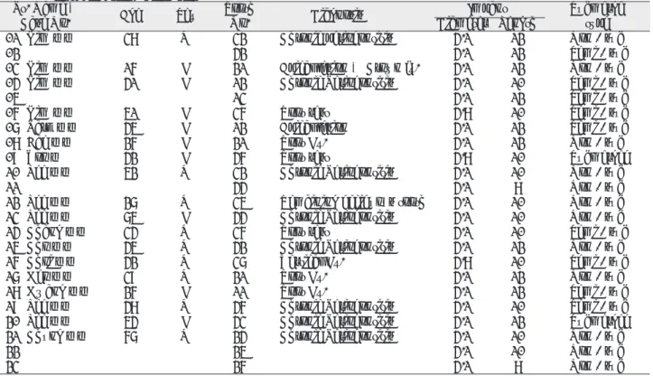
Fig. 1.Case of immediate implantation of upper left central incisor(#21).
A. Clinical view of #21 tooth just before extraction.
B. Clinical view of #21 tooth socket immediately after extraction shows some defects.
C. Xenogenic bone(Bio-Oss�) was grafted.
D. Absorbable membrane(Bio-gide�) was applied.
E. Sutures were placed with non-submerged method.
F. Final treatment outcome shows a pleasing result.
G. Intraoral radiograph after 1-year of loading shows sta- ble state of implant-bone level.
(A) (B)
(C) (D)
(E)
(G)
(F)
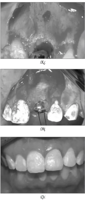
Fig. 2.Case of immediate implantation of lower right first molar(#46).
A. Clinical view of #46 tooth socket immediately after extraction.
B. Implant was placed into the socket and large bone defect was seen.
C. Autogenous bone and xenogenic bone(Bio-Oss�) was grafted.
D. Absorbable membrane(Bio-gide�) was applied.
E. Sutures were placed with submerged method.
F. Final treatment outcome shows a good result.
G. Panoramic radiograph after 1-year of loading shows well functioning state of implant.
(A) (B)
(C) (D)
(E)
(G)
(F)
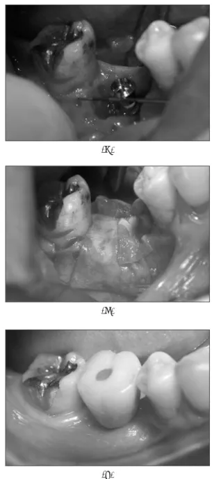
Fig. 3. Case of immediate implantation of lower lateral incisors(#32, 42).
A. Clinical view of #32~42 tooth just before extraction.
B. Implants were placed into the sockets of #32, 42 teeth.
C. Sutures were placed with non-submerged(#32) and semi-submerged method(#42).
D. Abutments connected with implants shows stable peri- odontal reaction.
E. Final treatment result shows no specific problems.
F. Clinical view at 1-year examination.
G. Panoramic radiograph at 1-year examination shows no abnormal peri-implant bone resorption.