INTRODUCTION
Based on the conventional Bra�nemark protocol, it has been recommended that any loading on dental implants should be avoided for a certain healing period. Early loading might cause formation of connective tissue between the implant and its surrounding bone. It has been reported that radi- ographic horizontal and vertical marginal bone loss were observed due to the overloading on dental implants.1
Dental implants with machined surface, traditionally, require six months of healing time in maxilla, and three months in mandible for successful osseointegration.2Albrektsson suggested that the first one month after implant placement was a critical period wherein overloading might lead to failure of the osseointegration due to imbalance between the bone formation
and resorption.3Recent implants with improved surface char- acteristics shorten the healing time with the increased contact between bone and implant.
Some randomized controlled trials supported immediate or early loading concept in the full-arch restorations for the suc- cess of implant osseointegration.4-6In a meta-analysis study of 13 prospective trials by Ioannidou et al., early implant load- ing, whether in partial or full arch, was not found to be asso- ciated with worse outcomes compared to the conventional load- ing.7It was also found in their study that immediate implant loading was associated with slightly, although not statistically significant, worse outcomes compared to the conventional load- ing. On the other hand, some prospective studies showed early-loaded implants occasionally rotated at the time of abutment connection.8,9
Effect of loading time on the survival rate of anodic oxidized implants: prospective multicenter study
Seok-Gyu Kim1, DDS, PhD, Pil-Young Yun2, DDS, PhD, Hyun-Sik Park3, DDS, PhD, June-Sung Shim4, DDS, PhD, Jung-Won Hwang5, DDS, PhD, Young-Kyun Kim6*, DDS, PhD
1Department of Prosthodontics, Samsung Medical Center, College of Medicine, Sungkyunkwan University, Seoul, Korea
2Department of Oral and Maxillofacial Surgery, Section of Dentistry, Seoul National University Bundang Hospital, Seongnam, Korea
3HEIM Dental Clinic, Seoul, 4Department of Prosthodontics, College of Dentistry, Yonsei University, Seoul, Korea
5Seoul Baruljung Dental Clinic, Seoul, 6Department of Oral and Maxillofacial Surgery, Section of Dentistry, Seoul National University Bundang Hospital, School of Dentistry, Seoul National University, Seongnam, Korea
PURPOSE. The purpose of this prospective study was to evaluate the effect of early loading on survival rate or clinical parameter of anodic oxidized implants during the 12- month postloading period. MATERIALS AND METHODS. Total 69 implants were placed in 42 patients. Anodic oxidized implants (GS II, Osstem Cor., Busan, Korea) placed on the posterior mandibles were divided into two groups, according to their prosthetic loading times: test group (2 to 6 weeks), and control group (3 to 4 months). The implant survival rates were determined during one- year postloading period and analyzed by Kaplan-Meier method. The radiographic peri-implant bone loss and periodontal parameters were also evaluated and statistically analyzed by unpaired t-test. RESULTS. Total 69 implants were placed in 42 patients. The cumulative postloading implant survival rates were 88.89% in test group, compared to 100% in control group (P<.05). Periimplant marginal bone loss (T: 0.27±0.54 mm, C: 0.40±0.55 mm) and periodontal parameters showed no significant difference between the groups (P>.05). CONCLUSION. Within the limitation of the present study, implant survival was affected by early loading on the anodic oxidized implants placed on posterior mandibles during one-year follow-up. Early implant loading did not influence peri-implant marginal bone loss, and periodontal parameters. [J Adv Prosthodont 2012;4:18-23]
KEY WORDS: Anodic oxidized; Loading time; Survival
Corresponding author: Young-Kyun Kim
Department of Oral and Maxillofacial Surgery, Section of Dentistry, Seoul National University Bundang Hospital
300 Gumi-dong, Bundang-gu, Seongnam, Korea Tel. 82 31 787 7541: e-mail, [email protected]
Received November 25, 2011 / Last Revison January 19, 2012 / Accepted February 1, 2012
ⓒ 2012 The Korean Academy of Prosthodontics
This is an Open Access article distributed under the terms of the Creative Commons Attribution Non-Commercial License (http://creativecommons.org/licenses/by- nc/3.0) which permits unrestricted non-commercial use, distribution, and reproduction in any medium, provided the original work is properly cited.
Marginal bone resorption around dental implants can jeop- ardize the stability of peri-implant tissue which may lead to peri- implantitis or unesthetic implant restorations. Vercruyssen and Quirynen showed, in their long-term study, that some factors such as smoking, guided bone regeneration, the presence of dehis- cence and bone quantity clearly showed a significant impact on the marginal bone loss around the dental implants.10There are few controlled studies evaluating the effect of loading time on marginal bone resorption around the dental implants.
The purpose of the present study was to investigate the effect of early implant loading on the implant survival rate. The null hypothesis was that there was no effect of early implant loading on implant survival. Anodic oxidized implants has been researched for expediting the osseointegration and reducing the healing time.11-17Thus, the present study examined the effect of early prosthetic loading on implant survival, periimplant bone loss, and periodontal parameters of anodic oxidized implants placed in posterior mandible.
MATERIALS AND METHODS Study design/sample
To address the research purpose, the investigators designed and implemented a prospective study that was conducted in one dental hospital and one private practice in Korea. The study pop- ulation was composed of fifty patients (24 males, 26 females, 17 to 75 years of age) presented for evaluation and man- agement of missing edentulous areas in the posterior mandible between November, 2007 and March, 2008. To be included in the study sample, patients had to have missing mandibular pre- molars or molars with the presence of occluding dentition.
Patients should have adequate width and height of the alveolar bone in the mandible to allow placement of implants with more than 3.5 mm in diameter and 7.0 mm in length, and be a non- smoker or smoker who signed to quit smoking during the study period. Simultaneous minor guided bony regeneration with implant placement can be allowed.
Patients were excluded as study subjects if they had radia- tion therapy in the maxillofacial area, pre-implantation bone grafts, uncontrolled systemic diseases such as hypertension or diabetes mellitus, or parafunctional habits.
Study design/variables
Anodic oxidized implants (GS II, Osstem Co., Busan, Korea) were prepared for insertion in the posterior mandibles of the patients who fulfilled the presurgical inclusion and exclusion criteria. The implant specimens were divided into two groups according to the prosthetic loading time: test (2 to 6 weeks) and control (3 to 4 months). Grouping was randomly done by the restorative dentist. Loading time was determined
as the time between placement of the dental implants and load- ing by the definitive implant prostheses. Each patient was informed that different loading times of implants were applied and signed a written informed consent form prior to the sur- gical procedure. The study protocol was approved by the institutional review board for clinical research of each dental hospital. The clinical and radiographic observations of the den- tal implants were performed during the following year. The implant survival rates, the peri-implant marginal bone loss, gin- gival inflammation index, plaque index, and width of keratinized gingiva were obtained and statistically analyzed.
Surgical procedure
Prophylatic antibiotics and gargling solutions (0.2% chlorhex- idine digluconate) were provided prior to the operations.
The surgery was performed under a local anesthesia (2%
lidocaine with 1: 100,000 epinephrine). A crestal incision was made and a full mucoperiosteal flap was raised. The implants were placed according to the manufacturer’s recommendation.
The primary stability of the dental implants was measured with Osstell Mentor (Integration Diagnostics AB, Go¨teberg, Sweden). The bone graft procedures were, if needed, performed with a xenograft (BioOss; Geistlich Pharma AG, Wolhusen, Switzerland) combined with or without a small amount of auto- graft. Submerged or nonsubmerged implant placements were determined based on the operator’s judgment. Mucoperiosteal flaps were closed with simple interrupted and horizontal mattress sutures. Postoperative gargling solutions (0.2%
chlorhexidine digluconate), antibiotics, and anti-inflammatory agents were given to patients for a week. Liquid diet was rec- ommended postoperatively for two weeks if patients have been wearing removable dentures. A second surgery was, if indicated, carried out.
Prosthetic procedure
At the time of final impression for implant prostheses, the implant stability (ISQ: implant stability quotient) on each implant was measured with Osstell Mentor. Only implants with ISQ values above 65 were prepared for the impression pro- cedures. Forty implants from twenty six patients were loaded early (2 to 6 weeks, test). Conventional loading (3 to 4 month, control) was applied to forty three implants in twen- ty four patients. Silicone rubber impressions were made with custom tray (open type). Full contour wax-up was made on the master cast. Metal frameworks were adapted on the implants after being cast and finished. Framework fit was confirmed with standard radiographs. Veneering porcelains were added on the framework, if needed. The definitive fixed implant prosthesis was cemented or screw-tightened to 30 Ncm with the torque controler (Osstem Implant Co., Busan, Korea).
Data collection methods
1) Implant Survival
The implant survival was evaluated during the 12-month post loading period. “Failed (not survived)”implants include implants with clinical mobility or pain on function as well as lost implants.18
2) Clinical Evaluation Procedure
Soft tissue conditions, such as plaque index and gingival inflam- mation index were evaluated on buccal and lingual gingivae, and the width of buccal keratinized mucosa was measured at the 12-month follow-up visit.
Plaque index evaluates the thickness of the plaque at the gin- gival margin as follows: plaque index 0: no plaque detected, 1: a little plaque detected by exploring around gingival mar- gin, 2: visible plaque detected, 3: much plaque detected.19,20
Gingival inflammation index evaluates the gingival status in clinical trials including redness, swelling, bleeding on prob- ing, and the degree of inflammation. It can be categorized into the followings: gingival inflammation index 0: no inflammation, redness, and bleeding of gingiva, 1: a little inflammation and redness, but no bleeding of gingiva, 2: moderate inflam- mation, redness, swelling, and bleeding on probing of gingi- va, 3: severe inflammation, redness, swelling, and spontaneous bleeding of gingiva.19,20
The width of buccal keratinized gingiva was measured as the midbuccal distance between the mucogingival junction and the most coronal aspect of the free gingival margin.21
The resonance frequency analysis values (Implant Stability Quotient) for evaluating the primary stability of implants were measured with Osstell Mentor (Integration Diagnostics AB ) at implant placement and second surgery.
3) Radiographic Evaluation Procedure10,22
Marginal bone loss was defined as the average radiograph- ic bone level changes in mesial and distal sides around
implants. It was measured as the vertical distance between the implant platform and the first bone-implant contact area.
Crestal bone level measured on the periapical radiograph taken immediately after the prosthetic loading was compared with the one taken at the 12 month postloading visit. The radi- ographs were taken using digital periapical radiography with paralleling cone technique (Rinn alignment system, Dentsply Rinn, Elgin, IL The magnification power was adjusted using the length of the placed implants. The mesial and distal sides were measured, and the mean value was calculated.
Statistical analyses
The implant survival rates were compared between two groups by Kaplan-Meier survival analysis. The mean values of the crestal bone loss, the gingival inflammation index, the plaque index, and the width of keratinized gingiva were also compared between two groups by unpaired t-test. Statistical analyses were done using SPSS 18.0 for Windows (SPSS Inc., Chicago, IL, USA). It was considered statistically significant for P values <.05.
RESULTS
From November, 2007 to March, 2008, eighty three implants placed in the posterior mandibles of fifty patients were enrolled in the study. Fourteen implants (4 from test group and 10 from control group) of eight patients were excluded from the final sample due to loss of follow-up. The final sam- ple was composed of forty two patients with a mean age of 53 and 48% were male. A total of sixty nine implants were placed (Table 1).
The distribution of implants in length and diameter is presented in Fig. 1 and 2. The non-submerged implant placement was dom- inant in test group, whereas the subemerged implant placement was dominant in control goup (Fig. 3). Based on the average value of the RFA, ISQ value was higher at the final impression
Fig. 1. Distribution of implant lengths.
Number of implant
12 10 8 6 4 2
0 7.0 8.5 10.0 11.5 13.0 (mm)
Implant length
test control
Fig. 2. Distribution of implant diameters.
Number of implant
20
15
10
5
0 3.5 4.0 4.5 5.0 (mm)
Implant diameter
test control
than the placement in all groups (Fig. 4).
The final implant prostheses in test group were 7 single implant crowns, 10 fixed partial dentures (FPDs) (21 implants) and 2 overdentures (4 implants). Control group had 11 single implant crowns and 10 FPDs (22 implants).
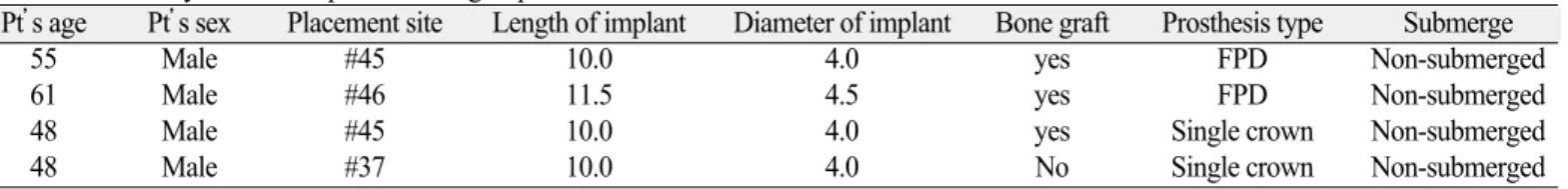
Implant survival and peri-implant condition were evaluated during 12-month prosthetic loading period. Test group had 4 failed implants during this time, while there was no failure in control group (P<.05) (Table 1). In test group, two single implant
crowns from two male patients showed clinical mobility at 6- month postloading visit, and were removed and replaced.
Another male patient had one implant presented with mobil- ity and the other with severe bone resorption, both of which were eliminated and replaced (Table 2).
Average bone resorption rate was 0.27±0.54 mm in test group, and 0.40±0.55 mm in control group. They revealed no sta- tistically significant differences (Table 3, P>.05). Gingival inflam- mation index, plaque index and the width of keratinized gin- giva among the 3 groups also revealed no statistically significant differences (Table 4, P>.05).
DISCUSSION
The present prospective clinical study investigated the effect of early loading on implant survival. The null hyposthesis claiming no influence was rejected because test group, 2 to 6 weeks loading group, presented 4 failed implants compared to control group showing no failures during the follow-up peri- Fig. 3. Distribution of submerged and non-submerged placement.
Number of implant
35 30 25 20 15 10 5 0
submerged non-submerged Type of implant placement
test control
Fig. 4. Average value of resonance frequency analysis at implant placement (ISQ-1) and at the final impression (ISQ-2).
Average value of resonance frequency analysis 80 78 76 74 72 70 68 66 64
ISQ-1 ISQ-2 RFA value
test control
Table 2. Summary of failed implants in test group
Pt’s age Pt’s sex Placement site Length of implant Diameter of implant Bone graft Prosthesis type Submerge
55 Male #45 10.0 4.0 yes FPD Non-submerged
61 Male #46 11.5 4.5 yes FPD Non-submerged
48 Male #45 10.0 4.0 yes Single crown Non-submerged
48 Male #37 10.0 4.0 No Single crown Non-submerged
Table 3. Amount of marginal bone loss around implant during 12-month prosthetic loading period
Test Control
Amount of bone loss (mm) 0.27±0.54 0.40±0.55 (P>.05)
Table 4. Periodontal evaluation around implant
Test Control
Gingival inflammation index 0.43±0.63a 0.63±0.71a
Plaque index 1.21±0.88b 1.25±0.92b
Width of keratinized 2.64±1.55c 2.10±1.40c gingiva (mm)
The same letters indicate mean values with no statistically significant dif- ferences (P>.05).
Table 1. Number and ages of patients, number of implants placed, and survival rate
Number of Average age Number of Survival patients of patients implants rate (%)
Test 22 50 36 88.89*
Control 20 52 33 100.00
*Kaplan-Meier survival analysis: test vs control (P<.05) (Log-Rank test)
od. This trial was also aimed for evaluating the effect of early prosthetic loading on the peri-implant marginal bone loss and some periodontal parameters. Early loading was not found to have any effect on the marginal bone loss or periodontal indices.
There were some different opinions about the time period of
‘early loading’, such as within 2 weeks, 35 days or 6 weeks from implant placement. In the present trial, the early loading period was set as 2 to 6 weeks.4,7,8,23According to the meta-analy- sis report of 13 prospective trials regarding immediate or early implant loading, survival rates of the implant under non-conventional loading such as immediate and early load- ing did not show any significant difference from the ones under conventional loading.7,8,23,24-28
There have been, nonetheless, some clinical studies report- ing immediate loading showed lower survival rates of the implant than conventional loading.29-31Cochran suggested that implant stability was summed up by the decreasing primary stability and the increasing secondary stability after placement. He pro- posed the total implant stability reached the lowest point during 2 to 4weeks after placement, when implant osseoin- tegration was likely to fail by any interfering forces.32
Regarding the association between the implant loading time and marginal bone loss around the implant, there were some different opinions. It was reported there was no significant dif- ference in marginal bone loss of the implants between imme- diately loaded and conventionally loaded.24,26,28Early loaded implants were also claimed to have no significant difference in peri-implant marginal bone loss compared to convention- ally loaded ones.23,27 Difference in periodontal parameters between early and conventionally loaded implants were also revealed to have no significance.4In contrast, in 1 year follow- up research about maxillary full arch implants of 24 patients, Fischer and Stenberg found that 2-week early loading showed more alveolar bone resorption than the conventional loading.6 It was noteworthy that in Fischer’s study they collected only maxillary implant cases, while previous researches claim- ing no differences had mandibular implant overdenture cases.
The present trial included rough-surfaced microthreaded implant cases placed only in mandible so that the results did not show any significant difference in marginal bone loss under dif- ferent loading times.33-35GS II implant, used in the present tri- al, was claimed to have a dual thread design composed of microthread and macrothread with anodized surfaces. The neck portion of the GS II implant with platform switching was claimed to have the effect of reducing marginal bone loss around implants.36-41
Some authors suggested that the implant placed in a non-recon- structed recipient site should survive better than the one placed in a reconstructed site.42Becktor et al.43discovered that bone graft in maxillary edentulous area generated a significant difference in the implant’s survival rate as 75.1% vs 84.0%.
Nonetheless, Woo et al. suggested that successful den- toalveolar reconstructive procedures were not an independent risk factor for implant failure.44Sbordone et al. reported sim- ilar implant cumulative survival rates were shown both in native and grafted sites.45In the present trial, three out of four failed implant cases in test group had minor bone graft procedures.
It may be because surgical damage from the graft procedure can change the blood circulation around the implant and has negative influence on the recovery of the soft tissues and the bony tissues.
This trial researched the implants placed only on the posterior mandible for the purpose of controlling the bone quality, but did not evaluate the bone quality of each case. The small sam- ple size from limited areas for implant placement was the lim- itation of this trial.
CONCLUSION
Within the limitations of the present clinical study, it was con- cluded that early implant loading could increase the possibility of implant failure in the posterior mandible. The periimplant marginal bone loss and periodontal parameters were not affected by early implant loading.
REFERENCES
1. Bernard JP, Belser UC, Martinet JP, Borgis SA. Osseointegration of Bra�nemark fixtures using a single-step operating technique.
A preliminary prospective one-year study in the edentulous mandible. Clin Oral Implants Res 1995;6:122-9.
2. Lazzara RJ, Porter SS, Testori T, Galante J, Zetterqvist L. A prospective multicenter study evaluating loading of osseotite im- plants two months after placement: one-year results. J Esthet Dent 1998;10:280-9.
3. Albrektsson T. Direct bone anchorage of dental implants. J Prosthet Dent 1983;50:255-61.
4. Payne AG, Tawse-Smith A, Duncan WD, Kumara R. Conventional and early loading of unsplinted ITI implants supporting mandibu- lar overdentures. Clin Oral Implants Res 2002;13:603-9.
5. Romeo E, Chiapasco M, Lazza A, Casentini P, Ghisolfi M, Iorio M, Vogel G. Implant-retained mandibular overdentures with ITI implants. Clin Oral Implants Res 2002;13:495-501.
6. Fischer K, Stenberg T. Early loading of ITI implants support- ing a maxillary full-arch prosthesis: 1-year data of a prospective, randomized study. Int J Oral Maxillofac Implants 2004;19:
374-81.
7. Ioannidou E, Doufexi A. Does loading time affect implant survival? A meta-analysis of 1,266 implants. J Periodontol 2005;76:1252-8.
8. Salvi GE, Gallini G, Lang NP. Early loading (2 or 6 weeks) of sandblasted and acid-etched (SLA) ITI implants in the posterior mandible. A 1-year randomized controlled clinical trial. Clin Oral Implants Res 2004;15:142-9.
9. Roccuzzo M, Bunino M, Prioglio F, Bianchi SD. Early loading of sandblasted and acid-etched (SLA) implants: a prospective split-mouth comparative study. Clin Oral Implants Res 2001;12:572-8.
10. Vercruyssen M, Quirynen M. Long-term, retrospective evalu- ation (implant and patient-centred outcome) of the two-im- plant-supported overdenture in the mandible. Part 2: marginal
bone loss. Clin Oral Implants Res 2010;21:466-72.
11. Larsson C, Thomsen P, Lausmaa J, Rodahl M, Kasemo B, Ericson LE. Bone response to surface modified titanium implants:
studies on electropolished implants with different oxide thick- nesses and morphology. Biomaterials 1994;15:1062-74.
12. Hall J, Lausmaa J. Properties of a new porous oxide surface on titanium implants. Appl Osseointegration Res 2000;1:5-8.
13. Sul YT, Johansson CB, Jeong Y, Wennerberg A, Albrektsson T. Resonance frequency and removal torque analysis of implants with turned and anodized surface oxides. Clin Oral Implants Res 2002;13:252-9.
14. Zechner W, Tangl S, Fu¨rst G, Tepper G, Thams U, Mailath G, Watzek G. Osseous healing characteristics of three different im- plant types. Clin Oral Implants Res 2003;14:150-7.
15. Salata LA, Burgos PM, Rasmusson L, Novaes AB, Papalexiou V, Dahlin C, Sennerby L. Osseointegration of oxidized and turned implants in circumferential bone defects with and without ad- junctive therapies: an experimental study on BMP-2 and auto- genous bone graft in the dog mandible. Int J Oral Maxillofac Surg 2007;36:62-71.
16. Rocci A, Martignoni M, Burgos PM, Gottlow J, Sennerby L.
Histology of retrieved immediately and early loaded oxidized implants: light microscopic observations after 5 to 9 months of loading in the posterior mandible. Clin Implant Dent Relat Res 2003;5:88-98.
17. Burgos PM, Rasmusson L, Meirelles L, Sennerby L. Early bone tissue responses to turned and oxidized implants in the rab- bit tibia. Clin Implant Dent Relat Res 2008;10:181-90.
18. Mangano C, Mangano F, Shibli JA, Tettamanti L, Figliuzzi M, d’Avila S, Sammons RL, Piattelli A. Prospective evaluation of 2,549 Morse taper connection implants: 1- to 6-year data. J Periodontol 2011;82:52-61.
19. Lo¨e H. The Gingival Index, the Plaque Index and the Retention Index Systems. J Periodontol 1967;38:610-6.
20. Silness J, Lo¨e H. Periodontal disease in pregnancy. II. Correlation between oral hygiene and periodontal condtion. Acta Odontol Scand 1964;22:121-35.
21. Wilson TG Jr, Kornman KS. Fundamentals of periodontics.
Chicago; Quintessence; 1996. p. 195-218.
22. Ricci G, Aimetti M, Stablum W, Guasti A. Crestal bone resorption 5 years after implant loading: clinical and radiologic results with a 2-stage implant system. Int J Oral Maxillofac Implants 2004;
19:597-602.
23. Tawse-Smith A, Payne AG, Kumara R, Thomson WM. Early load- ing of unsplinted implants supporting mandibular overden- tures using a one-stage operative procedure with two different implant systems: a 2-year report. Clin Implant Dent Relat Res 2002;4:33-42.
24. Randow K, Ericsson I, Nilner K, Petersson A, Glantz PO.
Immediate functional loading of Bra�nemark dental implants. An 18-month clinical follow-up study. Clin Oral Implants Res 1999;10:8-15.
25. Jo HY, Hobo PK, Hobo S. Freestanding and multiunit immediate loading of the expandable implant: an up-to-40-month prospec- tive survival study. J Prosthet Dent 2001;85:148-55.
26. Chiapasco M, Abati S, Romeo E, Vogel G. Implant-retained mandibular overdentures with Bra�nemark System MKII implants:
a prospective comparative study between delayed and immediate loading. Int J Oral Maxillofac Implants 2001;16:537-46.
27. R�ynesdal AK, Amundrud B, Hannaes HR. A comparative clinical investigation of 2 early loaded ITI dental implants supporting an overdenture in the mandible. Int J Oral Maxillofac Implants 2001;16:246-51.
28. Cannizzaro G, Leone M. Restoration of partially edentulous pa- tients using dental implants with a microtextured surface: a prospec- tive comparison of delayed and immediate full occlusal loading.
Int J Oral Maxillofac Implants 2003;18:512-22.
29. Schnitman PA, Wo¨hrle PS, Rubenstein JE, DaSilva JD, Wang NH. Ten-year results for Bra�nemark implants immediately loaded with fixed prostheses at implant placement. Int J Oral Maxillofac Implants 1997;12:495-503.
30. Ericsson I, Nilson H, Lindh T, Nilner K, Randow K. Immediate functional loading of Bra�nemark single tooth implants. An 18 months’clinical pilot follow-up study. Clin Oral Implants Res 2000;11:26-33.
31. Wolfinger GJ, Balshi TJ, Rangert B. Immediate functional loading of Bra�nemark system implants in edentulous mandibles:
clinical report of the results of developmental and simplified pro- tocols. Int J Oral Maxillofac Implants 2003;18:250-7.
32. Cochran DL. The evidence for immediate loading of implants.
J Evid Based Dent Pract 2006;6:155-63.
33. Shin YK, Han CH, Heo SJ, Kim S, Chun HJ. Radiographic eval- uation of marginal bone level around implants with different neck designs after 1 year. Int J Oral Maxillofac Implants 2006;21:
789-94.
34. Abrahamsson I, Berglundh T. Tissue characteristics at mi- crothreaded implants: an experimental study in dogs. Clin Implant Dent Relat Res 2006;8:107-13.
35. Nickenig HJ, Wichmann M, Schlegel KA, Nkenke E, Eitner S.
Radiographic evaluation of marginal bone levels adjacent to par- allel-screw cylinder machined-neck implants and rough-surfaced microthreaded implants using digitized panoramic radiographs.
Clin Oral Implants Res 2009;20:550-4.
36. Atieh MA, Ibrahim HM, Atieh AH. Platform switching for mar- ginal bone preservation around dental implants: a systematic review and meta-analysis. J Periodontol 2010;81:1350-66.
37. Wagenberg B, Froum SJ. Prospective study of 94 platform- switched implants observed from 1992 to 2006. Int J Periodontics Restorative Dent 2010;30:9-17.
38. Luongo R, Traini T, Guidone PC, Bianco G, Cocchetto R, Celletti R. Hard and soft tissue responses to the platform- switching technique. Int J Periodontics Restorative Dent 2008;
28:551-7.
39. Rodrl′guez-Ciurana X, Vela-Nebot X, Segala`-Torres M, Calvo- Guirado JL, Cambra J, Me′ndez-Blanco V, Tarnow DP. The ef- fect of interimplant distance on the height of the interimplant bone crest when using platform-switched implants. Int J Periodontics Restorative Dent 2009;29:141-51.
40. Jung RE, Jones AA, Higginbottom FL, Wilson TG, Schoolfield J, Buser D, Ha¨mmerle CH, Cochran DL. The influence of non-matching implant and abutment diameters on radiograph- ic crestal bone levels in dogs. J Periodontol 2008;79:260-70.
41. Cochran DL, Bosshardt DD, Grize L, Higginbottom FL, Jones AA, Jung RE, Wieland M, Dard M. Bone response to loaded im- plants with non-matching implant-abutment diameters in the ca- nine mandible. J Periodontol 2009;80:609-17.
42. Schliephake H, Neukam FW, Wichmann M. Survival analysis of endosseous implants in bone grafts used for the treatment of severe alveolar ridge atrophy. J Oral Maxillofac Surg 1997;55:
1227-33.
43. Becktor JP, Isaksson S, Sennerby L. Survival analysis of en- dosseous implants in grafted and nongrafted edentulous maxillae.
Int J Oral Maxillofac Implants 2004;19:107-15.
44. Woo VV, Chuang SK, Daher S, Muftu A, Dodson TB.
Dentoalveolar reconstructive procedures as a risk factor for implant failure. J Oral Maxillofac Surg 2004;62:773-80.
45. Sbordone L, Toti P, Menchini-Fabris G, Sbordone C, Guidetti F. Implant survival in maxillary and mandibular osseous onlay grafts and native bone: a 3-year clinical and computerized to- mographic follow-up. Int J Oral Maxillofac Implants 2009;24:
695-703.