Received:April 2, 2018, Revised:April 11, 2018, Accepted:April 11, 2018 Corresponding to:Eun Young Lee http://orcid.org/0000-0001-6975-8627
Division of Rheumatology, Department of Internal Medicine, Seoul National University College of Medicine, 101 Daehak-ro, Jongno-gu, Seoul 03080, Korea. E-mail:[email protected]
Copyright ⓒ 2018 by The Korean College of Rheumatology. All rights reserved.
This is a Open Access article, which permits unrestricted non-commerical use, distribution, and reproduction in any medium, provided the original work is properly cited.
Compound K Inhibits Interleukin-1β-induced Expression of Inflammatory Mediators and Matrix Metalloproteinases by Inhibiting Mitogen-activated Protein Kinase Activation in Chondrocytes
Eun Hye Park1, Ji Soo Kim1, Jeong Seok Lee1, Yun Jong Lee2, Yeong Wook Song1, Eun Young Lee1
Division of Rheumatology, Department of Internal Medicine, 1Seoul National University Hospital, Seoul, 2Seoul National University Bundang Hospital, Seongnam, Korea
Objective. This study examined the anti-inflammatory and chondroprotective effects of compound K (CK), a ginsenoside me- tabolite, on chondrocytes from osteoarthritis (OA) patients following stimulation with interleukin (IL)-1β. Methods. Articular cartilage samples were obtained from six OA patients undergoing total knee replacement surgery. Nitric oxide (NO) production was measured by the Griess reaction. Subsequently, the mRNA and protein levels of matrix metalloproteinases (MMPs), inducible NO synthase (iNOS), and mitogen-activated protein kinases (MAPKs) were examined by a reverse transcription-polymerase chain reaction and western blot analysis. Cartilage degradation was assessed using a glycosaminoglycan (GAG) assay. Results.
CK inhibited IL-1β-induced NO production and iNOS expression in a dose-dependent manner. In addition, it inhibited the IL-1 β-stimulated release of MMP-1, -3, and -13 and tissue inhibitor of matrix metalloproteinase-1 from OA patient chondrocytes.
In addition, CK effectively suppressed the IL-1β-induced phosphorylation of p38, extracellular signal-regulated kinase-1/2, and c-Jun N-terminal kinase MAPKs. Moreover, the IL-1β-mediated release of GAG was inhibited by CK in a dose-dependent manner. Conclusion. CK inhibited the IL-1β-induced expression of inflammatory mediators and MMPs by, at least in part, in- hibiting MAPK activation, and has potential as a therapeutic agent for the treatment of OA. (J Rheum Dis 2018;25:188-196) Key Words. Panax, Ginsenosides, Nitric oxide, Matrix metalloproteinases, Osteoarthritis
INTRODUCTION
Osteoarthritis (OA) is the most common form of arthri- tis and a leading cause of pain and disability in the grow- ing elderly population [1]. It is characterized by an active and complex process involving inflammatory, mechan- ical, and metabolic factors, which ultimately disrupt the structure of the synovial joint, leading to its failure [2].
The pathologic changes seen in OA joints include degra- dation of the articular cartilage, thickening of the sub- chondral bone, formation of osteophytes, varying degrees of synovial inflammation, and degeneration of ligaments [3].
Of the cytokines involved in the pathogenesis of OA, the
inflammatory cytokine interleukin (IL)-1β is considered key. Patients with OA have an elevated level of IL-1β in the synovial fluid, synovial membrane, cartilage, and sub- chondral bone [4,5]. Furthermore, expression of IL-1 re- ceptor type 1 has been reported to be increased on the sur- face of chondrocytes from OA patients compared to those from healthy individuals [6]. IL-1β plays a crucial role in the destruction of the cartilage matrix by upregulating the production of proteases, such as matrix metallopro- teinases (MMPs) and zinc-dependent endopeptidases, which are specifically controlled by tissue inhibitors of metalloproteinases (TIMPs) [7,8]. MMPs are synthesized and secreted by resident chondrocytes, particularly under
arthritic conditions. Among the various MMPs, collage- nases such as MMP-1, -8, and -13 are particularly im- portant, and are known to be upregulated during joint in- flammation and degeneration [8,9].
Collagenase expression in chondrocytes is regulated by the activation of mitogen-activated protein kinase (MAPK) family members, including c-Jun N-terminal kinase (JNK), p38 MAPK, and extracellular signal-regu- lated kinase (ERK), and several transcription factors, in- cluding nuclear factor-κB (NF-κB) [9-11]. IL-1β in- duces heightened inducible nitric oxide (NO) synthase (iNOS) and cyclooxygenase-2 (COX-2) expression in chondrocytes, leading to elevated production of NO and prostaglandin E2 (PGE2), which accelerate cartilage deg- radation by inhibiting proteoglycan biosynthesis [6,12].
Treatments for OA aim to relieve pain, decrease joint stiffness and swelling, prevent cartilage loss, and main- tain the patient’s quality of life [13]. Since targeting NO and PGE2 is beneficial in OA therapy, non-steroidal an- ti-inflammatory drugs (NSAIDs) have been used as treat- ments for this disease for several years. However, long-term use of NSAIDs is known to induce gastro- intestinal, renal, and cardiovascular toxicity [14]. Therefore, there remains a need for a safer, yet effective, treatment option for OA.
Ginseng, especially Panax ginseng Meyer, is one of the most widely used herbal medications globally, and is em- ployed in the treatment of various diseases [15]. It is tak- en as an oral supplement, and its biological activities de- rive from ginsenosides, its major pharmacologically ac- tive components [16]. Compound K (CK) (Supplementary Figure 1) is a bacterial metabolite of the ginsenoside Rb1.
The uptake and rate of absorption of CK in the human gastrointestinal tract, and ultimately its passage into sys- temic circulation, are rapid. It has been suggested that CK suppresses receptor activator of NF-κB ligand-induced osteoclastogenesis and tumor necrosis factor-α (TNF- α)-induced production of MMPs by fibroblast-like syno- viocytes in rheumatoid arthritis [17]. CK also attenuates collagen-induced arthritis via suppression of the humoral immune response and modulation of mediators of joint destruction [18].
However, very few studies have reported the effects of CK in OA. In this study, we evaluated the anti-in- flammatory and chondroprotective effects of CK on chon- drocytes from OA patients following IL-1β stimulation and examined the underlying mechanisms.
MATERIALS AND METHODS
Preparation of CK
CK was obtained from the Korea Ginseng Corporation (Daejeon, Korea). Its purity was higher than 97%, based on high-performance liquid chromatography analysis.
Chondrocyte culture
Articular cartilage samples were obtained from six OA patients who fulfilled the American College of Rheuma- tology criteria for OA [19] and were undergoing total knee replacement surgery. Primary chondrocytes were isolated from articular cartilage as described previously [20]. The cells were cultured in Dulbecco’s modified Eagle medium containing 10% fetal bovine serum at 37oC in the presence of 5% CO2. Cells from passages 1 to 3 were used in this study. The study was approved by Institutional Review Boards and ethics committees of Seoul National University Hospital (no. 1603-146-751), and was conducted in accordance with good clinical prac- tice guidelines and the Declaration of Helsinki.
Enzyme-linked immunosorbent assay (ELISA)
Chondrocytes were pretreated with CK or ginsenoside Rb1 1 hour prior to IL-1β (2 ng/mL) stimulation for 24 hours. Culture supernatants were collected and stored at−20oC until analysis. The concentrations of MMP-1, -3, and -13 and TIMP-1 in cell culture supernatants were measured using ELISA kits (R&D Systems, Minneapolis, MN, USA) according to the manufacturer’s instructions.
Measurement of NO
Chondrocytes were pretreated with CK or Rb1 1 hour prior to IL-1β (2 ng/mL) stimulation for 24 hours. The NO concentration in the culture supernatant was meas- ured using Griess reagent according to the manu- facturer’s instructions. Briefly, cell culture supernatant was collected and mixed with an equal volume of Griess reagent, and optical density at 540 nm was measured 30 minutes later.
Western blot analysis
Chondrocytes were pretreated with the indicated con- centrations of CK 1 hour prior to IL-1β (2 ng/mL) stim- ulation for 30 minutes. Nuclear and cytoplasmic proteins were extracted from the cells using NE-PER Nuclear and Cytoplasmic Extraction Reagent (Thermo Scientific, Waltham, MA, USA). Protein concentration was de-
termined with a Bradford assay (Bio-Rad Laboratories, Hercules, CA, USA). The samples were separated by so- dium dodecyl sulfate (SDS)-polyacrylamide gel electro- phoresis (PAGE) on a 12% gel and transferred to poly- vinylidene difluoride membranes. The membranes were then blocked with 5% non-fat milk at room temperature for 1 hour and probed with primary antibodies (all diluted 1:1,000) against the following proteins: iNOS, ERK1/2, phosphorylated (p-)ERK1/2, JNK, p-JNK, p38, and p-p38. After being washed three times, the membranes were incubated with horseradish peroxidase-conjugated secondary antibodies for 1 hour. Antibody binding was vi- sualized using a chemiluminescent substrate (SuperSignal West Femto; Thermo Scientific).
Reverse transcription-polymerase chain reaction (RT-PCR)
Total RNA was isolated with TRIzol reagent (Invitrogen) according to the manufacturer’s protocol. Complemen- tary DNA was synthesized by reverse transcription using 1 mg of DNase-treated total RNA and an iScript Reverse Transcriptase Kit (Bio-Rad Laboratories). RT-PCR was performed using primers targeting sequences encoding iNOS, MMP-1, -3, and -13, and TIMP-1. Actin was used as the internal control.
Zymography
Serum-starved cells were cultured in the presence of CK for 48 hours. Cell culture supernatant (10 μL) was mixed with 10 μL of sample buffer (0.5 M Tris-HCl, 20% glycer- ol, 10% SDS, and 0.1% bromophenol blue) and incubated for 10 minutes at room temperature. The samples were loaded on gelatin (1 mg/mL)-containing SDS-poly- acrylamide gels and electrophoresed under a constant voltage (125 V). Subsequently, the gels were incubated in renaturation buffer (2.5% Triton X-100) with gentle agi- tation for 30 minutes at room temperature. The gels were then equilibrated for 30 minutes in developing buffer (50 mM Tris base, 0.2 M NaCl, 5 mM CaCl2, and 0.02%
Brij-35), and incubated in fresh developing buffer over- night at 37oC. Zymographic activity was detected by staining the gels with 0.2% Coomassie blue, before de- staining them with a destaining solution (7.5% acetic acid in 20% ethanol). Areas affected by protease activity ap- peared as clear bands against a dark blue background, rep- resenting substrate digestion. Molecular weights were es- timated by comparison with prestained SDS-PAGE mark- ers and a human pro-MMP-2 standard (AnaSpec,
Fremont, CA, USA).
Glycosaminoglycan (GAG) assay
Cartilage degradation was assessed by measuring the amount of proteoglycan released into the culture me- dium, as described previously [21]. Briefly, culture me- dium was added to a solution of 1,9-dimethylmethylene blue (DMB) (Sigma, St. Louis, MO, USA), a meta- chromatic dye that binds sulfated GAG, a major compo- nent of proteoglycans. The quantity of GAG-DMB com- plex formed was measured in a 96-well plate using a plate reader (Infinite M200; TECAN, Männedorf, Switzerland) at a wavelength of 595 nm. Loss of GAG was calculated and expressed as the total GAG (μg) released per mg (wet weight) of cartilage.
Thiazolyl blue tetrazolium bromide (MTT) assay
An MTT assay was used to determine the effects of CK and Rb1 on chondrocyte viability. Briefly, chondrocytes were seeded in a 96-well plate at a density of 6×10³ per well. The cells were treated with 0 to 10 μM CK or 0 to 200 μM Rb1 for 1 hour, and stimulated with IL-1β (2 ng/mL) for 24 hours. Subsequently, 20 μL MTT (5 mg/mL) was added to each well, and the cells were in- cubated for an additional 4 hours. The supernatant was then removed, and the formazan crystals formed were dissolved using 150 μL of dimethyl sulfoxide. Absorbance at 570 nm was measured with a microplate reader (Bio-Rad Laboratories).Statistical analyses
Data are presented as the mean±standard error of the mean of three independent experiments. Statistical anal- ysis was carried out using the Kruskal–Wallis test or Wilcoxon signed-rank test, as appropriate, and p-values of 0.05 or less were considered statistically significant.
RESULTS
CK inhibited IL-1β-induced NO production
To investigate the anti-inflammatory effect of CK, its in- fluence on IL-1β-induced NO production was evaluated.Levels of NO in the supernatant increased after IL-1β treatment. However, CK inhibited IL-1β-induced NO production in a dose-dependent manner from a concen- tration of 1 μM. NO concentration was decreased by 95.8% when 5 μM CK was administered (Figure 1A).
Rb1 at and above a concentration of 5 μM also sup-
Figure 1. Interleukin (IL)-1β-induced nitric oxide (NO) production inhibited by (A) compound K (CK) and (B) Rb1. (C) Strong in- hibitory effect of CK on NO production is related to decreased expression of induced NO synthase (iNOS), (D) but the expression of iNOS was not inhibited by Rb1. The values presented are the means±standard error of mean. of three independent experiments.
*p<0.05, **p<0.005.
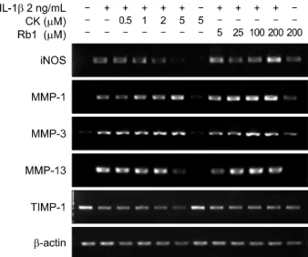
Figure 2. Effects of compound K (CK) and Rb1 on interleukin (IL)-1β-induced mRNA expression of induced nitric oxide synthase (iNOS), matrix metalloproteinases (MMPs), and tis- sue inhibitor of metalloproteinases-1 (TIMP-1). The cells were pretreated with different concentrations of CK or Rb1 for 1 hour before incubation with IL-1β (2 ng/mL). Following 24 hours of treatment, total RNA was isolated, and reverse tran- scription-polymerase chain reaction was performed using iNOS, MMP-1, MMP-3, MMP-13 and TIMP-1 primers.
pressed IL-1β-induced NO production (Figure 1B).
However, its inhibitory effect in this respect was far weak- er than that of CK. Rb1 resulted in a 41.0% decrease in NO release when used at a concentration of more than 20 times the highest dose of CK.
CK inhibited IL-1β-induced iNOS expression
The effect of CK on IL-1β-induced iNOS expression was tested by western blotting. CK (5 μM) significantly inhibited iNOS expression triggered by IL-1β (Figure 1C). However, Rb1 did not reduce iNOS levels, even when used at 200 μM (Figure 1D). Next, RT-PCR was performed to ascertain whether expression of iNOS mRNA was consistent with that of the corresponding protein. CK also suppressed IL-1β-induced iNOS mRNA expression in a dose-dependent manner (Figure 2).CK inhibited IL-1β-induced MAPK activation in chondrocytes
The effect of CK on MAPK activation resulting from IL-1 β stimulation was assessed by western blot analysis. CK suppressed IL-1β-induced activation of p38, ERK1/2, and JNK MAPKs in a dose-dependent fashion, and this
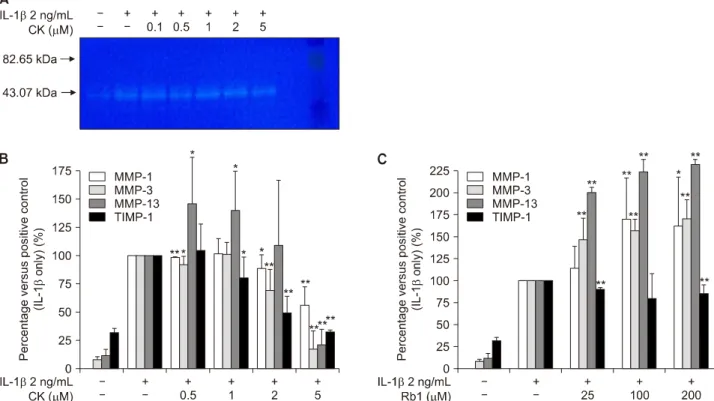
Figure 4. Activity of matrix metalloproteinases (MMPs) was inhibited by (A) compound K (CK) on the zymographic study.
Production of MMP-1, MMP-3, MMP-13, and tissue inhibitor of metalloproteinases-1 (TIMP-1) measured by enzyme-linked im- munosorbent assay was inhibited by (B) CK in a dose-dependent manner but not by (C) Rb1. IL: interleukin. The values presented are the means±standard error of mean of three independent experiments. *p<0.05, **p<0.005.
Figure 3. Compound K (CK) inhibits interleukin (IL)-1β-induced mitogen-activated protein kinase activation. ERK: extracellular signal-regulated kinase, JNK: c-Jun N-terminal kinase.
was particularly evident in the case of ERK1/2 (Figure 3).
Interestingly, the attenuation of IL-1β-induced phos- phorylation of MAPK signaling proteins by CK was evi- dent from a concentration of 2 μM, comparable to that at
which NO production began to be inhibited.
CK suppressed IL-1β-induced MMP expression and activity
Since MMPs are responsible for damaging the cartilage matrix, we examined the effects of CK on IL-1β-induced MMP expression and activity. CK suppressed IL-1β -induced MMP-13 mRNA expression (Figure 2).
However, both CK and Rb1 had little effect on mRNA lev- els of MMP-1 and MMP-3, and no effect on those of TIMP-1. IL-1β-induced MMP activity (Figure 4A) was al- so inhibited by CK, especially at a concentration of 5 μM.
CK inhibited IL-1β-induced MMP-1, -3, and -13 and TIMP-1 production
The effects of CK on IL-1β-induced production of MMP-1, -3, and -13 and TIMP-1 were evaluated by ELISA.
Treatment with CK reduced the elevated levels of MMP-13, -1, and -3 caused by IL-1β stimulation, espe- cially at a concentration of 5 μM (Figure 4B). Furthe- rmore, CK inhibited TIMP-1 production in a dose-de- pendent manner. In contrast, release of MMPs increased
Figure 6. Effects of (A) compound K (CK) and (B) Rb1 on the cell viability of chondrocytes. The cells were cultured with different concentrations of CK (0∼10 μM) or Rb1 (0∼200 μM) for 24 hours. The cell viability was determined by MTT assay. IL:
interleukin. The values are means±standard error of mean of three independent experiments.
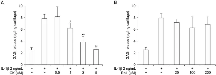
Figure 5. Effect of (A) compound K (CK) and (B) Rb1 on chondrocyte degeneration analyzed through the glycosaminoglycan (GAG) assay. IL: interleukin. The values presented are the means±standard error of mean of three independent experiments. *p<0.05, **p<
0.005.
as the dose of Rb1 was raised (Figure 4C).
CK inhibited chondrocyte degeneration
The net effect of CK on chondrocyte degeneration was analyzed using a GAG assay. CK dose-dependent sup- pression of IL-1β-mediated GAG release was observed (Figure 5A), whereas Rb1 failed to prevent GAG loss, even at higher doses (Figure 5B).
Effects of CK on cell viability
The cytotoxicity of CK to chondrocytes was evaluated with an MTT assay. Cell viability was not affected by CK at con- centrations up to 10 μM (Figure 6). Therefore, the effects of CK on chondrocytes were not attributable to cytotoxicity.
DISCUSSION
In this study, CK effectively suppressed the IL-1β -mediated release of the inflammatory mediator NO and MMPs including MMP-1, -3, and -13, at least in part by blocking MAPK activation. CK decreased IL-1β-induced NO production by attenuating the expression of its up- stream molecule, iNOS, at both the mRNA and protein level. In addition, CK was capable of inhibiting MMP pro- duction and activity triggered by IL-1β.
It is widely accepted that excess production of in- flammatory cytokines plays an important role in the pathogenesis of OA. Of such cytokines, IL-1β has been shown to have a pivotal function in this disease due to its
upregulation of MMP production, which leads to de- creased synthesis of proteoglycan and collagen [22]. IL-1β also induces the expression of iNOS and COX-2, resulting in elevated production of NO and PGE2, respectively [6].
IL-1β-mediated overproduction of NO has been re- ported to be an important proinflammatory factor in the development of OA due to its association with chon- drocyte and synoviocyte death [23]. NO also upregulates the production of MMPs and other inflammatory cyto- kines [24]. In the present study, CK effectively inhibited chondrocyte NO production and iNOS expression in- duced by IL-1β, suggesting that it has an anti-in- flammatory effect on IL-1β-stimulated chondrocytes.
MMPs are capable of degrading all of the components of the extracellular matrix (ECM) [25]. In particular, colla- genases, including MMP-1, -8, and -13, specifically de- grade type II collagen and proteoglycans through other MMPs in the ECM of cartilage, generating a micro- environment favoring the development of OA [8].
Whereas MMP-1 is expressed ubiquitously, MMP-13 is preferentially expressed by articular chondrocytes and is more closely linked to the degradation of type II collagen.
The latter has thereby long been regarded as a key media- tor of cartilage degradation in joint disorders [9]. MMP-3, responsible for the degradation of non-collagenous ma- trix components in OA, has been specifically linked to proteoglycan loss [26]. Our study demonstrated that CK effectively suppressed IL-1β-stimulated MMP-1, -3, and -13 release by chondrocytes, which was associated with the inhibition of MMP mRNA expression, MMP-13 in particular. This indicates that CK treatment has a sig- nificant impact on cartilage homeostasis. CK admin- istration had a greater effect on the release of MMP-13 than that of MMP-1, and also inhibited TIMP-1 production.
Notably, the balance between MMPs and TIMPs has been reported to be disturbed in OA [8,27].
A growing body of evidence indicates that MAPK signal- ing pathways are critical in the regulation of in- flammatory mediators [11,28]. Furthermore, they have been found to play a distinct role in cartilage matrix ho- meostasis, and alterations to such signaling, especially the p38 and ERK1/2 pathways, are associated with chon- drocyte dysfunction as part of the pathogenesis and pro- gression of OA [29]. Stimulating chondrocytes with IL-1 β results in phosphorylation and activation of ERK1/2, JNK, and p38 MAPKs [30]. In our study, CK inhibited the IL-1β-induced activation of these proteins, implying that attenuation of MAPK activity partly accounts for the in-
hibitory effects of CK on the expression of genes encoding MMPs and iNOS, although other signaling pathways also exert an important influence on OA-related gene expression.
Ginsenoside Rb1 has been reported to inhibit TNF-α production in lipopolysaccharide-stimulated RAW264.7 macrophages [31], suppress activation of NF-κB [32], and reduce IL-1β-induced NO production [33]. However, the results of our study revealed the anti-inflammatory effect of Rb1 to be minimal, even at much higher concen- trations than those at which CK was used. Moreover, the dose of Rb1 required to achieve an anti-inflammatory ef- fect may not be feasible in real-world clinical settings.
Rb1 and CK are structurally similar but differ in the num- ber of sugar moieties, and their effects on chondrocytes also vary.
CK is thought to have numerous biological and pharma- cological properties, including anti-cancer, anti-diabetic, anti-tumor, anti-inflammatory, and anti-oxidant activity [34]. Previous studies have reported that the anti-in- flammatory effects of CK are mainly brought about by re- ductions in iNOS, COX-2, and proinflammatory cytokine levels [32,35]. CK hinders inflammatory responses by controlling both ROS generation and MAPK, NF-κB, and activator protein-1 activity [35]. The anti-inflammatory activities of CK have also been explored in a variety of ani- mal models of inflammation-associated disease [32,36].
In the present study, compared to Rb1, CK exerted a greater anti-inflammatory effect on IL-1β-stimulated chondrocytes at much lower concentrations, suggesting that it may constitute a therapeutic option for OA patients.
Certain aspects of the subject under investigation merit further study. First, the mechanism connecting NO in- hibition to the maintenance of cartilage homeostasis needs to be clarified, since Bezerra et al. [37] have re- ported that NOS reduces inflammation while paradoxi- cally enhancing cartilage damage. Furthermore, given that previous attempts targeting inflammation, such as anti-TNF treatment, were not successful in OA treatment [38], whether the anti-inflammatory activity of CK on IL-1β-stimulated osteoarthritic chondrocytes leads to a successful OA treatment also needs to be elucidated.
However, CK possesses not only anti-inflammatory ef- fects but also chondroprotective effects, which were pro- ven by its ability to suppress the IL-1β-mediated release of MMP-1, -3, and -13, and inhibit chondrocyte degener- ation as analyzed by GAG assay. Hence, CK may serve as
a potential therapeutic agent in the treatment of OA; fur- ther in vivo studies are warranted. We demonstrated here that CK suppressed IL-1β-induced MAPK activation;
however, the key MAPK signaling pathway factors in- volved remain to be elucidated. hypoxia-inducible factor (HIF)-2α is a transcription factor with catabolic effects in the osteoarthritic process, and acts as a central regu- lator of endochondral bone formation through direct reg- ulation of collagen X, MMP-13, and vascular endothelial growth factor expression [39]. Overexpression of HIF-2 α is known to directly cause progressive cartilage dam- age by upregulating the expression of various degradative enzymes, including MMP-1, -3, and -13 and ADAM met- allopeptidase with thrombospondin type 1 motif 4 [40].
Further investigation to establish whether CK inhibits HIF-2α expression would therefore also be beneficial and interesting. Finally, additional studies are warranted to test the effect of CK during the various stages of OA.
CONCLUSION
The results of this study demonstrate that CK suppresses IL-1β-induced expression of NO, iNOS, and MMP-1, -3, and -13 in chondrocytes from OA patients. Moreover, this effect is mediated, at least in part, by the regulation of MAPK signaling pathways. Considering its anti-in- flammatory and chondroprotective effects on IL-1β- stimulated osteoarthritic chondrocytes, CK may serve as a therapeutic agent in the treatment of this disease.
ACKNOWLEDGMENTS
This study was supported by grant of Seoul National University Hospital (no. 0420060350).
CONFLICT OF INTEREST
No potential conflict of interest relevant to this article was reported.
SUPPLEMENTARY DATA
Supplementary data can be found with this article online at https://doi.org/10.4078/jrd.2018.25.3.188.
REFERENCES
1. Centers for Disease Control and Prevention (CDC).
Prevalence of doctor-diagnosed arthritis and arthritis-at- tributable activity limitation --- United States, 2007-2009.
MMWR Morb Mortal Wkly Rep 2010;59:1261-5.
2. Liu-Bryan R. Inflammation and intracellular metabolism:
new targets in OA. Osteoarthritis Cartilage 2015;23:
1835-42.
3. Loeser RF, Goldring SR, Scanzello CR, Goldring MB.
Osteoarthritis: a disease of the joint as an organ. Arthritis Rheum 2012;64:1697-707.
4. Melchiorri C, Meliconi R, Frizziero L, Silvestri T, Pulsatelli L, Mazzetti I, et al. Enhanced and coordinated in vivo ex- pression of inflammatory cytokines and nitric oxide syn- thase by chondrocytes from patients with osteoarthritis.
Arthritis Rheum 1998;41:2165-74.
5. Farahat MN, Yanni G, Poston R, Panayi GS. Cytokine ex- pression in synovial membranes of patients with rheuma- toid arthritis and osteoarthritis. Ann Rheum Dis 1993;
52:870-5.
6. Martel-Pelletier J, McCollum R, DiBattista J, Faure MP, Chin JA, Fournier S, et al. The interleukin-1 receptor in nor- mal and osteoarthritic human articular chondrocytes.
Identification as the type I receptor and analysis of binding kinetics and biologic function. Arthritis Rheum 1992;
35:530-40.
7. Bian Q, Wang YJ, Liu SF, Li YP. Osteoarthritis: genetic fac- tors, animal models, mechanisms, and therapies. Front Biosci (Elite Ed) 2012;4:74-100.
8. Burrage PS, Mix KS, Brinckerhoff CE. Matrix metal- loproteinases: role in arthritis. Front Biosci 2006;11:
529-43.
9. Vincenti MP, Brinckerhoff CE. Transcriptional regulation of collagenase (MMP-, MMP-13) genes in arthritis: in- tegration of complex signaling pathways for the recruitment of gene-specific transcription factors. Arthritis Res 2002;4:
157-64.
10. Berenbaum F. Signaling transduction: target in osteoa- rthritis. Curr Opin Rheumatol 2004;16:616-22.
11. Sondergaard BC, Schultz N, Madsen SH, Bay-Jensen AC, Kassem M, Karsdal MA. MAPKs are essential upstream sig- naling pathways in proteolytic cartilage degrada- tion--divergence in pathways leading to aggrecanase and MMP-mediated articular cartilage degradation. Osteoar- thritis Cartilage 2010;18:279-88.
12. Hwang HS, Park IY, Kim DW, Choi SY, Jung YO, Kim HA.
PEP-1-FK506BP12 inhibits matrix metalloproteinase ex- pression in human articular chondrocytes and in a mouse carrageenan-induced arthritis model. BMB Rep 2015;48:
407-12.
13. Hochberg MC, Altman RD, April KT, Benkhalti M, Guyatt G, McGowan J, et al. American College of Rheumatology 2012 recommendations for the use of nonpharmacologic and pharmacologic therapies in osteoarthritis of the hand, hip, and knee. Arthritis Care Res (Hoboken) 2012;64:
465-74.
14. Farkouh ME, Greenberg JD, Jeger RV, Ramanathan K, Verheugt FW, Chesebro JH, et al. Cardiovascular outcomes in high risk patients with osteoarthritis treated with ibupro- fen, naproxen or lumiracoxib. Ann Rheum Dis 2007;66:
764-70.
15. Yuan HD, Kim JT, Kim SH, Chung SH. Ginseng and dia- betes: the evidences from in vitro, animal and human
studies. J Ginseng Res 2012;36:27-39.
16. Hasegawa H. Proof of the mysterious efficacy of ginseng: ba- sic and clinical trials: metabolic activation of ginsenoside:
deglycosylation by intestinal bacteria and esterification with fatty acid. J Pharmacol Sci 2004;95:153-7.
17. Choi YS, Kang EH, Lee EY, Gong HS, Kang HS, Shin K, et al.
Joint-protective effects of compound K, a major ginsenoside metabolite, in rheumatoid arthritis: in vitro evidence.
Rheumatol Int 2013;33:1981-90.
18. Lee YJ, Song KY, Lee EY, Kang HS, Song YW. Compound K, a metabolite of ginsenosides, attenuates collagen-induced arthritis in mice. J Rheum Dis 2015;22:154-66.
19. Altman R, Asch E, Bloch D, Bole G, Borenstein D, Brandt K, et al. Development of criteria for the classification and re- porting of osteoarthritis. Classification of osteoarthritis of the knee. Diagnostic and Therapeutic Criteria Committee of the American Rheumatism Association. Arthritis Rheum 1986;29:1039-49.
20. Cheng AW, Stabler TV, Bolognesi M, Kraus VB.
Selenomethionine inhibits IL-1β inducible nitric oxide synthase (iNOS) and cyclooxygenase 2 (COX2) expression in primary human chondrocytes. Osteoarthritis Cartilage 2011;19:118-25.
21. Huang CY, Hung LF, Liang CC, Ho LJ. COX-2 and iNOS are critical in advanced glycation end product-activated chon- drocytes in vitro. Eur J Clin Invest 2009;39:417-28.
22. Kobayashi M, Squires GR, Mousa A, Tanzer M, Zukor DJ, Antoniou J, et al. Role of interleukin-1 and tumor necrosis factor alpha in matrix degradation of human osteoarthritic cartilage. Arthritis Rheum 2005;52:128-35.
23. Chen WP, Wang YL, Tang JL, Hu PF, Bao JP, Wu LD. Morin inhibits interleukin-1β-induced nitric oxide and prosta- glandin E2 production in human chondrocytes. Int Immunopharmacol 2012;12:447-52.
24. Sasaki K, Hattori T, Fujisawa T, Takahashi K, Inoue H, Takigawa M. Nitric oxide mediates interleukin-1-induced gene expression of matrix metalloproteinases and basic fi- broblast growth factor in cultured rabbit articular chondrocytes. J Biochem 1998;123:431-9.
25. Lewis EJ, Bishop J, Bottomley KM, Bradshaw D, Brewster M, Broadhurst MJ, et al. Ro 32-3555, an orally active collage- nase inhibitor, prevents cartilage breakdown in vitro and in vivo. Br J Pharmacol 1997;121:540-6.
26. Bonassar LJ, Frank EH, Murray JC, Paguio CG, Moore VL, Lark MW, et al. Changes in cartilage composition and phys- ical properties due to stromelysin degradation. Arthritis Rheum 1995;38:173-83.
27. Wilusz RE, Sanchez-Adams J, Guilak F. The structure and function of the pericellular matrix of articular cartilage.
Matrix Biol 2014;39:25-32.
28. Liacini A, Sylvester J, Li WQ, Huang W, Dehnade F, Ahmad M, et al. Induction of matrix metalloproteinase-13 gene ex- pression by TNF-alpha is mediated by MAP kinases, AP-,
and NF-kappaB transcription factors in articular chondrocytes. Exp Cell Res 2003;288:208-17.
29. Thalhamer T, McGrath MA, Harnett MM. MAPKs and their relevance to arthritis and inflammation. Rheumatology (Oxford) 2008;47:409-14.
30. Akhtar N, Haqqi TM. Epigallocatechin-3-gallate suppresses the global interleukin-1beta-induced inflammatory re- sponse in human chondrocytes. Arthritis Res Ther 2011;
13:R93.
31. Rhule A, Navarro S, Smith JR, Shepherd DM. Panax noto- ginseng attenuates LPS-induced pro-inflammatory media- tors in RAW264.7 cells. J Ethnopharmacol 2006;106:121-8.
32. Joh EH, Lee IA, Jung IH, Kim DH. Ginsenoside Rb1 and its metabolite compound K inhibit IRAK-1 activation--the key step of inflammation. Biochem Pharmacol 2011;82:278-86.
33. Jia P, Chen G, Li R, Rong X, Zhou G, Zhong Y. Ginsenoside Rb1 reduces nitric oxide production via inhibition of nu- clear factor-κB activation in interleukin-1β-stimulated SW1353 chondrosarcoma cells. Trop J Pharm Res 2014;
13:1071.
34. Jia L, Zhao Y, Liang XJ. Current evaluation of the millen- nium phytomedicine- ginseng (II): Collected chemical enti- ties, modern pharmacology, and clinical applications ema- nated from traditional Chinese medicine. Curr Med Chem 2009;16:2924-42.
35. Park JS, Shin JA, Jung JS, Hyun JW, Van Le TK, Kim DH, et al. Anti-inflammatory mechanism of compound K in acti- vated microglia and its neuroprotective effect on ex- perimental stroke in mice. J Pharmacol Exp Ther 2012;
341:59-67.
36. Lee JY, Shin JW, Chun KS, Park KK, Chung WY, Bang YJ, et al. Antitumor promotional effects of a novel intestinal bac- terial metabolite (IH-901) derived from the proto- panaxadiol-type ginsenosides in mouse skin. Carcinogen- esis 2005;26:359-67.
37. Bezerra MM, Brain SD, Greenacre S, Jerônimo SM, de Melo LB, Keeble J, et al. Reactive nitrogen species scavenging, rather than nitric oxide inhibition, protects from articular cartilage damage in rat zymosan-induced arthritis. Br J Pharmacol 2004;141:172-82.
38. Chevalier X, Ravaud P, Maheu E, Baron G, Rialland A, Vergnaud P, et al. Adalimumab in patients with hand osteo- arthritis refractory to analgesics and NSAIDs: a randomised, multicentre, double-blind, placebo-controlled trial. Ann Rheum Dis 2015;74:1697-705.
39. Saito T, Fukai A, Mabuchi A, Ikeda T, Yano F, Ohba S, et al.
Transcriptional regulation of endochondral ossification by HIF-2alpha during skeletal growth and osteoarthritis development. Nat Med 2010;16:678-86.
40. Yang S, Kim J, Ryu JH, Oh H, Chun CH, Kim BJ, et al.
Hypoxia-inducible factor-2alpha is a catabolic regulator of osteoarthritic cartilage destruction. Nat Med 2010;16:687-93.
Supplementary Figure 1. Chemical structure of (A) compound K (CK) and (B) ginsenoside Rb1.