I. 서론
Nitric Oxide (NO)는 대식세포와 같은 면역세포에 의해 생성되어 각종 생리 및 병리적 과정에 있어 중 요한 역할을 하는 것으로 알려져 있다1). NO는 각종 의 조직에 존재하는 nitric oxide synthase (NOS)에 의해 L-arginine으로부터 생성된다2). Neuronal NOS, endothelial NOS, 그리고 inducible NOS (iNOS) 등 3 종의 NOS가 존재하며2), 이 중 NOS-2라고도 불리는 iNOS는 세균 감염 시 세균내독소와 각종 염증성 싸 이토카인 등의 염증성 자극에 의해 대식세포를 포함 한 다양한 세포에서 발현된다3,4). iNOS가 일단 발현 되면 장기간 다량의 NO가 생성되어 염증 시의 세포 독성에 관여하는 것으로 여겨진다1).
NO는 새로운 유형의 염증 매개물질로 주목받고 있다1). NOS 활성과 NO 생성을 억제함으로써 골관 절염, 사구체 신염, 그리고 대장염 등 염증성 질환의 진행과 심도를 억제할 수 있음이 밝혀진 바도 있다
5,6). NO는 다른 염증성 질환에서와 마찬가지로 염증 성 치주질환에 있어서도 중요한 역할을 하는 것으로 여겨진다. 치주질환 시 NO의 생성이 증가하며7), 치
주질환 병인균주의 하나인 Actinobacillus actino- micetemcomitrans의 세균내독소가 대식세포에서 NO의 생성을 유발함이 보고된 바 있다8,9). 또한, 치 주 질 환 병 인 균 주 인 Prevotella intermedia와 Prevotella nigrescens의 세균내독소는 대식세포에서 NO 생성과 iNOS의 발현을 유발하였다10,11). 한편, 만 성치주염에 이환된 치은조직 내에는 iNOS 단백질과 mRNA가 건강한 치주조직에 비해 고농도로 존재하
며12-15), 치주조직 내에서의 iNOS의 주 공급원으로는
대식세포, 다형핵 백혈구, 섬유아세포, 그리고 혈관 내피세포 등을 들 수 있다12-15).
Interleukin-6 (IL-6)는 조직손상과 감염에 대한 숙 주의 반응에 있어 중요한 역할을 한다16,17). IL-6는 골 흡수의 촉진 등 다양한 생물학적 활성을 가지고 있 으며18), 치주염을 포함한 다양한 염증성 질환의 발병 에 있어 중요한 역할을 하는 것으로 알려져 있다19). 실제로, 치주질환 시 IL-6의 생성이 증가되며20), 치주 질환 병인균주에 의한 자극에 의해 치은섬유아세포 에서 IL-6의 생성이 유발된다21).
NO와 IL-6 등의 염증 매개물질의 생성을 억제함으 로써 치주질환과 같은 염증성 질환의 처치가 가능할
괴화 추출물이 대식세포에서의 nitric oxide와 interleukin-6의 생성에 미치는 영향
이지은1ㆍ이주연1ㆍ최점일1ㆍ김종관2ㆍ김성조1
1부산대학교 치과대학 치주과학교실
2연세대학교 치과대학 치주과학교실
대한치주과학회지 : Vol. 35, No. 1, 2005
* 이 연구는 한국 보건복지부 (03-PJ1-PG1-CH08-0001) 지원으로 수행되었음.
교신저자: 김성조, 부산광역시 서구 아미동 1-10 부산대학교 치과대학 치주과학교실, 우편번호: 602-739
수 있다. 본 연구에서는, 염증성 치주질환의 처치에 있어 괴화의 활용 가능성을 평가하기 위해, 괴화 추 출물이 대식세포에서의 NO와 IL-6의 생성에 미치는 영향 등을 분석하여 다소의 지견을 얻었기에 이를 보고하는 바이다.
II. 연구재료 및 방법
1. 괴화 추출물의 준비부산광역시 소재 광명당에서 건조된 괴화 (꽃봉오 리)를 구입하여, 100% 메탄올로 3회 추출하고, 4℃ 이 하에서 감압 하에 증발시킨 후, -20℃ 에 보관하였다.
2. 균주 및 배양 조건
Prevotella intermediaATCC 25611을 통법에 따라 1㎍/ml menadione과 5㎍/ml hemin을 포함하고 있 는 enriched trypticase soy agar 또는 GAM broth (Nissui, Tokyo, Japan)를 이용하여 37℃ 의 혐기성 조건 (5% H2/5% CO2/90% N2)에서 배양하였다. 액 체배지에서 24시간 배양한 early stationary phase의 균주를 4℃ 에서 12,000 x g로 20분 원심분리하여 회 수하고, phosphate buffered saline (PBS, pH 7.2)으 로 3회 세척한 후 동결건조 하였다.
3. 세균내독소의 분리
Westphal과 Jann22)의 hot phenol-water 방법에 의 거하여 동결건조한 균주로부터 세균내독소를 추출 하였다. 간략히 소개하면, 균주를 소독된 증류수에 녹인 후 90% phenol을 가하고, 68℃에서 20분간 2회 추출하여 냉각한 후, 7,000 x g에서 15분간 원심분리 하여 aqueous phase를 수집하고, 4℃에서 증류수로 철저히 투석하였다. 투석 후 105,000 x g에서 3시간 원심분리하여 동결건조한 세균내독소를 0.1 M Tris (pH 8.0)에 녹 인 DNase (25 μ g/ml; Sigma Chemical, St. Louis, Mo, USA)와 RNase (25 μg/ml;
Sigma)로 37℃에서 밤새 배양하여 핵산을 제거하였
으며, proteinase K (50 μg/ml)를 첨가하여 60℃ 에 서 1시간 가열하고 37℃ 에서 밤새 배양하여 오염된 단백질을 제거하였다. 순수분리한 세균내독소의 단 백질 함량은 Markwell 등23)의 방법에 의해 측정한 바에 의하면 0.1% 미만이었다. 또한, sodium dode- cyl sulfate (SDS)-polyacrylamide gel에 과량의 분리 한 세균내독소를 가하여 전기영동한 후 Coomassie blue로 염색한 결과 단백질 밴드는 보이지 않았다 (자료제시 않함).
4. RAW 264.7 세포의 배양
마우스의 macrophage-like cell line인 RAW264.7 (American Type Culture Collection, Rockville, MD.) 을 37℃ 의 5% CO2/95% air 배양기에서 10% [v/v]
heat-inactivated fetal bovine serum (FBS), 100 U/ml penicillin, 100 μg/ml streptomycin, 10 mM HEPES, 2 mM L-glutamine, 0.2% NaHCO3, 그리고 1 mM sodi- um pyruvate이 포함된 Dulbecco's modified Eagle's medium (DMEM)으로 배양하였다. Confluence 상태 에서 배지와 비부착세포를 제거하고 새로운 배지를 가하여 24시간 배양한 후, rubber policeman으로 세 포를 모아 3회 수세하고, 생존세포의 수를 세었다.
Well 당 1×10⁶의 세포수로 24 well에 분주한 후, 2시 간 이상 배양하여 plate에 세포가 부착되도록 하였다.
그 후 1 μg/ml의 세균내독소와 다양한 농도의 괴화 추출물을 가하여 24시간 배양하고, 상층액을 수집하 여 NO 및 IL-6 의 정량을 위해 -70℃ 에 보관하였다.
5. 세포독성의 분석
괴화 추출물의 세포독성을 MTT assay를 이용하여 분석하였다. 이 assay는 mitochondrial dehydroge- nases에 의해 3-(4,5-dimethylthiazol-2-yl)-2,5- diphenyltetrazolium bromide (MTT)가 formazan으 로 전환되는 것에 의거한다. 세포를 1 μg/ml의 세균 내독소와 다양한 농도의 괴화 추출물을 가하여 24시 간 배양하고, 최종 농도가 0.5 mg/ml가 되도록 MTT 를 배양액에 가하였다. 37℃ 의 5% CO2에서 2시간
배양한 후, 상층액을 제거하고 세포를 dimethyl sul- foxide (DMSO)로 용해하였다. 그 후 Spectra Max 250 ELISA Reader (Molecular Devices, USA)를 이용 하여 570 nm에서의 흡광도를 측정하여 MTT가 for- mazan로 전환되는 정도를 정량하였다. 세포의 생존 도는 대조군에 대한 백분율을 구하여 평가하였다.
6. NO의 정량
NO의 농도는 배양 상층액 내의 nitrite (NO2-) 농도 를 측정하여 결정하였다24). 간략히 소개하면, 96- well flat-bottomed microtiter plate에서 배양 상층액 100 μl와 동일 부피의 Griess reagent (1% sulfanil- amide, 0.1% naphthylethylene diamine dihydrochlo- ride, and 2.5% phosphoric acid) (Sigma)를 혼합한 후 실온에서 10분간 방치하고, Spectra Max 250 ELISA Reader (Molecular Devices, USA)를 이용하여 540 nm에서 흡광도를 측정하였다. Nitrite의 농도는 배양액으로 연속 희석한 NaNO2(Sigma)로 제작한 표준 곡선을 이용하여 결정하였다.
7. IL-6의 정량
Mouse IL-6 ELISA set (BD Biosciences Pharmingen, San Diego, CA)을 이용하여 배양액 상 층액 내의 IL-6농도를 결정하였다. 제조자의 지시에 의거하여 monoclonal antibody로 microtiter plate를 coating하고, quantitative solid-phase sandwich enzyme immunoassay를 시행하여, 배양액 ml 당 IL- 6의 양을 결정하였다.
8. iNOS의 immunoblot 분석
Plate당 5×10⁶의 세포수로 T-25 culture plate에 세 포를 분주하여, 1 μg/ml의 세균내독소와 다양한 농도 의 괴화 추출물을 가하여 24 시간 배양하였다. 그 후, 세포를 PBS로 3번 세척하고, protease 억제제 (1 mM PMSF, 5 mg/ml aprotinin, 5 mg/ml pepstatin A, 그리 고 5 mg/ml leupeptin)를 포함하고 있는 lysis buffer
(50 mM Tris-Cl [pH 8.0], 150 mM NaCl, 0.002% sodi- um azide, 0.1% SDS, 1% Nonidet P-40) 200 μl를 가하 여 얼음 중탕 하에 30분간 배양하여 세포를 용해시켰 다. 불용성 물질을 제거하기 위해 10,000 x g에서 10분 간 원심분리하였고, 단백질 농도는 bicinchoninic acid (BCA) protein assay reagents (Pierce, USA)를 이용하 여 제조사의 지시에 따라 측정되었다. Cell lysate 내의 단백질들을 SDS-PAGE에 의해 분리하고, nitrocellu- lose paper에 transfer하였다. 5% skim milk (in PBS- Tween-20)에서 1시간 동안 membrane을 blocking한 후 anti-iNOS antibody (Santa Cruz Biotechnology, Santa Cruz, CA, USA)와 배양하였다. 그 후 PBS- Tween-20으로 3회 세척하고, 2,000배로 희석한 horse- radish peroxidase-conjugated goat anti-rabbit sec- ondary antibody (Santa Cruz Biotechnology)와 30분 배양한 후, enhanced chemiluminescence detection system (ECL) (Amersham Pharmacia Biotech, USA)을 이용하여 antibody-specific protein을 관찰하였다.
Protein-specific band의 강도는 densitometric software 를 이용하여 평가하였다.
9. Reverse Transcriptase-Polymerase Chain Reaction과 PCR product의 분석
세포 (2×10⁷cells/dish)를 100 mm tissue culture dish에 분주하고, 1 μg/ml의 세균내독소와 다양한 농도의 괴화 추출물을 가하여 24 시간 배양하였다.
배양 후 세포를 PBS로 2회 수세하고, 원심분리하여 회수하였다. 제조사의 지시에 따라 RNeasy Mini Kit (Qiagen, Valencia, CA, USA)을 활용하여 total RNA 를 분리하였다. AccuPower RT/PCR Premix kit (Bioneer, Korea)과 thermal cycler (GeneAmp PCR system 2400; PE Applied Biosystems, USA)를 이용하 여, 추출한 RNA로부터 cDNA를 합성하고, reverse transcription-polymerase chain reaction (RT-PCR)을 수행하여 cDNA를 증폭하였다25). Internal control로 는β-actin을 활용하였다. Nonsaturating PCR condi- tion을 위한 cycle 수는 예비실험을 통해 결정하였다.
iNOS와 IL-6를 위한 PCR 증폭은 95℃ 에서 1분간, 62
℃ 에서 1분간, 그리고 72℃ 에서 1분간, 35 cycle로 수행되었다. 사용된 oligonucleotide primer는 다음 과 같다: iNOS, 5、-CACTGGGACAGCACAGAAT-3、
(sense) and 5、-TGTGTCTGCAGATGTGCTGA-3、
(antisense) (corresponding to positions 348-367 and 857-838, respectively, of the published mouse iNOS mRNA sequence), yielding a 510-bp product; IL-6, 5'-AGAGGAGACTTCACAGAGGA-3、(sense) and 5、-ATCTCTCTGAAGGACTCTGG-3、(antisense) (corresponding to positions 82-101 and 423-404, respectively, of the published mouse IL-6 mRNA sequence), yielding a 342-bp product.; β-actin, 5、- TCCTTCGTTGCCGGTCCACA-3、(sense) and 5、- CGTCTCCGGAGTCCATCACA-3、(antisense) (corre- sponding to positions 44-63 and 553-534, respective- ly, of the published mouse iNOS mRNA sequence), yielding a 508-bp product. 증폭된 PCR 산물을 ethidium bromide를 포함하고 있는 1.5% agarose gel에서 전기 영동하여 자외선 하에서 관찰하였다.
Gel 사진 상의 PCR band의 강도는 densitometry를 이용하여 평가하였다.
10. 통계 분석
통계 분석은 Student's paired t-test에 의거하였다.
자료는 4회의 독립적인 실험의 평균과 표준편차로 표시하였다.
III. 연구 결과
1. 괴화 추출물이 NO 생성에 미치는 영향
1 μg/ml의P. intermedia세균내독소와 다양한 농 도의 괴화 추출물을 가하여 24시간 RAW264.7 세포 를 배양하고, 배양 상층액 내의 NO 농도를 측정하였 다. 괴화 추출물은 20 μg/ml에서 400 μg/ml의 농도 범주에서 세균내독소에 의한 대식세포에서의 NO 생 성을 농도 의존적으로 억제하여, 200 μg/ml과 400 μ Figure 1. Effect of methanol extract of Sophorae Flos on P. intermedia LPS-induced NO production from
RAW264.7 cells. Cells were treated with LPS (1 μg/ml) and indicated concentrations of methanol extract of Sophorae Flos for 24 h, after which culture supernatants were collected and assayed for NO
80
70
60
50
40
control 0 20 50
Sophorae Flos (µg/ml)100 200 300 400 30
20
10
0 NO2- (µM)
g/ml로 괴화 추출물을 첨가하였을 때 NO 생성이 약 74%와 91% 각각 억제되었다 (Figure 1). 괴화 추출물 은 세포독성을 초래하지는 않았다 (자료 제시 않함).
2. 괴화 추출물이 iNOS 단백질 발현에 미치는 영향
NO 생성에 대한 괴화 추출물의 억제효과가 iNOS 단백질 발현의 억제에 의한 것인지를 확인하기 위하 여, 괴화 추출물이 세균내독소에 의한 iNOS 단백질의 발현에 미치는 영향을 평가하였다. 세균내독소를 가 하지 않은 RAW264.7 세포는 iNOS 단백질을 발현하 지 않았다. 1 μg/ml의 세균내독소로 자극된 대식세 포에서는 iNOS에 대한 특이 항체와 반응하는 약 130 kDa의 단백질이 발현되었다 (Figure 2). 그러나, 괴화 추출물은 20 μg/ml에서 400 μg/ml의 농도 범주에서 세균내독소에 의한 대식세포에서의 iNOS 단백질의 발현을 농도 의존적으로 억제하여, 400 μg/ml로 괴화 추출물을 첨가하였을 때 iNOS의 발현이 약 81% 억제
되었다 (Figure 2).
3. 괴화 추출물이 iNOS mRNA 발현에 미치는 영향
괴화 추출물이 세균내독소에 의한 iNOS 전사와 iNOS mRNA의 발현에 미치는 영향을 RT-PCR에 의해 확인하였다. RAW264.7 세포를 1 μg/ml의 세균내독 소에 노출시켰을 때 iNOS mRNA가 발현되었다 (Figure 3). 그러나, 괴화 추출물은 20 μg/ml에서 400 μg/ml의 농도 범주에서 세균내독소에 의한 대식세포 에서의 iNOS mRNA의 발현을 억제하여, 400 μg/ml 로 괴화 추출물을 첨가하였을 때 iNOS mRNA의 발현 이 약 79% 억제되었다 (Figure 3).
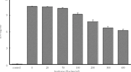
4. 괴화 추출물이 IL-6 생성에 미치는 영향
1 μg/ml의 세균내독소와 다양한 농도의 괴화 추 출물을 가하여 24시간 RAW264.7 세포를 배양하고, Figure 2. Effect of methanol extract of Sophorae Flos on iNOS protein expression in RAW264.7 cells stimulat- ed with P. intermedia LPS. Cells were treated with LPS (1 μg/ml) and indicated concentrations of methanol extract of Sophorae Flos for 24 h. iNOS protein synthesis was measured by immunoblot analysis of cell lysates using iNOS-specific antibody, and iNOS protein levels were quantified by densitometry
80 100 iNOS
60
40
control 0 20 50
Sophorae Flos (µg/ml)
100 200 300 400
20
iNOS protein expression (% of maximun) 0
배양 상층액 내의 IL-6 농도를 측정하였다. 괴화 추출 물은 20 μg/ml에서 400 μg/ml의 농도 범주에서 세 균내독소에 의한 대식세포에서의 IL-6 생성을 농도 의존적으로 억제하여, 괴화 추출물을 200 μg/ml과 400 μg/ml로 첨가하였을 때 IL-6의 생성이 약 26%와 41% 각각 억제되었다 (Figure 4).
5. 괴화 추출물이 IL-6 mRNA 발현에 미치는 영향
괴화 추출물이 세균내독소에 의한 IL-6 mRNA의 발현에 미치는 영향을 RT-PCR에 의해 확인하였다.
RAW264.7 세포를 1 μg/ml의 세균내독소에 노출시 켰을 때 IL-6 mRNA가 발현되었다(Figure 5). 그러나, 괴화 추출물은 20 μg/ml에서 400 μg/ml의 농도 범 주에서 세균내독소에 의한 대식세포에서의 IL-6 mRNA의 발현을 억제하여, 400 μg/ml로 괴화 추출 물을 첨가하였을 때 IL-6 mRNA의 발현이 약 62% 억
제되었다 (Figure 5).
IV. 고찰
치주질환은 숙주와 병원균 간의 상호 작용의 결과 이다. 치주질환 병인균주는 직접적으로 조직파괴를 초래하거나, 숙주의 반응을 조절하여 간접적으로 병 리작용을 발휘한다. 전통적인 치주처치는 각종의 비 외과적 및 외과적 방법에 의한 치태 및 치태 축적 촉 진 요소의 제거 즉, 항균요법에 중점을 두어왔다. 그 러나, 이러한 처치 방식은 해부학적으로 복잡한 부위 에서의 접근의 문제 등으로 인한 불완전한 처치로 질환의 재발을 초래하기도 한다. 이러한 점을 보완 하기 위해 각종의 항균제가 보조적으로 사용되어 왔 으나, 이 중 항생제는 내성균주의 출현 및 과민반응 의 가 능 성 등 으 로 인 한 문 제 점 이 있 으 며26), chlorhexidine 같은 합성제재는 장기간 사용 시 몇몇 Figure 3. Effect of methanol extract of Sophorae Flos on iNOS mRNA expression in RAW264.7 cells stimulat- ed with P. intermedia LPS. Cells were treated with LPS (1 μg/ml) and indicated concentrations of methanol extract of Sophorae Flos for 24 h. See Materials and methods for further details
90 80 70 60 100 β-actin
iNOS
50 40 30
control 0 20 50
Sophorae Flos (µg/ml)
100 200 300 400
20 10 0
iNOS mRNA expression (% of actin)
Figure 4. Effect of methanol extract of Sophorae Flos on P. intermedia LPS-induced IL-6 production from RAW264.7 cells. Cells were treated with LPS (1 μg/ml) and indicated concentrations of methanol extract of Sophorae Flos for 24 h, after which culture supernatants were collected and assayed for IL-6
12
9
6
control 0 20 50
Sophorae Flos (µg/ml)100 200 300 400 3
0
IL-6 (ng/ml)
Figure 5. Effect of methanol extract of Sophorae Flos on IL-6 mRNA expression in RAW264.7 cells stimulated with P. intermedia LPS. Cells were treated with LPS (1 μg/ml) and indicated concentrations of methanol extract of Sophorae Flos for 24 h. See Materials and methods for further details
control 0 20 50
Sophorae Flos (µg/ml)100 200 300 400 90
80 70 60 100
50 40 30 20 10 0
IL-6 mRNA expression (% of actin)
β-actin IL-6
합병증을 초래하기도 한다27-29).
한편, 숙주의 염증반응을 조절하는 것은 치주질환 의 처치에 있어 유용한 방법이 될 수 있다. 이는 치주 질환 병인균주에 대한 숙주의 염증반응을 조절함으 로써 치주질환의 빈도와 심도를 변화시킬 수 있기 때문이다. 이러한 점에서 비스테로이드성 소염제의 활용은 치주질환의 처치에 있어 유용할 수 있는데, 이는 이 약제가 arachidonic acid의 대사를 차단하여 염증을 억제하고, 치주질환과 관련된 치조골 파괴 등 을 억제하여 치주조직을 강화시킬 수 있기 때문이다
30-34).
치주질환의 처치에 있어 전신적 또는 국소적 보조 요법으로 유용할 수 있는 것으로 이미 보고된 약제 로는 flurbiprofen, ibuprofen, mefenamic acid, 그리 고 naproxen 등이 있으나, 이들의 장기적 사용과 관 련하여 몇몇 문제점들이 제시되고 있다35,36). 그러나, 천연생약 제재의 소염제의 경우에는 이러한 문제점 들이 최소일 수 있다는 장점이 있다. 이에 약물의 구 강 내 사용과 관련하여, 우수한 소염효과를 가지면서 도 여타의 합성약제에서 흔히 발생하는 부작용이 없 어, 치주질환의 처치 및 예방에 있어 유용할 수 있는 천연생약제의 개발 필요성이 있다.
괴화 (Sophorae Flos)는 콩과 식물인 회화나무 (Sophora japonica L.)의 꽃봉오리로서, rutin과 quercetin 등 의 flavonoid, sophoradiol 등 의 isoflavonoid, 그리고 betulin 등의 triterpenoid 성분 을 포함하고 있다37). 괴화는 소염, 수렴, 지혈, 그리 고 이뇨 등의 효과가 있는 것으로 알려져 있다37).
본 연구는 염증성 치주질환의 처치에 있어 괴화의 활용 가능성을 평가하기 위해 수행되었다. 본 연구 의 결과에 의하면, P.intermedia 세균내독소에 의한 대식세포에서의 NO 생성과 iNOS의 발현 그리고 IL- 6의 생성과 IL-6 mRNA의 발현이 괴화 추출물에 의 해 현저하게 억제되었다. 이는 괴화 추출물이, NO와 IL-6 등 염증 매개물질의 생성을 억제함으로써, 치주 질환을 포함한 염증성 질환의 처치에 있어 유용할 수 있음을 의미한다. 활성성분의 분리 및 생물학적 활성 분석, 활성성분이 실험적 치주질환에 미치는 영 향, 그리고 실제 치주처치에서의 활용 가능성 등에
대한 연구가 추후 수행되어야 할 것으로 사료된다.
V. 결론
NO와 IL-6는 다른 염증성 질환에서와 마찬가지로 염증성 치주질환에 있어서도 중요한 역할을 하는 것 으로 여겨지며, NO와 IL-6 등의 염증 매개물질의 생 성을 억제함으로써 치주질환의 처치가 가능할 수 있 다. 본 연구는 염증성 치주질환의 처치에 있어 괴화 의 활용 가능성을 평가하기 위해 수행되었다. 본 연 구의 결과에 의하면, P.intermedia 세균내독소에 의 한 대식세포에서의 NO 생성과 iNOS의 발현 그리고 IL-6의 생성과 IL-6 mRNA의 발현이 괴화 추출물에 의해 현저하게 억제되었다. 이는 괴화 추출물이, NO 와 IL-6 등 염증 매개물질의 생성을 억제함으로써, 치 주질환을 포함한 염증성 질환의 처치에 있어 유용할 수 있음을 의미한다. 활성성분의 분리 및 생물학적 활성 분석, 활성성분이 실험적 치주질환에 미치는 영 향, 그리고 실제 치주처치에서의 활용 가능성 등에 대한 연구가 추후 수행되어야 할 것으로 사료된다.
VI. 참고문헌
1. Moncada S, Palmer RMJ, Higgs EA. Nitric oxide:
physiology, pathology, and pharmacology.
Pharmacol Rev 1991;43:109-142.
2. Nathan C, Xie QW. Nitric oxide synthases: roles, tolls and controls. Cell 1994;78:915-918.
3. Geller DA, Nussler AK, Di Silvio M et al.
Cytokines, endotoxin, and glucocorticoids regu- late the expression of inducible nitric oxide syn- thase in hepatocytes. Proc Natl Acad Sci USA 1993;90:522-526.
4. Nathan C, Xie QW. Regulation of biosynthesis of nitric oxide. J Biol Chem 1994;269:13725-13728.
5. Southey A, Tanaka S, Murakami T et al.
Pathophysiological role of nitric oxide in rat experimental colitis. Int J Immunopharmacol 1997;19:669-676.
6. Weinberg JB, Granger DL, Pisetsky DS et al. The role of nitric oxide in the pathogenesis of spon- taneous murine autoimmune disease: increased nitric oxide production and nitric oxide synthase expression in MRL-lpr/lpr mice, and reduction of spontaneous glomerulonephritis and arthritis by orally administered NG-monomethyl-L-arginine.
J Exp Med 1994;179:651-660.
7. Matejka M, Partyka L, Ulm C, Solar P, Sinzinger H. Nitric oxide synthesis is increased in peri- odontal disease. J Periodont Res 1998;33:517- 518.
8. Blix IJ, Helgeland K. LPS from Actinobacillus actinomycetemcomitansand production of nitric oxide in murine macrophages J774. Eur J Oral Sci 1998;106:576-581.
9. Sosroseno W, Barid I, Herminajeng E, Susilowati H. Nitric oxide production by a murine macrophage cell line (RAW264.7) stimulated with lipopolysaccharide from Actinobacillus actinomycetemcomitans. Oral Microbiol Immunol 2002;17:72-78.
10. Kim SJ, Ha MS, Choi EY, Choi JI, Choi IS.
Prevotella intermedia lipopolysaccharide stimu- lates release of nitric oxide by inducing expres- sion of inducible nitric oxide synthase. J Periodont Res 2004;39;424-431.
11. Kim SJ, Ha MS, Choi EY, Choi JI, Choi IS. Nitric oxide production and inducible nitric oxide syn- thase expression induced by Prevotella nigrescenslipopolysaccharide. FEMS Immunol Med Microbiol. 2005;43:51-58.
12. Batista AC, Silva TA, Chun JH, Lara VS. Nitric oxide synthesis and severity of human periodon- tal disease. Oral Diseases 2002;8:254-260.
13. Hirose M, Ishihara K, Saito A et al. Expression of cytokines and inducible nitric oxide synthase in inflamed gingival tissue. J Periodontol 2001;72:590-597.
14. Kendall HK, Haase HR, Li H, Xiao Y, Bartold PM. Nitric oxide synthase type-II is synthesized by human gingival tissue and cultured human gingival fibroblasts. J Periodont Res 2000;35:194- 200.
15. Lappin DF, Kjeldsen M, Sander L, Kinane DF.
Inducible nitric oxide synthase expression in periodontitis. J Periodont Res 2000;35:369-373.
16. Kishimoto T. The biology of interleukin-6.
Blood 1989;74:1-10.
17. Hirano T, Akira S, Taga T, Kishimoto T.
Biological and clinical aspects of interleukin-6.
Immunol Today 1990;11;443-449.
18. Ishimi Y, Miyaura C, Jin CH et al. IL-6 is pro- duced by osteoblasts and induce bone resorp- tion. J Immuol 1990;145:3897-3903.
19. Takahashi K, Takashiba S, Nagai A et al.
Assessment of interleukin-6 in the pathogenesis of periodontal disease. J Periodontol 1994;65:147-153.
20. Guillot JL, Pollock SM, Johnson RB. Gingival interleukin-6 concentration following phase 1 therapy. J Periodontol 1990;66:667-672.
21. Takada H, Mihara J, Morisaki I, Hamada S.
Production of cytokines by human gingival fibroblast, 1991:265-276. In S Hamada, SC Holt, and JR McGhee (ed.), Periodontal disease:
pathogenesis and host immune response.
Quintessence, Tokyo.
22. Westphal O, Jann K. Bacterial lipopolysaccha- rides: extraction with phenol-waterand further applications of the procedure. In: RL Whistler eds. Methods in carbohydrate chemistry. New York: Academic Press, 1965:83-91.
23. Markwell MA, Haas SM, Bieber LL, Tolbert NE.
A modification of the Lowry procedure to sim- plify protein determination in membrane and lipoprotein samples. Anal Biochem 1978;87:206- 210.
24. Green LC, Wagner DA, Glogowski J et al.
Analysis of nitrate, nitrite, and [15N]nitrate in bio- logical fluids. Anal Biochem 1982;126:131-138.
25. Rappolee DA, Wang A, Mark D, Werb Z. Novel method for studying mRNA phenotypes in sin- gle or small numbers of cells. J Cell Biochem 1989;39:1-11.
26. Goodson JM, Tanner A. Antibiotic resistance of the subgingival microbiota following local tetra- cycline therapy. Oral Microbiol Immunol 1992;7:113-117.
27. Lang NP, Hotz P, Graf H et al. Effects of super- vised chlorhexidine mouthrinses in children. A longitudinal clinical trial. J Periodontal Res.
1982;17:101-111
28. Löe H. Does chlorhexidine have a place in the prophylaxis of dental diseases? J Periodontal Res Suppl. 1973;12:93-99.
29. Eriksen HM, Nordbo H, Kantanen H, Ellingsen JE. Chemical plaque control and extrinsic tooth discoloration. J Clin Periodontol 1985;12:345- 350.
30. Vogel RI, Schneider L, Goteiner D. The effects of a topically-active nonsteroidal anti-inflammatory drug on ligature-induced periodontal disease in the squirrel monkey. J Clin Periodontol 1986;13:139-144.
31. Heasman PA, Seymour RA, Boston PF. The
effect of a topical non-steroidal anti-inflammato- ry drug on the development of experimental gingivitis in man. J Clin Periodontol 1989;16:353- 358.
32. Williams RC, Jeffcoat MK, Howell TH et al.
Altering the progression of human alveolar bone loss with the non-steroidal anti-inflammatory drug flurbiprofen. J Periodontol 1989;60: 485- 490.
33. Williams RC, Jeffcoat MK, Howell TH et al.
Topical flurbiprofen treatment of periodontitis in beagles J Periodont Res 1988;23:166-169.
34. Howell TH, Jeffcoat MK, Goldhaber P et al.
Inhibition of alveolar bone loss in beagles with the NSAID naproxen. J Periodont Res 1991;26:498-501.
35. Bjarnason I, Hayllar J, MacPherson AJ, Russell AS. Side effects of nonsteroidal anti-inflammato- ry drugs on the small and large intestine in humans. Gastroenterol 1993;104:1832-1847.
36. Sikes DH, Agrawal NM, Zhao WW et al.
Incidence of gastroduodenal ulcers associated with valecoxib compared with that of ibuprofen and diclofenac in patients with osteoarthritis. Eur J Gastroenterol Hepatol 2002;14:1101-1111.
37. 생약학연구회. 현대생약학, 학창사 1994:317- 321.
-Abstract-
Suppression of nitric oxide and interleukin-6 production by methanol extract of Sophorae Flos in macrophage cells
Ji-Eun Lee1, Ju-Youn Lee1, Jeom-Il Choi1, Chong-Kwan Kim2, Sung-Jo Kim1
1Department of Periodontology, College of Dentistry, Pusan National University
2Department of Periodontology, College of Dentistry, Yonsei University
Both nitric oxide (NO) and interleukin-6 (IL-6) have been thought to have a role in the pathogenesis of inflammatory periodontal disease as it does in other inflammatory diseases, and the inhibitors of NO and IL-6 production have been considered as potential anti-inflammatory agents. In this study, we evaluated methanol extract of Sophorae Flosfor inhibition of NO and IL-6 production in Prevotella intermediaLPS-induced mouse macrophages RAW264.7 cells. Dried Sophorae Flos was sliced, and extracted with 100% methanol. LPS from P. intermedia ATCC 25611 was prepared by the standard hot phenol-water method. NO production was assayed by measuring the accumulation of nitrite in culture supernatants and IL-6 was measured using mouse IL-6 ELISA kit. Western blot analysis of iNOS and analysis of reverse transcription (RT)-PCR products were car- ried out. The methanol extract of Sophorae Flos concentration-dependently reduced the production of NO and the expression of iNOS protein and mRNA in RAW264.7 cells treated with P.intermediaLPS. Sophorae Flos also suppressed IL-6 production and the expression of IL-6 mRNA in RAW264.7 cells stimulated by P.interme- diaLPS. The inhibition of NO and IL-6 production by Sophorae Flos may be useful in the therapy of inflamma- tory diseases such as periodontitis. This hypothesis, however, remains to be tested.
Key words : Sophorae Flos, Prevotella intermediaLPS, RAW264.7 cells, nitric oxide, interleukin-6