© 2016 The Korean Ophthalmological Society
This is an Open Access article distributed under the terms of the Creative Commons Attribution Non-Commercial License (http://creativecommons.org/licenses /by-nc/3.0/) which permits unrestricted non-commercial use, distribution, and reproduction in any medium, provided the original work is properly cited.
Original Article
Longitudinal Changes in Retinal Nerve Fiber Layer Thickness after Intravitreal Anti-vascular Endothelial Growth Factor Therapy
Young-Joon Jo1,2, Woo-Jin Kim1, Il-Hwan Shin1, Jung-Yeul Kim1,2
1Department of Ophthalmology, Chungnam National University College of Medicine, Daejeon, Korea
2Research Institute for Medical Science, Chungnam National University College of Medicine, Daejeon, Korea
Purpose: To determine the effects of intravitreal anti-vascular endothelial growth factor (VEGF) on thickness of the retinal nerve fiber layer (RNFL) in patients with age-related macular degeneration.
Methods: Twenty eyes of 20 patients diagnosed with age-related macular degeneration who underwent intrav- itreal anti-VEGF injection were studied. Postinjection RNFL thickness was measured using optical coherence tomography. Average thickness, four-quadrant RNFL thicknesses, and intraocular pressure (IOP) in affected eyes were measured before and 6 and 12 months after anti-VEGF injection for comparison. RNFL thickness and IOP in affected and normal fellow eyes were also compared. Given that macular lesions can affect RNFL thickness, the changes in thickness were evaluated by dividing the 12 clock-hour RNFL into the pathologic ar- eas adjacent to the lesion and the non-pathologic area.
Results: The mean clock-hour segment in the pathologic area was 4.8 hours. A significantly thicker RNFL was exhibited in temporal quadrants and pathologic areas (p = 0.043 and 0.048, respectively) in affected eyes before injection compared to the baseline RNFL thickness in normal eyes. No significant differences were found in RNFL thickness or IOP between affected and normal eyes after injection. The changes over time in the temporal and pathologic areas were statistically significant at 6 and 12 months after injection compared to baseline data (p < 0.05). No significant differences were displayed in RNFL thickness in the other three quad- rants or in non-pathologic areas in either affected or normal eyes. Sequential changes in RNFL thickness in affected eyes were not significant.
Conclusions: Repeat intravitreal anti-VEGF treatment did not have a significant effect on RNFL thickness.
RNFL thickness significantly decreased with time in the pathologic areas and in the temporal segment adja- cent to exudative macular lesions. The reduction in RNFL thickness was most likely associated with changes in the macular lesion rather than with anti-VEGF injection.
Key Words: Anti-vascular endothelial growth factor, Optical coherence tomography, Retinal nerve fiber layer
Vascular endothelial growth factor (VEGF) is involved in angiogenesis and vascular permeability and plays a cru- cial role in inducing exudative age-related macular degen- eration (AMD) by promoting choroidal neovascularization.
Anti-VEGF therapy is widely used in various eye disor- ders, including AMD, because it is effective for suppress-
Received: May 19, 2015 Accepted: July 13, 2015
Corresponding Author: Jung-Yeul Kim, MD. Department of Ophthal- mology, Chungnam National University Hospital, #282 Munhwa-ro, Jung-gu, Daejeon 35015, Korea. Tel: 82-42-280-8433, Fax: 82-42-255- 3745, E-mail: [email protected]
ing angiogenesis and macular edema [1-3].
The efficacies of the anti-VEGF agents ranibizumab (Lucentis; Genentech, San Francisco, CA, USA) and beva- cizumab (Avastin, Genentech) have been illustrated in many studies, although some complications have been re- ported [4-7]. Elevated intraocular pressure is a common complication following intravitreal injection of anti-VEGF.
In some studies, intraocular pressure increased steeply fol- lowing injection but declined to normal levels within 10 to 30 minutes [8-11]. In other studies, intraocular pressure re- mained elevated for some time following injection [12-14].
Some studies have also suggested that blocking VEGF with antagonists leads to nerve damage, given the role of VEGF in neurophysiological processes [15-17].
Because the effects of intravitreally injected VEGF medications are of limited duration, repeated injections are required for long-term VEGF-inhibiting effects. Repeat in- travitreal injection might result in sustained intraocular pressure, and blocking VEGF might cause optic nerve damage. Therefore, the effects of repeat intravitreal an- ti-VEGF injection on the optic nerve were evaluated in pa- tients with AMD, based on spectral domain optical coher- ence tomography (OCT) retinal nerve fiber layer (RNFL) thickness measurements.
Materials and Methods
Study population
This was a prospective cohort study. The protocol was approved by the institutional review board of Chungnam National University Hospital. All participants signed in- formed consent forms, and the study adhered to the tenets of the Declaration of Helsinki.
The study initially included 24 eyes of 24 patients who presented to the retina clinic at the hospital, were diag- nosed with unilateral exudative AMD, and underwent an- ti-VEGF treatment with intravitreal ranibizumab. Normal fellow eyes served as the control group. All subjects re- ceived three intravitreal injections of ranibizumab at monthly intervals from the diagnosis date. Additional in- jections were administered if necessary, depending on the presence of macular edema or hemorrhage.
Subjects were excluded if they had an eye disorder that could affect RNFL thickness, such as glaucoma or retinal
vessel obstruction, a history of photodynamic treatment for macular degeneration, bilateral exudative AMD, or a history of intraocular surgery such as vitrectomy. In addi- tion, four of the initial patients were excluded because of poor data quality, resulting in a total of 20 eyes of 20 pa- tients included for final analysis.
All subjects underwent intraocular pressure measure- ment, slit-lamp examination, and fundus examination. The average and four-quadrant RNFL thicknesses were mea- sured in the affected and normal counterpart eyes using a Cirrus spectral domain OCT (Carl Zeiss Meditec, Dublin, CA, USA).
Measurement of retinal nerve fiber layer thickness RNFL thickness was measured before and 6 and 12 months after intravitreal injection with the optic disc cube mode of Cirrus spectral domain OCT. OCT was performed by an experienced examiner. Poor-quality images with a signal strength less than 5 and any scans with visible eye movements, blinking artifacts, or poor centration were ex- cluded. Also, images with missing parts, misplacement of boundaries between retinal layers, or images showing seemingly distorted anatomy that resulted in readings of zero or otherwise abnormally low values were discarded to prevent algorithm segmentation failure. All eyes were scanned twice to evaluate reproducibility, and good repro- ducibility was achieved based on intraclass correlation co- efficients of 0.941 to 0.989. The Optic Disc Cube 200 × 200 scanning program (Cirrus HD-OCT, Carl Zeiss Meditec) obtains 200 A-scans from 200 linear B-scans evenly dis- tributed in a 6 mm × 6 mm area centered over the optic nerve. This scanning mode was used to measure the aver- age and four-quadrant RNFL thicknesses.
RNFL thicknesses were measured in affected eyes be- fore and 6 and 12 months after intravitreal injection for se- quential comparison. RNFL thickness in affected and nor- mal eyes was also compared. RNFL thickness was divided into 12 segments of 30 degrees for 12 clock-hour RNFL thickness measurements.
Under the assumption that macular lesions affect RNFL thickness, changes in average thickness were evaluated af- ter dividing the 12 clock-hour RNFL thickness measure- ments into pathologic area, including the lesion, and non-pathologic area of the affected eye and the corre- sponding area of the normal fellow eye (Fig. 1A and 1B).
Measurement of intraocular pressure
Intraocular pressure measurements were obtained using a Goldmann applanation tonometer before and 6 and 12 months after intravitreal injection for sequential compari- son. Intraocular pressure in affected and normal eyes was also compared.
Statistical analysis
Statistical analysis was performed using PASW Statistics ver. 18.0 (SPSS Inc., Chicago, IL, USA). Sequential time-re- lated changes in RNFL thickness in affected and normal eyes were analyzed using Wilcox signed-ranks tests. The comparison of RNFL thickness between affected and nor- mal eyes at different time points was conducted using a Mann-Whitney U-test. A p-value less than 0.05 was con- sidered statistically significant.
Results
Twenty eyes of 20 patients were enrolled, of whom 13 were male and seven were female with a median age of
67.1 ± 8.9 years. The average best-corrected visual acuity was 0.5 ± 0.6 (logarithm of the minimum angle of resolu- tion), and the mean intraocular pressure was 15.3 ± 2.6 mmHg. The mean number of anti-VEGF injections was 5.0
± 1.0, and the mean clock-hour segment in the pathologic area was 4.8 ± 0.5 in the 12 clock-hour RNFL thickness analysis (Table 1).
Average and quadrant retinal nerve fiber layer thick- ness
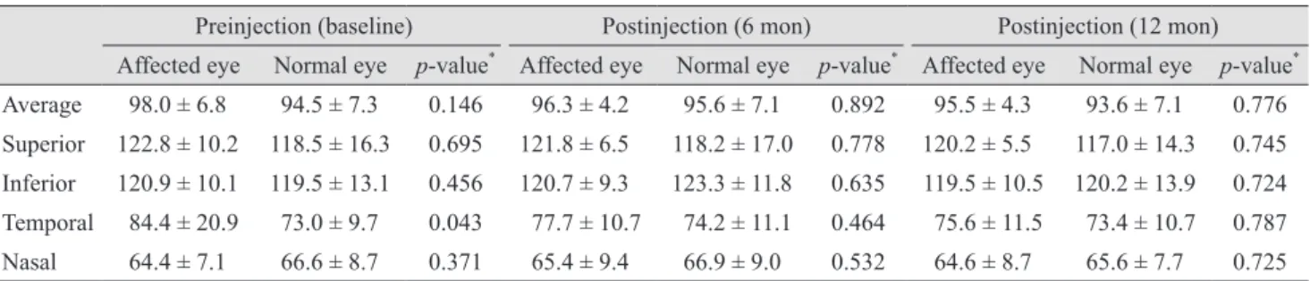
The overall average RNFL thickness at baseline was 98.0 ± 6.8 µm in affected eyes and 94.5 ± 7.3 µm in normal eyes (p = 0.146). Postinjection differences in RNFL thick- ness between the two groups were not significant at 6 or 12 months (p = 0.892 and 0.776, respectively). The average RNFL thickness in affected eyes was 96.3 ± 4.2 µm at 6 months and 95.5 ± 4.3 µm at 12 months after intravitreal injection, showing no significant difference from baseline (p = 0.153 and 0.070, respectively).
Segmental RNFL thickness in the superior, inferior, and nasal quadrants did not exhibit significant changes in the affected or normal eyes at 6 and 12 months postinjection compared to baseline RNFL thickness (p > 0.05). Postin- jection changes in RNFL thickness were not significant in affected eyes over time (p > 0.05).
Temporal RNFL thickness at baseline was 84.4 ± 20.9 µm in affected eyes, which was significantly thicker than the 73.0 ± 9.7 µm observed in normal eyes (p = 0.043).
However, postinjection differences in temporal RNFL thickness between the two groups were not significant at 6
125 139 69
49
53 90 159
111 76
107
176 118
119 144 77
60
78 153 107
68 51
48
109 125
Fig. 1. Fundus photograph and retinal nerve fiber layer thickness analysis of (A) the right affected eye and (B) the left normal fel- low eye of a patient with age-related macular degeneration. The red and black arrows represent the pathologic and non-pathologic areas in the affected eye, respectively. The green and blue arrows represent the same area in the unaffected eye, respectively.
125 139 69
49
53 90 159
111 76
107
176 118
119 144 77
60
78 153 107
68 51
48
109 125
A
B
Table 1. Patient demographics
Variable Value
No. of patients (eyes) 20 (20)
Age (yr) 67.1 ± 8.9
Sex (male / female) 13 / 7
Laterality (OD / OS) 7 / 13
BCVA (logMAR) 0.5 ± 0.6
Intraocular pressure (mmHg) 15.3 ± 2.6
No. of injections (average) 5.0 ± 1.0
Range of pathologic RNFL area (clock-hour) 4.8 ± 0.5 Values are presented as number or mean ± standard deviation.
OD = right eye; OS = left eye; BCVA = best-corrected visual acuity; logMAR = logarithm of the minimum angle of resolution;
RNFL = retinal nerve fiber layer.
or 12 months (p = 0.464 and 0.787, respectively). Time-re- lated changes in temporal RNFL thickness were signifi- cant in affected eyes 6 and 12 months after injection com- pared to baseline (p = 0.012 and 0.006, respectively) (Table 2 and Fig. 2).
Clock-hour thickness in pathologic and non-pathologic areas
The overall average RNFL thickness at baseline was 85.8 ± 21.2 µm in the pathologic area of affected eyes, which was significantly greater than the 74.0 ± 10.0 µm measured in normal eyes (p = 0.048). However, postinjec- tion differences in RFNL thickness were not significant between affected and normal eyes at 6 or 12 months (p = 0.607 and 0.665, respectively). Longitudinal changes in RNFL thickness in the pathologic areas were significant in
affected eyes at 6 and 12 months postinjection compared to baseline (p = 0.011 and 0.005, respectively).
The average RNFL thickness in the non-pathologic area was not significantly different between affected and nor- mal eyes ( p > 0.05). The average postinjection RNFL thickness did not change significantly in affected eyes over time (Table 3 and Fig. 3).
Average intraocular pressure
The average intraocular pressure at baseline was 15.3 ± 2.6 mmHg in affected eyes and 16.0 ± 2.9 mmHg in nor- mal eyes, showing no significant difference (p = 0.473).
Postinjection differences in intraocular pressure between the two groups were not significant at 6 or 12 months (p = 1.000 and 0.534, respectively) (Table 4).
Discussion
Intravitreal anti-VEGF injection is commonly used in the treatment of a wide variety of retinal diseases, includ- ing exudative AMD. However, intravitreal injection can result in complications, including endophthalmitis, intra- ocular hemorrhage, and ocular hypertension. Several stud- ies have addressed these complications [4-7].
Temporary ocular hypertension following intravitreal anti-VEGF injection is caused by increased intraocular volume [8,9,11]. Falkenstein et al. [9] reported that the aver- age intraocular pressure soared to 36.3 mmHg from a baseline of 15.2 mmHg 3 minutes after intravitreal injec- tion of 0.05 mL bevacizumab in 70 patients. However, the mean intraocular pressure decreased to 24.6 mmHg at 10 minutes, and intraocular pressure in all eyes decreased to
Table 2. Changes in average and quadrant retinal nerve fiber layer thickness in affected and normal counterpart eyes Preinjection (baseline) Postinjection (6 mon) Postinjection (12 mon) Affected eye Normal eye p-value* Affected eye Normal eye p-value* Affected eye Normal eye p-value* Average 98.0 ± 6.8 94.5 ± 7.3 0.146 96.3 ± 4.2 95.6 ± 7.1 0.892 95.5 ± 4.3 93.6 ± 7.1 0.776 Superior 122.8 ± 10.2 118.5 ± 16.3 0.695 121.8 ± 6.5 118.2 ± 17.0 0.778 120.2 ± 5.5 117.0 ± 14.3 0.745 Inferior 120.9 ± 10.1 119.5 ± 13.1 0.456 120.7 ± 9.3 123.3 ± 11.8 0.635 119.5 ± 10.5 120.2 ± 13.9 0.724 Temporal 84.4 ± 20.9 73.0 ± 9.7 0.043 77.7 ± 10.7 74.2 ± 11.1 0.464 75.6 ± 11.5 73.4 ± 10.7 0.787 Nasal 64.4 ± 7.1 66.6 ± 8.7 0.371 65.4 ± 9.4 66.9 ± 9.0 0.532 64.6 ± 8.7 65.6 ± 7.7 0.725 Values are presented as mean ± standard deviation.
*Comparison between affected eyes and normal counterpart eyes in each period.
140 120 100 80 60 40 20
0 Average Superior Inferior Temporal Nasal
RNFL (μm)
Baseline 6 mon 12 mon
**
Fig. 2. Longitudinal changes in average and quadrant retinal nerve fiber layer (RNFL) thickness in affected eyes. The differ- ences between baseline and postinjection at 6 months (p = 0.012) and 12 months (p = 0.006) were statistically significant in the temporal areas. *p < 0.05, Wilcoxon signed-rank test.
less than 30 mmHg after 15 minutes. Sharei et al. [11] also reported that intraocular pressure increased to more than 40 mmHg immediately after intravitreal injection of 0.05 mL ranibizumab in 71.1% of 45 patients but decreased to 21 mmHg at 10 minutes.
On the other hand, some studies have observed a longer duration of ocular hypertension. Kahook et al. [12] report- ed six cases of chronic ocular hypertension after one-time or repeat injection of bevacizumab and cited aqueous out- flow induced by an accumulated trabecular meshwork and
inflammatory responses as the cause of sustained ocular hypertension [15].
In our study, intraocular pressure measurements of af- fected and normal eyes were obtained 6 and 12 months af- ter intravitreal injection for comparison. The difference in mean intraocular pressure was not significantly different between the affected and normal eyes.
In addition, anti-VEGF drugs can have adverse effects on neuronal cells due to the blockage of VEGF [16-19].
Considering that VEGF is involved in the survival of neu- ronal cells and has neuroprotective and neurotrophic ac- tions in addition to its angiogenesis role [16,17], anti-VEGF treatment can affect the neurophysiologic role of VEGF and cause damage to optic nerves.
Because the effects of anti-VEGF agents are of limited duration, repeated intravitreal injections are required in many cases. As a result, brief ocular hypertension, in- creased intraocular pressure fluctuations, changes in ocu- lar blood flow, and adverse effects on the optic nerve can ensue.
Seth et al. [20] analyzed changes in the cup-to-disc ratio in 23 eyes over 9 months following repeat intravitreal in- jection of pegaptanib. They observed no statistically sig- nificant changes in the ratio between treated and normal eyes. Horsley et al. [19] retrospectively assessed changes in RNFL thickness in patients with AMD following repeat intravitreal anti-VEGF injection using stratus OCT and re- ported no significant postinjection changes in RNFL thick-
120 100 80 60 40 20
0 Pathologic area Non-pathologic area
RNFL (μm)
Baseline 6 mon 12 mon
**
Fig. 3. Longitudinal changes in average retinal nerve fiber layer (RNFL) thickness in the pathologic and non-pathologic areas of affected eyes. The differences between baseline and post-injec- tion at 6 months (p = 0.011) and 12 months (p = 0.005) were sta- tistically significant in the pathologic areas. *p < 0.05, Wilcoxon signed-rank test.
Table 3. Changes in pathologic and non-pathologic area retinal nerve fiber layer thickness in affected and normal eyes Preinjection (baseline) Postinjection (6 mon) Postinjection (12 mon) Affected eye Normal eye p-value* Affected eye Normal eye p-value* Affected eye Normal eye p-value* Pathologic area 85.8 ± 21.2 74.0 ± 10.0 0.048 80.6 ± 13.5 78.0 ± 14.4 0.607 79.0 ± 13.7 77.2 ± 14.1 0.665 Non-pathologic
area† 101.1 ± 6.9 100.4 ± 11.6 0.724 100.8 ± 6.1 100.9 ± 11.1 0.745 99.8 ± 6.5 98.7 ± 11.0 0.957 Values are presented as mean ± standard deviation.
*Comparison between area in affected eyes and corresponding area in normal eyes in each period; †Average clock-hour retinal nerve fiber layer thickness, excluding areas adjacent to macular lesion.
Table 4. Changes in IOP in affected and normal counterpart eyes
Preinjection (baseline) Postinjection (6 mon) Postinjection (12 mon) Affected eye Normal eye p-value* Affected eye Normal eye p-value* Affected eye Normal eye p-value* IOP (mmHg) 15.3 ± 2.6 16.0 ± 2.9 0.473 16.2 ± 2.8 16.2 ± 3.1 1.000 16.4 ± 2.7 16.9 ± 2.6 0.534 Values are presented as mean ± standard deviation.
IOP = intraocular pressure.
*Comparison between affected eyes and normal eyes in each period.
ness throughout the follow-up period. Another study as- sessed RNFL thickness in 49 eyes of 49 patients with AMD using stratus OCT for 12 months after an average of 4.8 intravitreal injections of an anti-VEGF agent [21]. They found that average RNFL was significantly thinner at 12 months compared to baseline values. They also found that RNFL thickness significantly decreased in the superior, inferior, and temporal quadrants at 12 months compared to baseline data. However, there was no significant change in nasal RNFL thickness.
In the present study, RNFL thickness at baseline was significantly greater in the temporal and pathologic areas of affected eyes compared to normal eyes. However, no significant difference was displayed between the two groups after injection. RNFL thickness in the temporal and pathologic areas of affected eyes did, however, display significant changes at sequential time points after injec- tion. Longitudinal changes in RNFL thickness were not significant in quadrants other than the temporal quadrant in non-pathologic areas of affected eyes. Therefore, the re- duction in RNFL thickness in the pathologic area is more likely to be due to a change in the macular lesion rather than a result of increased pressure due to anti-VEGF ad- ministration. That is, RNFL thickness was increased at baseline due to an exudative lesion, edema, or hemorrhage in the macular area [22], and RNFL thickness was reduced 6 and 12 months after injection because the exudative le- sion improved after repeat injection. This conclusion is supported by the findings of greater RNFL thickness in the temporal quadrant in affected eyes at baseline com- pared to that of normal eyes, without a significant differ- ence in postinjection RNFL thickness between the two groups. The mean clock-hour RNFL segment in the patho- logic area was 4.8 hours. This area overlapped that of the temporal quadrant, leading to similar changes in RNFL thickness.
Hwang et al. [22] described two reasons for increased RNFL thickness in diabetic macular edema (DME) pa- tients. First, an increase in temporal sector RNFL thick- ness in patients with acutely worsening DME might be re- lated to a change in macular tomography due to macular edema. Second, the breakdown of the inner blood retinal barrier in RNFL causes RNFL edema, which then results in increased RNFL thickness in all sectors in patients with chronic DME. The present study targeted AMD patients rather than DME patients. Thus, a similar increase in tem-
poral sector RNFL thickness at baseline in patients with AMD might be related to changes in macular tomography due to macular edema.
This prospective study represents an improvement over previous studies. Spectral domain-OCT was used, which offers better reproducibility than time-domain OCT.
Four-quadrant RNFL thickness measures were conducted in addition to an overall assessment of average RNFL thickness. Additionally, pathologic and non-pathologic ar- eas close to and distant from the macular lesions were sep- arately evaluated.
In conclusion, repeat intravitreal anti-VEGF treatment did not have a significant effect on overall RNFL thick- ness. However, RNFL thickness significantly decreased with time in the temporal and pathologic areas. It can be concluded that the reduction in RNFL thickness was more associated with changes in the macular lesion rather than with anti-VEGF injection. Long-term prospective studies with more subjects are needed to investigate the long-term effects of anti-VEGF treatment in various disease states.
Conflict of Interest
No potential conflict of interest relevant to this article was reported.
References
1. Rosenfeld PJ, Brown DM, Heier JS, et al. Ranibizumab for neovascular age-related macular degeneration. N Engl J Med 2006;355:1419-31.
2. Gunther JB, Altaweel MM. Bevacizumab (Avastin) for the treatment of ocular disease. Surv Ophthalmol 2009;54:372- 400.
3. el Matri L, Chebil A, Kort F, et al. Intravitreal injection of triamcinolone combined with bevacizumab for choroidal neovascularization associated with large retinal pigment epithelial detachment in age-related macular degeneration.
Graefes Arch Clin Exp Ophthalmol 2010;248:779-84.
4. Jager RD, Aiello LP, Patel SC, Cunningham ET Jr. Risks of intravitreous injection: a comprehensive review. Retina 2004;24:676-98.
5. Angulo Bocco MC, Glacet-Bernard A, Zourdani A, et al.
Intravitreous injection: retrospective study on 2028 injec-
tions and their side effects. J Fr Ophtalmol 2008;31:693-8.
6. Sampat KM, Garg SJ. Complications of intravitreal injec- tions. Curr Opin Ophthalmol 2010;21:178-83.
7. Day S, Acquah K, Mruthyunjaya P, et al. Ocular complica- tions after anti-vascular endothelial growth factor therapy in Medicare patients with age-related macular degenera- tion. Am J Ophthalmol 2011;152:266-72.
8. Hollands H, Wong J, Bruen R, et al. Short-term intraocular pressure changes after intravitreal injection of bevacizum- ab. Can J Ophthalmol 2007;42:807-11.
9. Falkenstein IA, Cheng L, Freeman WR. Changes of intra- ocular pressure after intravitreal injection of bevacizumab (Avastin). Retina 2007;27:1044-7.
10. Kim JE, Mantravadi AV, Hur EY, Covert DJ. Short-term intraocular pressure changes immediately after intravitreal injections of anti-vascular endothelial growth factor agents.
Am J Ophthalmol 2008;146:930-4.e1.
11. Sharei V, Hohn F, Kohler T, et al. Course of intraocular pressure after intravitreal injection of 0.05 mL ranibizum- ab (Lucentis). Eur J Ophthalmol 2010;20:174-9.
12. Kahook MY, Kimura AE, Wong LJ, at al. Sustained eleva- tion in intraocular pressure associated with intravitreal bevacizumab injections. Ophthalmic Surg Lasers Imaging 2009;40:293-5.
13. Bakri SJ, McCannel CA, Edwards AO, Moshfeghi DM.
Persisent ocular hypertension following intravitreal ranibi- zumab. Graefes Arch Clin Exp Ophthalmol 2008;246:955- 8.
14. Adelman RA, Zheng Q, Mayer HR. Persistent ocular hy- pertension following intravitreal bevacizumab and ranibi- zumab injections. J Ocul Pharmacol Ther 2010;26:105-10.
15. Jalil A, Fenerty C, Charles S. Intravitreal bevacizumab (Avastin) causing acute glaucoma: an unreported complica- tion. Eye (Lond) 2007;21:1541.
16. Sondell M, Lundborg G, Kanje M. Vascular endothelial growth factor has neurotrophic activity and stimulates ax- onal outgrowth, enhancing cell survival and Schwann cell proliferation in the peripheral nervous system. J Neurosci 1999;19:5731-40.
17. Zachary I. Neuroprotective role of vascular endothelial growth factor: signalling mechanisms, biological function, and therapeutic potential. Neurosignals 2005;14:207-21.
18. Nishijima K, Ng YS, Zhong L, et al. Vascular endothelial growth factor-A is a survival factor for retinal neurons and a critical neuroprotectant during the adaptive response to ischemic injury. Am J Pathol 2007;171:53-67.
19. Horsley MB, Mandava N, Maycotte MA, Kahook MY.
Retinal nerve fiber layer thickness in patients receiving chronic anti-vascular endothelial growth factor therapy.
Am J Ophthalmol 2010;150:558-61.e1.
20. Seth RK, Salim S, Shields MB, Adelman RA. Assessment of optic nerve cup-to-disk ratio changes in patients receiv- ing multiple intravitreal injections of antivascular endothe- lial growth factor agents. Retina 2009;29:956-9.
21. Martinez-de-la-Casa JM, Ruiz-Calvo A, Saenz-Frances F, at al. Retinal nerve fiber layer thickness changes in patients with age-related macular degeneration treated with intrav- itreal ranibizumab. Invest Ophthalmol Vis Sci 2012;53:6214- 8.
22. Hwang DJ, Lee EJ, Lee SY, et al. Effect of diabetic macular edema on peripapillary retinal nerve fiber layer thickness profiles. Invest Ophthalmol Vis Sci 2014;55:4213-9.