© 2016 The Korean Ophthalmological Society
This is an Open Access article distributed under the terms of the Creative Commons Attribution Non-Commercial License (http://creativecommons.org/licenses /by-nc/3.0/) which permits unrestricted non-commercial use, distribution, and reproduction in any medium, provided the original work is properly cited.
Original Article
Is Retinal Nerve Fiber Layer Thickness Change Related to Headache Lateralization in Migraine?
Alime Gunes1, Seden Demirci2, Levent Tok1, Ozlem Tok1, Serpil Demirci2, Süleyman Kutluhan2
1Department of Ophthalmology, Süleyman Demirel University Faculty of Medicine, Isparta, Turkey
2Department of Neurology, Süleyman Demirel University Faculty of Medicine, Isparta, Turkey
Purpose: To evaluate retinal nerve fiber layer (RNFL) thickness in migraine patients with unilateral headache.
Methods: A total of 58 patients diagnosed with migraine headache consistently occurring on the same side and 58 age- and sex-matched healthy subjects were evaluated in this cross-sectional study. RNFL thickness was measured using spectral-domain optical coherence tomography, and the side with the headache was com- pared with the contralateral side as well as with the results of healthy subjects.
Results: The mean patient age was 33.05 ± 8.83 years, and that of the healthy subjects was 31.44 ± 8.64 years (p = 0.32). The mean duration of disease was 10.29 ± 9.03 years. The average and nasal RNFL thicknesses were significantly thinner on the side of headache and on the contralateral side compared to control eyes (p
< 0.05, for all). Thinning was higher on the side of the headache compared to the contralateral side; however, this difference was not statistically significant.
Conclusions: The RNFL thicknesses were thinner on the side of the headache compared to the contralateral side in the migraine patients with unilateral headache, but this difference was not statistically significant.
Key Words: Migraine, Retinal nerve fiber layer thickness, Unilateral headache
Migraine is a common neurological disorder with epi- sodic headaches and is most common in females between the ages of 20 and 45 years [1,2]. One of the typical criteria for migraine is a unilateral character of the headache, which is more common than the bilateral type [3-5].
The pathophysiology of migraine is not clearly under- stood [6]. The complex pathophysiology of the disease en- tails proposed vascular and neuronal mechanisms [7]. Cor- tical spreading depression, first described by Leao [8], is a
key event characterized by dramatic changes in cortical potentials, significant transient increases in extracellular ions and neurotransmitters, and transient increases in cor- tical blood flow followed by sustained flow decreases [9].
This focal regional cerebral blood flow decrease, reported especially in migraine with aura, begins in the posterior circulation [10]. Reduced blood flow and vasospasm are usually limited to one hemisphere [11]. Rarely, hypoperfu- sion originates from other regions of the brain or even the retina and might also result in retinal infarction due to ret- inal artery occlusion [12,13]. Although vasospasm of cere- bral and retinal blood vessels is transient, recurrent mi- graine attacks can cause permanent structural changes in the retina.
Optical cohorence tomography (OCT) is a non-invasive
Received: February 27, 2015 Accepted: May 19, 2015
Corresponding Author: Alime Gunes, MD. Department of Ophthalmol- ogy, Süleyman Demirel University Faculty of Medicine, Isparta 32260, Turkey. Tel: 90-5054828345, Fax: 90-2462112830, E-mail: dralimesefer@
hotmail.com
and objective cross-sectional imaging technique that pro- vides quantitative in vivo information about the retinal nerve fiber layer (RNFL) [14]. This measurement sensitive- ly evaluates ganglion cell and retinal nerve fiber damage.
Several studies have evaluated RNFL thickness in mi- graine patients using OCT [15-21]. However, there has been no study evaluating the relationship between RNFL thick- ness and headache lateralization in migraine patients.
Therefore, the aim of the present study was to evaluate RNFL thickness on the side of the headache and the con- tralateral side of migraine patients who experience head- aches that are almost always on the same side and to com- pare migraine patients with healthy subjects.
Materials and Methods
The study was an observational, cross-sectional case se- ries that included 58 consecutive patients diagnosed with migraine headaches consistently located on the same side and 58 age- and sex-matched healthy subjects. All partici- pants were Caucasian. The study was approved by the Sü- leyman Demirel University Department of Medical Sci- ences ethics committee and was performed according to the tenets of the Declaration of Helsinki.
All participants were aged between 18 years and 45 years. Patients with glaucoma, intraocular pressure higher than 21 mmHg, refractive error greater than ±3 spherical diopters, optic neuropathy, optic disc anomaly, cataract, vitreo-retinal diseases, history of ocular trauma, ocular surgery or ocular laser, diabetes mellitus, hypertension, hyperlipidemia, cardiovascular or renal disease, history of central nervous system disorders including epilepsy, en- cephalitis, brain tumors, infarction, head trauma, any type of headache except for migraine, and users of drugs, alco- hol, and cigarettes were excluded from the study. All mea- surements were performed during attack-free periods in migraine patients.
A detailed neurological examination was performed for each patient. Migraine was diagnosed according to the International Classification of Headache Disor der second edition [3]. The migraine patients with headaches that con- sistently occur on the same side were included in the study.
Clinical and demographic characteristics of the patients were noted and included gender, age, migraine variables such as severity of headache, duration of headache, dura-
tion of attack, side of headache, and frequency of attacks.
The severity of headache was assessed by the Migraine Disability Assessment questionnaire that evaluates head- ache-related disability [22].
Detailed ophthalmological examinations were performed containing spherical equivalent, best-corrected visual acui- ty, slit lamp biomicroscopy, intraocular pressure with Goldmann applanation tonometry, central corneal thick- ness, anterior chamber depth, and axial length (PacScan 300AP, Biometric/Pachymeter; Sonomed, New York, NY, USA). RNFL thickness measurements were performed us- ing spectral-domain OCT (Spectral OCT SLO; OPKO/OTI Instrumentation, Miami, FL, USA). Average, temporal, nasal, inferior, and superior quadrant peripapillary RNFL thicknesses were recorded. Both eyes of migraine patients and the right eyes of the control group were included in this study. The findings in the migraine group were com- pared with the findings in healthy subjects. Additionally, in migraine patients, the mean RNFL thicknesses were com- pared between the side of headache and the contralateral side.
SPSS ver. 15.0 (SPSS Inc., Chicago, IL, USA) was used to analyze data. The normality of distribution for continu- ous variables was tested with the Kolmogorov-Smirnov test. Categorical variables were shown as frequency and percentages. For the comparisons of parametric data of two groups, Student’s t-test was used for parametric vari- ables and Mann-Whitney U-test was used for non-para- metric variables. Categorical variables were compared be- tween the groups by the chi-square test. Pearson’s correlation was used to determine the strength of the rela- tionship between the variables. Comparisons of parametric data of the three groups were performed with one-way ANOVA testing. Levene’s test was used to determine ho- mogeneity of variances and in case of homogeneity of variance, post hoc Tukey’s test was used. Data were pre- sented as mean ± standard deviation. A p-value less than 0.05 was considered to be significant.
Results
The demographic and clinical characteristics of all sub- jects are summarized in Table 1. There were no statistically significant differences between the migraine patients and healthy subjects in terms of age, sex, visual acuity, spheri-
cal equivalent, intraocular pressure, axial length, anterior chamber depth, and central corneal thickness. The mean age of the patients and controls were 33.05 ± 8.83 years and 31.44 ± 8.64 years, respectively (p = 0.32).
The mean RNFL thicknesses in each quadrant of the migraine and control groups are given in Table 2. In mi-
graine patients, the average and nasal RNFL thicknesses were significantly thinner compared to healthy subjects (102.4 and 80.0 µm vs. 108.1 and 93.9 µm, respectively).
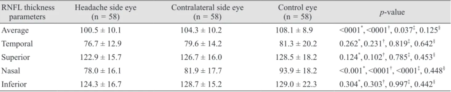
The mean RNFL thicknesses on the two sides of migraine patients and healthy subjects are given in Table 3. The av- erage and nasal RNFL thicknesses in both sides of the mi- graine patients were significantly thinner compared to the controls (p < 0.05, for all). In migraine patients, RNFL thicknesses were thinner on the side of headache than the contralateral side, but this difference was not statistically significant (Table 3).
The correlations between RNFL thicknesses and mi- graine headache variables are shown in Table 4. Two sig- nificant correlations were found between the duration of headache and superior RNFL thickness (r = -0.21, p = 0.02) and frequency of attacks and temporal-RNFL thickness (r
= -0.19, p = 0.03). We also observed no significant differ- ences in RNFL thicknesses between patients with aura and without aura (Table 5).
Discussion
In the present study, we evaluated the RNFL thickness on the headache side and contralateral side of headache in migraine patients with unilateral headache. We observed that RNFL thicknesses were significantly thinner bilateral- ly in migraine patients compared to healthy subjects. Al- though, this thinning was higher on the side of the head- ache compared to the contralateral side, this difference was not statistically significant.
RNFL thinning was previously shown in migraine pa- tients using OCT [16-21]. However, to our knowledge, this is the first study to investigate the relationship between migraine headache lateralization and RNFL thickness in migraine patients with unilateral headache.
Migraine is a common, chronic neurological disorder characterized by episodic headaches of unknown patho- physiology [1,6]. There are several theories with regards to the etiology of optic nerve damage in migraine patients in- cluding vascular abnormalities such as vasospasm or focal ischemia [15]. Supporting this hypothesis, Kara et al. [23]
reported that arterial vessel resistances in central retinal artery and posterior ciliary artery were higher in migraine patients in periods with no headaches using color Doppler sonography.
Table 1. Demographic and clinical characteristics in migraine patients and healthy controls
Migraine
(n = 58) Control
(n = 58) p-value Age (yr) 33.05 ± 8.83 31.44 ± 8.64 0.32*
Female 50 (86.2) 48 (82.7) 0.61†
SE (diopter) -0.72 ± 1.48 -0.57 ± 1.29 0.43* BCVA (logMAR) 0.01 ± 0.13 0.00 ± 0.00 0.17‡ IOP (mmHg) 15.13 ± 3.11 14.83 ± 3.14 0.45‡ Axial length (mm) 22.93 ± 2.08 23.33 ± 0.86 0.05‡ ACD (mm) 3.39 ± 0.31 3.43 ± 0.37 0.36*
CCT (µm) 541 ± 36 547 ± 39 0.24*
Migraine with aura 21 (36.2) - -
Headache duration (yr) 10.29 ± 9.03 - - Attack frequency
(mon) 4.36 ± 2.39 - -
Attack duration (hr) 32.06 ± 21.02 - - Headache severity
(MIDAS) 16.86 ± 10.23 - -
Headache side (right) 28 (48.2) - -
Values are presented as mean ± SD or number (%).
SE = spherical equivalent; BCVA = best-corrected visual acuity;
logMAR = logarithm of the minimal angle of resolution; IOP
= intraocular pressure; ACD = anterior chamber depth; CCT = central corneal thickness; MIDAS = Migraine Disability Assess- ment.
*Independent t-test; †Chi-square test; ‡Mann-Whitney U-test.
Table 2. Comparison of the RNFL thickness in migraine pa- tients and controls
RNFL thickness
parameters Migraine Control p-value*
Average 102.4 ± 10.3 108.1 ± 8.9 <0.001 Temporal 78.2 ± 13.6 81.3 ± 20.2 0.17 Superior 124.8 ± 15.9 128.5 ± 18.2 0.09 Nasal 80.0 ± 16.9 93.9 ± 18.2 <0.001 Inferior 126.5 ± 16.0 129.0 ± 22.3 0.34 Values are presented as mean ± SD.
RNFL = retinal nerve fiber layer.
*Independent t-test.
Tan et al. [15] evaluated RNFL thickness in patients with migraine using a scanning laser polarimetry. The research- ers reported that there was no differences in RNFL thick- ness in migraine patients compared to healthy subjects.
Several studies have also evaluated RNFL in migraine pa- tients by OCT [16-21], which is a more advanced application of RNFL evaluation and produces higher resolution images [14]. Martinez et al. [16] used OCT to measure the RNFL and reported reduced RNFL thickness in the temporal quadrant in migraine patients. Also, different results have been reported in other studies [17-21]. Sorkhabi et al. [17]
detected a significant thinning in the nasal quadrant-RN- FL in migraine patients. Gipponi et al. [18] and Kirbas et al. [20] found a significant thinning in the superior quad- rant RNFL in migraine patients. Another study reported that only the average RNFL thickness was significantly thinner in migraine patients [19]. These distinct results may be explained with the use of different methods and sample sizes, racial differences, and the lack of standard- ized patients in terms of migraine variables such as severi- ty of headache, duration of headache, duration of attack, and frequency of attacks. Ekinci et al. [21] evaluated sub- groups of migraine with aura and without aura. The re- searchers reported a significant thinning of RNFL in all quadrants in migraine patients with aura, but no signifi- cant differences in migraine patients without aura. Cere- bral hypoperfusion commonly occurs in the posterior re- gion of one hemisphere during the aura period [16].
Therefore, RNFL thicknesses may be more affected in mi- graine patients with aura compared to migraine patients without aura. In the present study, we found that the aver- age and nasal RNFL thicknesses were significantly thinner in patients with and without aura compared to healthy subjects. Although RNFL thicknesses were thinner in pa- Table 3. RNFL thickness in eyes of patients with migraine and control eyes
RNFL thickness
parameters Headache side eye
(n = 58) Contralateral side eye
(n = 58) Control eye
(n = 58) p-value
Average 100.5 ± 10.1 104.3 ± 10.2 108.1 ± 8.9 <0001*,<0001†, 0.037‡, 0.125§ Temporal 76.7 ± 12.9 79.6 ± 14.2 81.3 ± 20.2 0.262*,0.231†, 0.819‡, 0.642§ Superior 122.9 ± 15.7 126.7 ± 16.0 128.5 ± 18.2 0.124*,0.102†, 0.785‡, 0.453§ Nasal 78.0 ± 16.1 81.9 ± 17.7 93.9 ± 18.2 <0.001*,<0001†, <0001‡, 0.448§ Inferior 124.3 ± 16.7 128.7 ± 15.2 129.0 ± 22.3 0.304*,0.303†, 0.997‡, 0.442§ Values are presented as mean ± SD.
RNFL = retinal nerve fiber layer.
*One-way analysis of variance; †Comparison between control eyes and ipsilateral eyes by post hoc test; ‡Comparison between control eyes and contralateral eyes by post hoc test; §Comparison between ipsilateral eyes and contralateral eyes by post hoc test.
Table 4. The correlations between the retinal nerve fiber layer thickness and migraine headache variables
RNFL average RNFL temporal RNFL superior RNFL nasal RNFL inferior
r p-value r p-value r p-value r p-value r p-value
Duration of headache (yr) -0.14 0.13 -0.15 0.10 -0.21 0.02 -0.03 0.67 -0.05 0.56
Frequency of attacks (mon) -0.10 0.27 -0.19 0.03 -0.05 0.53 -0.03 0.75 -0.11 0.20
Duration of attack (hr) -0.09 0.31 -0.07 0.39 -0.10 0.27 -0.14 0.12 -0.11 0.21
Severity of headache (MIDAS) -0.08 0.38 -0.05 0.58 -0.01 0.87 -0.01 0.86 0.16 0.07 RNFL = retinal nerve fiber layer; MIDAS = Migraine Disability Assessment.
Table 5. The RNFL thickness in migraine subgroups RNFL
thickness Migraine with aura
(n = 21) Migraine without
aura (n = 37) p-value* Average 101.7 ± 10.4 103.5 ± 10.0 0.38 Temporal 78.0 ± 14.9 78.4 ± 11.0 0.90 Superior 123.9 ± 16.4 126.3 ± 15.1 0.44 Nasal 79.4 ± 16.5 80.9 ± 17.9 0.64 Inferior 125.4 ± 16.6 128.6 ± 15.0 0.30 Values are presented as mean ± SD.
RNFL = retinal nerve fiber layer.
*Independent t-test.
tients with aura compared to patients without aura, these differences were not statistically significant. This result may be due to small sample size and a lack of standardized patients. Selective nasal RNFL involvement might be ex- plained pathophysiologically by a differential vulnerability of retinal axons to ischemia. Moreover, RNFL thicknesses were thinner on the headache side compared to the contra- lateral side in all quadrants, but these differences were not statistically significant. During a migraine, reduced blood flow and vasospasm was reported to usually occur in one hemisphere, but other areas of the brain and even the reti- na may also be influenced by hypoperfusion [11,12]. There- fore, the bilateral RNFL involvement we observed might be a reflection of diffuse involvement of the brain and neuronal structures in migraine.
Additionally, Martinez et al. [16] reported a significant correlation between RNFL thickness and Migraine Dis- ability Assessment scores and duration of migraine dis- ease. On the contrary, Gipponi et al. [18] relate the RFNL thickness just to the presence of migraine and put forth that it does not depend on illness duration and frequency.
In our study, a significant correlation was found between headache duration and superior RNFL thickness, and at- tack frequency and temporal RNFL thickness. However, relationships between the RNFL thickness and migraine headache variables should be studied further.
In addition, there are some studies of unilateral involve- ment in migraine patients [24,25]. Friberg et al. [24] found reduced blood flow velocities in the middle cerebral artery on the affected side during migraine episodes in seven pa- tients. Hougaard et al. [25] studied 20 patients with unilat- eral headache and investigated the relationship between migraine with aura and structural gray matter abnormali- ties. Although the researchers found no differences in gray matter structure with regards to aura symptoms in mi- graine patients they identified thinning of the ipsilateral hemisphere cortical thickness in the inferior frontal gyrus.
The finding of more prominent RNFL thinning in head- ache side might be related with lateralized structural ab- normalities in the cortex, indicating that the hemisphere on the side of pain is affected more.
As noted above, a handful of literature reported contro- versial findings of RNFL thickness in migraine while some reports show no change and some reported thinning in different quadrants. In line with findings by Sorkhabi et al. [17] and Yulek et al. [19], we found average and nasal
thinning of RNFL in migraine patients.
This study has some limitations. It is a single-center study and has a relatively small sample size. Furthermore, these findings need to be confirmed in a larger patient group.
In conclusion, the average and nasal RNFL thicknesses were significantly thinner in migraine patients compared to healthy subjects and the RNFL thicknesses were thinner in the side of headache compared to the contralateral side, but this finding was not statistically significant. The RNFL thinning related to migraine headache lateralization may depend on the focal retinal blood flow decrease theory in migraine.
Conflict of Interest
No potential conflict of interest relevant to this article was reported.
References
1. Pietrobon D, Striessnig J. Neurobiology of migraine. Nat Rev Neurosci 2003;4:386-98.
2. Waters WE, O’Connor PJ. Prevalence of migraine. J Neu- rol Neurosurg Psychiatry 1975;38:613-6.
3. Headache Classification Subcommittee of the International Headache Society. The International Classification of Headache Disorders: 2nd edition. Cephalalgia 2004;24 Suppl 1:1-160.
4. Kudrow L. Current aspects of migraine headache. Psycho- somatics 1978;19:48-57.
5. Bhatia MS, Gupta R. Migraine: clinical pattern and psychi- atric comorbidity. Ind Psychiatry J 2012;21:18-21.
6. Silberstein SD. Migraine pathophysiology and its clinical implications. Cephalalgia 2004;24 Suppl 2:2-7.
7. Dalkara T, Zervas NT, Moskowitz MA. From spreading depression to the trigeminovascular system. Neurol Sci 2006;27 Suppl 2:S86-90.
8. Leao AA. Spreading depression of activity in the cerebral cortex. J Neurophysiol 1944;7:359-90.
9. Friberg L, Olesen J, Lassen NA, et al. Cerebral oxygen ex- traction, oxygen consumption, and regional cerebral blood flow during the aura phase of migraine. Stroke 1994;25:974- 9.
10. Bolay H, Reuter U, Dunn AK, et al. Intrinsic brain activity triggers trigeminal meningeal afferents in a migraine mod- el. Nat Med 2002;8:136-42.
11. Wang SJ. Epidemiology of migraine and other types of headache in Asia. Curr Neurol Neurosci Rep 2003;3:104-8.
12. Killer HE, Forrer A, Flammer J. Retinal vasospasm during an attack of migraine. Retina 2003;23:253-4.
13. Abdul-Rahman AM, Gilhotra JS, Selva D. Dynamic focal retinal arteriolar vasospasm in migraine. Indian J Ophthal- mol 2011;59:51-3.
14. Huang D, Swanson EA, Lin CP, et al. Optical coherence to- mography. Science 1991;254:1178-81.
15. Tan FU, Akarsu C, Gullu R. Retinal nerve fiber layer thickness is unaffected in migraine patients. Acta Neurol Scand 2005;112:19-23.
16. Martinez A, Proupim N, Sanchez M. Retinal nerve fibre layer thickness measurements using optical coherence to- mography in migraine patients. Br J Ophthalmol 2008;92:
1069-75.
17. Sorkhabi R, Mostafaei S, Ahoor M, Talebi M. Evaluation of retinal nerve fiber layer thickness in migraine. Iran J Neurol 2013;12:51-5.
18. Gipponi S, Scaroni N, Venturelli E, et al. Reduction in reti- nal nerve fiber layer thickness in migraine patients. Neurol
Sci 2013;34:841-5.
19. Yulek F, Dirik EB, Eren Y, et al. Macula and retinal nerve fiber layer in migraine patients: analysis by spectral domain optic coherence tomography. Semin Ophthalmol 2015;30:
124-8.
20. Kirbas S, Tufekci A, Turkyilmaz K, et al. Evaluation of the retinal changes in patients with chronic migraine. Acta Neurol Belg 2013;113:167-72.
21. Ekinci M, Ceylan E, Cagatay HH, et al. Retinal nerve fibre layer, ganglion cell layer and choroid thinning in migraine with aura. BMC Ophthalmol 2014;14:75.
22. Stewart WF, Lipton RB, Dowson AJ, Sawyer J. Develop- ment and testing of the Migraine Disability Assessment (MIDAS) Questionnaire to assess headache-related disabil- ity. Neurology 2001;56(6 Suppl 1):S20-8.
23. Kara SA, Erdemoglu AK, Karadeniz MY, Altınok D. Color Doppler sonography of orbital and vertebral arteries in mi- graineurs without aura. J Clin Ultrasound 2003;31:308-14.
24. Friberg L, Olesen J, Iversen HK, Sperling B. Migraine pain associated with middle cerebral artery dilatation: reversal by sumatriptan. Lancet 1991;338:13-7.
25. Hougaard A, Amin FM, Hoffmann MB, et al. Structural gray matter abnormalities in migraine relate to headache lateralization, but not aura. Cephalalgia 2015;35:3-9.