248
Korean J Ophthalmol 2011;25(4):248-251 DOI: 10.3341/kjo.2011.25.4.248 pISSN: 1011-8942 eISSN: 2092-9382
Original Article
Attenuated Age-Related Thinning of Peripapillary Retinal Nerve Fiber Layer in Long Eyes
Eun-Ji Kim, Samin Hong, Chan Yun Kim, Eun Suk Lee, Gong Je Seong
Institute of Vision Research, Department of Ophthalmology, Yonsei University College of Medicine, Seoul, Korea
Purpose: To assess the impact of axial length on the age-related peripapillary retinal nerve fiber layer (RNFL) thinning.
Methods: This cross-sectional observational comparative case series included 172 eyes from 172 healthy Korean subjects. Peripapillary RNFL thickness was measured using an Optic Disc Cube 200 × 200 scan of spectral do- main Cirrus HD OCT and the axial length was measured using IOL Master Advanced Technology. In age groups based on decade, the normal ranges of peripapillary RNFL thickness for average, quadrant, and clock-hour sec- tors were determined with 95% confidence intervals. After dividing the eyes into two groups according to axial length (cut-off, 24.50 mm), the degrees of age-related RNFL thinning were compared.
Results: Among the eyes included in the study, 53 (30.81%) were considered to be long eyes (axial length, 25.04 ± 0.48 μm) and 119 (69.19%) were short-to-normal length eyes (axial length, 23.57 ± 0.60 μm). The decrease in average RNFL thickness with age was less in long eyes (negative slope, -0.12 μm/yr) than in short-to-normal length eyes (negative slope, -0.32 μm/yr) (p < 0.001).
Conclusions: Age-related thinning of peripapillary RNFL thickness is attenuated in long eyes compared to short-to-normal length eyes.
Key Words:Axial length, Glaucoma, Optical coherence tomography, Retinal ganglion cell
ⓒ2011 The Korean Ophthalmological Society
This is an Open Access article distributed under the terms of the Creative Commons Attribution Non-Commercial License (http://creativecommons.org/licenses /by-nc/3.0/) which permits unrestricted non-commercial use, distribution, and reproduction in any medium, provided the original work is properly cited.
Received: July 12, 2010 Accepted: October 6, 2010
Corresponding Author: Gong Je Seong, MD, PhD. Institute of Vision Research, Department of Ophthalmology, Yonsei University College of Medicine, #712 Eonjuro, Gangnam-gu, Seoul 135-720, Korea. Tel: 82-2- 2019-3441, Fax: 82-2-3463-1049, E-mail: [email protected]
Glaucoma is a progressive optic neuropathy and the retinal nerve fiber layer (RNFL) is a sensitive indicator of early glaucomatous damage [1,2]. It is well known that older pa- tients have an increased risk for glaucoma. Histologic studies have reported a linear decay of ganglion cell axons as age in- creases [3]. Myopia has been reported as a risk factor for glaucoma, but, there is also controversy regarding the influ- ence of myopia on peripapillary RNFL thickness.
Budenz et al. [4] found that a decreaseing mean RNFL thickness is correlated with increasing age, axial length and smaller optic disc area measured by optical coherence to- mography (OCT). By contrast, Vernon et al. [5] did not find any statistically significant correlation between axial length, refractive error or age and mean RNFL thickness. There have been some studies on Korean eyes to determine the changes
in peripapillary RNFL thickness according to the degree of myopia and the effects of age on global and sectoral peri- papillary RNFL thicknesses [6-8]. To our knowledge, an as- sociation of axial length with age-related peripapillary RNFL thinning in myopic Koreans has never been reported. The purpose of this study was to assess the impact of axial length on age-related peripapillary RNFL thinning measured in healthy Korean eyes by a Cirrus HD OCT.
Materials and Methods
Subjects
After obtaining the approval of our Institutional Review Board for this study, we enrolled 172 healthy Korean sub- jects (age, 26 to 65 years) who visited the Health Promotion Center of our institute. They underwent a comprehensive medical examination including ophthalmologic examination and their clinical records were also reviewed. The subjects were excluded if they had any history of ocular trauma, or in- traocular surgical or laser treatment. All participants with diabetes or any other systemic disease or medication affect-
EJ Kim, et al. Age-Related RNFL Thinning in Long Eyes
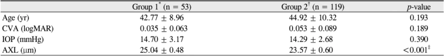
249 Table 1. Characteristics of long and short-to-normal length eyes
Group 1* (n = 53) Group 2† (n = 119) p-value
Age (yr) 42.77 ± 8.96 44.92 ± 10.32 0.193
CVA (logMAR) 0.035 ± 0.063 0.053 ± 0.089 0.189
IOP (mmHg) 14.70 ± 3.17 14.29 ± 2.68 0.390
AXL (μm) 25.04 ± 0.48 23.57 ± 0.60 <0.001‡
Values are presented as mean ± SD.
CVA = corrected visual acuity; IOP = intraocular pressure; logMAR = logarithm of the minimal angle of resolution; AXL = axial length.
*Long eyes, 24.5 < AXL ≤ 26.0 mm; †Short-to-normal eyes, 22.0 < AXL ≤ 24.5 mm; ‡p < 0.05.
Table 2. Retinal nerve fiber layer thickness in long and short-to-normal length eyes
Group 1* (n = 53) Group 2† (n = 119) p-value
Average RNFL thickness (μm) 96.32 ± 8.47 98.87 ± 10.01 0.109
Superior quadrant RNFL thickness (μm) 122.17 ± 14.50 123.81 ± 17.04 0.544
Nasal quadrant RNFL thickness (μm) 67.13 ± 9.74 70.73 ± 10.20 0.032‡
Inferior quadrant RNFL thickness (μm) 121.94 ± 14.08 132.02 ± 18.80 <0.001‡ Temporal quadrant RNFL thickness (μm) 74.21 ± 14.82 68.71 ± 10.92 0.007‡ Values are presented as mean ± SD.
RNFL = retinal nerve fiber layer; AXL = axial length.
*Long eyes, 24.5 < AXL ≤ 26.0 mm; †Short-to-normal eyes, 22.0 < AXL ≤ 24.5 mm; ‡p < 0.05.
ing the visual field or RNFL were also excluded.
Ophthalmologic examination
Their ophthalmologic exam included the corrected visual acuity (CVA), intraocular pressure (IOP), spherical and cy- lindrical refractive errors (Auto Ref-Keratometer RK-3;
Canon Inc., Tokyo, Japan), axial length (AXL; IOLMaster Advanced Technology, software ver. 5.02; Carl Zeiss Meditec Inc., Dublin, CA, USA), nonmydriatic fundus and optic disc photographs (Fundus Camera VX-10; Kowa Company, Tokyo, Japan), and peripapillary RNFL thickness measure- ments obtained with a spectral domain Cirrus HD OCT (Model 4000, software ver. 3.0.0.64; Carl Zeiss Meditec Inc.). The IOP was checked with a noncontact pneumo- tonometry (Tonometer TX-10, Canon Inc.), and then re-checked with a Goldmann applanation tonometry when the measure- ments were repeatedly higher than 21 mmHg.
The peripapillary RNFL thickness was measured by an Optic Disc Cube 200 × 200 scan of the Cirrus HD OCT. The Cirrus HD OCT measures RNFL thickness along a circle with a 1.73-mm radius around the optic disc. The scanning radius was not adjusted by other factors such as optic disc size. The same instrument was used by the same operator.
Measurements were performed without pupil dilation and as previously described elsewhere [9]. Scans with blinks or with a low signal strength (<6) were excluded from the analysis.
Only healthy eyes with a CVA of 20 / 30 or better, IOP of less than 21 mmHg, spherical refractive error within +/- 4.00 diopters and cylinder refractive error within +/- 3.00 diop- ters, 22.00 < AXL ≤ 26.00 mm, and normal appearance of optic nerve head, RNFL and fundus were included in the
study. Both eyes of each subject were included if they sat- isfied the entry criteria.
Statistical analysis
After dividing the study population into two groups ac- cording to their AXL (group 1, long eye group, 24.50 < AXL
≤ 26.00 mm; group 2, short-to-normal length eye group, 22.00 < AXL ≤ 24.50 mm) [10], the mean RNFL thickness and the correlation coefficient (β) of simple linear regression with age were determined for each scanned sector and com- pared between two groups. A comparison between the β val- ues was performed using the MedCalc ver. 9.6.4.0 (MedCalc Software, Mariakerke, Belgium) and all other statistical anal- yses were performed using the SPSS ver. 12.0.1 (SPSS Inc., Chicago, IL, USA).
Results
One hundred seventy-two eyes of 172 healthy Korean sub- jects were analyzed and their characteristics are shown at Table 1. According to their AXL, 53 eyes (30.81%) were clas- sified as long eyes (mean axial length ± SD, 25.04 ± 0.48 μm) and 119 eyes (69.19%) were classified as short-to-normal length eyes (mean axial length ± SD, 23.57 ± 0.60 μm). Age, CVA, and IOP were similar between the two groups.
Table 2 shows comparisons of the RNFL thickness between the groups. The mean average RNFL thicknesses of long eyes and short-to-normal length eyes were 96.32 ± 8.47 μm and 98.87 ± 10.01, respectively. The RNFLs were thinner in the long eyes than in the short-to-normal length eyes in the nasal and inferior quadrants (p = 0.032 and p < 0.001, respectively).
However, the temporal quadrant was thicker in the long eyes
Korean J Ophthalmol Vol.25, No.4, 2011
250
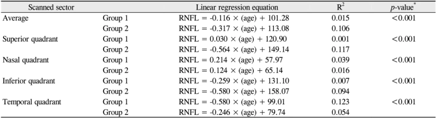
Table 3. Linear regression equation between age and retinal nerve fiber layer thickness in long (group 1) and short-to- normal (group 2) length eyes
Scanned sector Linear regression equation R2 p-value*
Average Group 1 RNFL = -0.116 × (age) + 101.28 0.015 <0.001
Group 2 RNFL = -0.317 × (age) + 113.08 0.106
Superior quadrant Group 1 RNFL = 0.030 × (age) + 120.90 0.001 <0.001
Group 2 RNFL = -0.564 × (age) + 149.14 0.117
Nasal quadrant Group 1 RNFL = 0.214 × (age) + 57.97 0.039 <0.001
Group 2 RNFL = 0.124 × (age) + 65.14 0.016
Inferior quadrant Group 1 RNFL = -0.259 × (age) + 131.10 0.007 <0.001
Group 2 RNFL = -0.580 × (age) + 158.07 0.094
Temporal quadrant Group 1 RNFL = -0.580 × (age) + 99.01 0.123 <0.001
Group 2 RNFL = -0.246 × (age) + 79.74 0.054
RNFL = retinal nerve fiber layer.
*p < 0.05.
200
160
120 80
40
0
200
160
120 80
40
0 20 30 40 50 60 70
Age (yr)
20 30 40 50 60 70 Age (yr)
A B
Fig. 1. Relationship between age and average peripapillary retinal nerve fiber layer (RNFL) thickness in long (A) and short-to-normal length (B) eyes.
than in the short-to-normal length eyes (p = 0.007).
Table 3 shows the linear regression of the effects of age on RNFL thickness. The decrease in average RNFL thick- ness with increasing age in long eyes (negative slope, -0.12 μm/yr) was less than that in short-to-normal length eyes (negative slope, -0.32 μm/yr) (p < 0.001). In all quadrants, age-related RNFL thinning was significantly different be- tween the two groups (all p < 0.001) (Table 3 and Fig. 1). In detail, for superior and inferior quadrants, the RNFL thick- ness in long eyes declined more with age than RNFL thick- ness in short-to-normal length eyes. Meanwhile, for the temporal quadrant, the RNFL thickness in long eyes showed faster thinning with age compared to the other groups. And, in long eyes, the RNFL thicknesses on the in- ferior and temporal quadrants decreased according to age;
in short-to-normal length eyes, the RNFL thicknesses de- creased in age in three quadrants, not including the nasal quadrant.
Discussion
Our results show a statistically significant linear decrease in average RNFL thickness with age, with a negative slope of 0.12 μm/yr for long eyes and 0.32 μm/yr for short-to-normal length eyes. The decrease in average RNFL thickness with increasing age in long eyes was less than that in short-to-normal length eyes. With respect to quadrants, the RNFL thicknesses for the superior and inferior quadrants in long eyes declined more with age than the short-to-normal length eyes. Further, in long eyes, the RNFL thicknesses in the inferior and tempo- ral quadrants decreased according to age; in short-to-normal length eyes, there was a decrease in three quadrants, not in- cluding the nasal quadrant. Our data are somewhat different from those of previous reports; Parikh et al. [11] found the maximum decay in the superior and temporal quadrants and Sung et al. [12] observed the steepest slopes in the superior, inferior, and nasal quadrants using the Stratus OCT. The ex- act reasons for these differences are unclear, but they could
EJ Kim, et al. Age-Related RNFL Thinning in Long Eyes
251 be due to different sample sizes and age distributions or eth-
nic populations. The precise mechanism underlying our find- ing is not fully understood yet. It is possible that the dis- tribution of a similar volume of RNFL in a larger area could cause the average RNFL thickness to be thinner in long eyes compared to short-to-normal length eyes. If the rate of abso- lute volume of RNFL loss is similar in both eye groups, the rate of RNFL thinning may seem more attenuated in long eyes than in other eyes.
In addition, in our study, RNFLs were thinner in the long eyes than in the short-to-normal length eyes, except for in the temporal quadrant. The temporal quadrant RNFL was thick- er in long eyes. Several studies [6,13] have reported that a high myopia group has significantly thicker RNFLs in the temporal quadrants, which is similar to our findings. In long eyes, the elongation of the globe leads to retinal dragging to- ward the temporal horizon. Thicker RNFLs in the temporal quadrant could be related to retinal dragging. There have been several studies reporting a significant association be- tween myopia and RNFL thickness [14,15], but our present study did not find a significant association between myopia and RNFL thickness, This difference might be due to the fact that we did not include very long eyes and highly myopic eyes. Further studies including very long eyes and highly myopic eyes are needed to establish the association between axial length and age-related peripapillary RNFL thinning and to confirm our findings in glaucomatous eyes as well as healthy eyes.
In conclusion, we found that age-related thinning of peri- papillary RNFL thickness is attenuated in long eyes com- pared to short-to-normal length eyes. Therefore, we suggest the need for carefully interpretation of OCT results in pa- tients with mild degrees of myopia.
Conflict of Interest
No potential conflict of interest relevant to this article was reported.
References
1. Quigley HA, Dunkelberger GR, Green WR. Chronic human
glaucoma causing selectively greater loss of large optic nerve fibers. Ophthalmology 1988;95:357-63.
2. Airaksinen PJ, Alanko HI. Effect of retinal nerve fibre loss on the optic nerve head configuration in early glaucoma. Graefes Arch Clin Exp Ophthalmol 1983;220:193-6.
3. Balazsi AG, Rootman J, Drance SM, et al. The effect of age on the nerve fiber population of the human optic nerve. Am J Ophthalmol 1984;97:760-6.
4. Budenz DL, Anderson DR, Varma R, et al. Determinants of normal retinal nerve fiber layer thickness measured by Stratus OCT. Ophthalmology 2007;114:1046-52.
5. Vernon SA, Rotchford AP, Negi A, et al. Peripapillary retinal nerve fibre layer thickness in highly myopic Caucasians as measured by Stratus optical coherence tomography. Br J Ophthalmol 2008;92:1076-80.
6. Choi SW, Lee SJ. Thickness changes in the fovea and peri- papillary retinal nerve fiber layer depend on the degree of myopia. Korean J Ophthalmol 2006;20:215-9.
7. Kim JW, Kim YY. Changes in RNFL thickness according to the myopia in patients with glaucoma and ocular hypertension.
J Korean Ophthalmol Soc 2008;49:1634-40.
8. Ha SW, Rho SH. Age-related differences of optical coherence tomography data in Koreans. J Korean Ophthalmol Soc 2005;
46:2037-44.
9. Budenz DL, Fredette MJ, Feuer WJ, Anderson DR. Reproducibility of peripapillary retinal nerve fiber thickness measurements with stratus OCT in glaucomatous eyes. Ophthalmology 2008;
115:661-666.e4.
10. Hoffer KJ. Intraocular lens implant power calculation, se- lection, and ocular biometry. In: Steinert RF, editor. Cataract surgery: techniques, complications, and management. 2nd ed.
Philadelphia: Elsevier Science; 2004. p.37-8.
11. Parikh RS, Parikh SR, Sekhar GC, et al. Normal age-related decay of retinal nerve fiber layer thickness. Ophthalmology 2007;114:921-6.
12. Sung KR, Wollstein G, Bilonick RA, et al. Effects of age on optical coherence tomography measurements of healthy retinal nerve fiber layer, macula, and optic nerve head. Ophthalmology 2009;116:1119-24.
13. Kim MJ, Lee EJ, Kim TW. Peripapillary retinal nerve fibre layer thickness profile in subjects with myopia measured us- ing the Stratus optical coherence tomography. Br J Ophthalmol 2010;94:115-20.
14. Rauscher FM, Sekhon N, Feuer WJ, Budenz DL. Myopia af- fects retinal nerve fiber layer measurements as determined by optical coherence tomography. J Glaucoma 2009;18:501-5.
15. Leung CK, Mohamed S, Leung KS, et al. Retinal nerve fiber layer measurements in myopia: an optical coherence tomog- raphy study. Invest Ophthalmol Vis Sci 2006;47:5171-6.