162
책임저자: 박진우, 충북 청주시 흥덕구 개신동 62번지, 361-711, 충북대학교 의과대학 외과학교실
Tel: 043-269-6033, Fax: 043-266-6037, E-mail: [email protected] 접수일:2008년 5월 13일, 게재승인일:2008년 6월 4일
갑상선암 세포주에서 Na-4-Phenylbutyrate의 세포 증식 억제 및 분화 유도 효과
을지대학교 의과대학 외과학교실, 1충북대학교 의과대학 외과학교실, 2Department of Surgery, University of California
최영진ㆍ박진우 1 ㆍ장이찬 1 ㆍ최재운 1 ㆍOrlo H. Clark 2
The Antiproliferative and Redifferentiative Effects of Na-4-Phenylbutyrate in Human Thyroid Cancer Cell Lines
Young Jin Choi, M.D., Jin-Woo Park, M.D.
1, Lee-Chan Jang, M.D.1,Jae-Woon Choi, M.D.
1, Orlo H. Clark, M.D.2Department of Surgery, Eulji University School of Medicine, Daejeon,
1Chungbuk National University School of Medicine, Cheongju, Korea,
2
Department of Surgery, University of California, UCSF/Mount Zion Medical Center, San Francisco, USA
Purpose: Sodium-4-phenylbutyrate (Na-4-PB) is an analogue of phenylacetate, which is a well-known redifferen-
tiating agent. In vitro and in vivo studies on this agent have been done and the clinical relevance of Na-4-PB has been studied in other malignancies, but not in thyroid cancer. We investigated the effect of Na-4-PB on cell proliferation and differentiation in thyroid cancer cell lines.Methods: We used 5 thyroid cancer cell lines: TPC-1, FTC-133, FTC-236, FTC-238 and XTC-1. MTT assay
and flowcytometry were used to measure the agent's antiproliferative effects and the cell cycle change. We evaluated the PPARγ expression via western blotting and the mRNA expressions of NIS, Tg and CD 97 were determined by performing RT-PCR. Troglitazone, a potent PPARγ agonist, was used in combined treatment with Na-4-PB.Results: Na-4-PB inhibited cell proliferation in a dose and time dependent manner in all 5 thyroid cancer cell
lines. By performing flowcytometry in the FTC-133 and TPC-1 cell lines, we identified that the antiproliferative effect of Na-4-PB was associated with an increased apoptotic cell population. Treatment with Na-4-PB upregulated the PPARγ expression, but the combined treatment of Na-4-PB with troglitazone did not seem to be synergistic for the antiproliferative effect. Treatment with Na-4-PB downregulated the CD97 mRNA expression and it upregulated the NIS and Tg mRNA expressions in both the FTC-133 and TPC-1 cell lines.Conclusion: Na-4-PB inhibited thyroid cancer cell proliferation by inducing apoptosis in a dose dependent manner.
Treatment with Na-4-PB increased the expression of PPARγ and it upregulated such differentiation markers as NIS and Tg, and it downregulated CD97, a dedifferentiation marker. Na-4-PB should be further evaluated as a new potential therapeutic agent for patients with thyroid cancer. (J Korean Surg Soc 2008;75:162-170)
Key Words: Na-4-PB, Histone deacetylase inhibitor, Thyroid cancer cell lines, CD 97, NIS
중심 단어: Na-4-PB, Histone deacetylase 억제제, 갑상선암 세포주, CD 97, NIS
서 론
대부분의 소포세포 기원 분화 갑상선암은 천천히 자라 고, 수술적 치료만으로도 완치될 가능성이 높다. 또한 소포 세포의 분화된 기능을 비교적 오랫동안 유지하여 갑상선 자극 호르몬(thyroid stimulating hormone, TSH) 억제 요법이 나 방사성 요오드 치료 등과 같은 술 후 보조 요법에도 잘 반응하여, 매우 양호한 예후를 보인다. 그러나 갑상선암의 10∼15%는 공격적인 생물학적 특성을 가지며, 이런 양호한 경과를 따르지 않는다. 갑상선암의 암화과정은 분화된 기 능을 잃어가는 탈분화(dedifferentiation)의 과정이라고 할 수 있는데, 잔존 또는 재발 갑상선암의 약 30%에서 방사성 요 오드를 흡착하고 thyroglobulin (Tg)을 생성하는 능력이 감소 하게 된다. 따라서 전통적인 방법만으로 치료할 수 없고, 재 발을 진단하거나, 종양의 부하(tumor burden)를 측정하기 어 렵게 된다.(1) 재분화요법(redifferentiation therapy)은 탈분화 된 종양세포가 다시 분화된 기능을 회복하게 하여 기존의 치료가 다시 유용해지도록 하는 것으로, 여러 약제들의 효 과가 증명되어 왔다.(2,3)
갑상선암은 재분화요법의 중요한 대상의 하나인데, phe- nylacetate와 phenylbutyrate 같은 방향성 지방산은 중요한 재 분화요법제(redifferentiating agents)의 하나로 이미 오래 전 부터 요소회로 대사장애 환자에서 치료제로 사용되어 왔 다.(4) 이 약제들은 전립선암, 유방암, 대장암, 폐암 등의 다 양한 암종에서 세포 증식을 억제하고 분화된 기능을 유도 하는 것으로 보고되어왔다.(5,6) Phenylacetate는 갑상선암 세포주를 이용한 실험에서 세포 증식을 억제시키고, so- dium/iodide symporter (NIS) mRNA의 발현을 증가시키며, 방사성 요오드 흡착을 증가시킨다고 보고되었다.(3) 그러나 유효 농도가 너무 높고, 경구 투여에 어려움이 있어 직접 임상에 적용하는 데 큰 제약이 있었다. 최근 phenylbutyrate 의 변형인 Sodium-4-phenylbutyrate (Na-4-PB)가 FDA 공인을 받아 요소회로 대사장애 환자에서 안전하게 경구 투여가 가능하게 되었다. Na-4-PB는 인체 여러 암종에서 세포 증식 을 억제하는 것으로 보고되며, 현재 임상시험이 진행되고 있다. 그러나 갑상선암 세포에서의 Na-4-PB 투여 효과는 보 고가 거의 없다.(3)
본 연구는 임상시험의 전 단계로써, in vitro 실험을 통해 5종류의 갑상선암 세포주를 이용하여 Na-4-PB가 갑상선암 세포의 증식과 분화에 미치는 영향을 알아보는 데 목적을
두고 있다.
방 법
1) 세포주와 배양 조건
모두 5종류의 갑상선암 세포주를 사용하였다. 갑상선암 세포주는 Dr. Clark (University of California at San Francisco- Mt. Zion Medical Center, San Francisco, CA, USA)으로부터 제공받았다. TPC-1 세포주는 유두상암에서 기원하였고, FTC-133, FTC-236, FTC-238는 소포암 세포주로 각각 원발 종양, 전이성 림프절, 전이성 폐결절에서 기원하였으며, XTC-1은 휘틀세포암에서 기원하였다. 세포주의 유지배양액 은 Dulbecco's modified Eagle's medium (DMEM: Life Technologies, NY, USA)을 기본으로 하는 10% fetal bovine serum (FBS), penicilline (10,000 U/ml), streptomycin (10,000 U/ml), fungizone (250 mg/ml), glutamine (12.5 mg/l)을 섞어 만들었다. 세포주는 가습배양기를 이용하여 37oC, 5% CO2, 95% O2의 환경에서 배양하였다. 모든 실험은 1% FBS 혈청 이 포함된 배양액(실험용 배양액)을 사용하여 이루어졌는 데, 실험 시작 24시간 전에 유지배양액을 제거하고 교체하 였다.
2) MTT assay를 통한 세포증식 억제효과의 평가
갑상선암 세포주를 96-well plate에 각 조건 당 6 well로, well 당 세포 수가 3∼5×10³이 되도록 분주하였다. 처음 24 시간은 유지 배양액으로 세포가 well에 안착할 수 있도록 하였고, 실험 시작 24시간 전에 실험용 배양액으로 바꾸어 주었다. 치료 시작일에 각각 0, 2.5, 5, 7.5, 10mM 농도의 Na-4-PB로 처리하고, 각각 0, 3, 5일에 MTT assay를 시행하 였다.
MTT assay는 배양액을 제거하는 것으로 시작하고, 각 well에 100μl의 배양액을 넣고, 5 mg/ml의 MTT 용액을 20 μl씩 넣어 준 뒤, 가습 배양기에서 차광 상태로 2시간 동안 두었다. 이후 각 well에 0.04 N HCl/iso-propanol/3% SDS 혼 합물 150μl를 첨가하고, 충분히 섞은 뒤 차광 상태에서 상 온에 1시간 동안 방치하였다. ELISA microplate reader를 이 용하여 595 nm에서 각각의 optical density (OD)를 측정하였 다. 세포증식의 분율은 치료군의 OD 값을 대조군의 OD 값 으로 나누어 나타냈다. 각각의 실험은 최소 세 번 시행하여 결과를 얻었다.
Na-4-PB와 peroxisome proliferator activator receptor-gamma
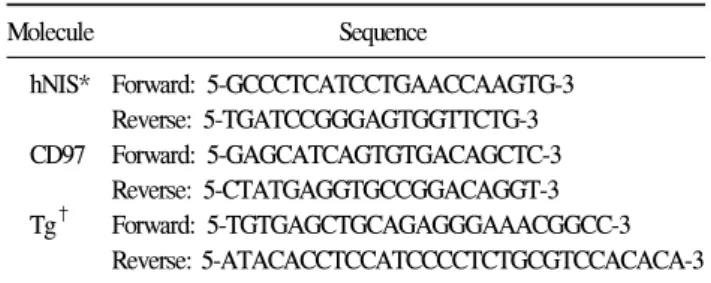
Table 1. Primers used for PCR reactions
Molecule Sequence
hNIS* Forward: 5-GCCCTCATCCTGAACCAAGTG-3 Reverse: 5-TGATCCGGGAGTGGTTCTG-3 CD97 Forward: 5-GAGCATCAGTGTGACAGCTC-3
Reverse: 5-CTATGAGGTGCCGGACAGGT-3 Tg† Forward: 5-TGTGAGCTGCAGAGGGAAACGGCC-3
Reverse: 5-ATACACCTCCATCCCCTCTGCGTCCACACA-3
*hNIS = sodium-iodide symporter; †Tg = thyroglobulin.
(PPARγ) 촉진제인 troglitazone의 병합 투여에서는 각각 0, 2.5 mM Na-4-PB, 5 uM troglitazone, 2.5 mM Na-4-PB와 5 uM troglitazone의 병합 투여군으로 나누고 0, 2, 4일에 각각 MTT assay를 시행하였다. Na-4-PB와 troglitazone은 Merck in Korea에서 구매하였다.
3) 유세포 분석(Flow cytometry)을 이용한 세포 주기 분 석(cell cycle analysis)
FTC-133 세포주와 TPC-1 세포주를 실험용 배양액에서 각각 0, 2.5, 5 mM 농도의 Na-4-PB로 48시간 치료한 후, 1×Trypsin/EDTA 용액을 이용하여, 세포를 수확하였다.
5×10⁵개의 세포를 200μl 1×PBS로 재부유한 뒤, 차가운 70% ethanol 2 ml로 20oC에서 30분간 고정하였다. 고정한 FTC-133, TPC-1 세포를 다시 원침하여 상층액을 버리고, 0.8 ml의 1×PBS로 재부유한 뒤, 1 mg/ml RNase (BIOMOL Research Laboratories, Inc. Plymouth Meeting, PA)와 400μg/
ml의 Propidium iodide (Sigma Chemical Co., St. Louis, MO) 를 각각 0.1 ml씩 넣고 충분히 혼합하여 빛을 피해 37oC에서 유세포 분석을 시행하였다.
유세포 분석은 Becton Dickness사의 FACScan을 이용하였 다. 자료 분석은 CELLQuest software를 이용하여, 분석할 세 포를 선택하여(gating) 각각의 sample마다 10,000 gated event 가 일어날 때까지 유세포 분석을 지속하였다. 세포 주기의 분석은 Modfit software (Verity Software House, Inc.)를 이용 하였다.
4) Western blotting을 이용한 PPARγ 발현 분석
갑상선암 세포주에서 Na-4-PB가 PPARγ 단백 발현에 미 치는 영향에 대해서는 각각 FTC-133 세포주와 TPC-1 세포 주에서 western blotting을 이용하여 알아보았다. FTC-133 세 포주와 TPC-1 세포주를 각각 농도 0, 2.5, 5 mM 의 Na-4-PB 와 10 ng/ml EGF, 각각 5, 10 uM/L의 troglitazone으로 처리하 였다. 48시간이 지난 후, 각 세포에서의 전체 단백질을 추출 하여, 단백질의 농도는 제조사의 설명서에 따라 Bio-Rad protein assay reagent (Bio-Rad, Richmond, CA)로 측정하였다.
20% SDS-PAGE (Ready Gels J; Bio-Rad)를 이용하여 50μg 의 단백을 분리하여 sample buffer와 혼합하고 95oC에서 3분 간 가열하고, 얼음에 식힌 다음 12%의 SDS-PAGE gel (Biorad, Hercules, CA)에서 전기영동 하였다. 단백을 nitro- cellulose membrane (Amersham Life Science, Inc., Piscataway, NJ)으로 전이시킨 다음, 밤새 5% 탈지분유(10 mM Tris-
NaCl buffer)를 이용하여 비특이적 결합을 막았다. PPARγ 단백의 발현을 알아보고자, 상온에서 1시간 동안 human PPARγ 단 클론 항체(Santa Cruz, Santa Cruz, CA)를 처리하 고 반응시켰다. Tris NaCl buffer를 이용하여 3회 세척 후 다 시 한 시간 동안 1:1000으로 희석한 horseradish peroxidase (HRP)가 결합한 이차 항체(Sigma Chemical Co., St. Louis, MO)에 노출시켰다. 3회 세척 후, 1분간 ECL detection re- agents (Amersham International)와 접촉시키고 나서, 방사선 촬영을 통해 단백질 발현을 알아보았다.
5) RT-PCR을 통한 CD97, Tg, NIS mRNA의 측정
FTC-133 세포주와 TPC-1 세포주를 각각 6 well plate에 3∼
5×10³ cells/well의 농도로 나누어 파종하고, 각각 0, 5, 10 mM의 Na-4-PB를 처리하였다. 24시간 동안 처리한 후, 배양 된 세포를 PBS로 3회 씻은 후, 상층액을 제거하고, 여기에 1 ml TRIzol reagent (GIBCO BRL Life Technologies, Mary- land, USA)를 넣어 세포를 녹여서 Total RNA를 분리하였다.
RNA pellet은 DEPC treated water에 약 0.5μg/μl에서 1.0μg/
μl의 농도로 녹여서 70oC에서 보관하였다. 준비된 RNA 의 정량과 정성은 260 nm와 280 nm의 투과도를 이용하여 분석하였다. 총 RNA 1μg과 random primer p(dN)6 (최후 농 도 1mM)를 12.5μl의 DEPC-treated water에 넣고 2분간 70oC 에 내버려둔 후 수 분간 식혔다. cDNA 합성은 recombinant MMLV reverse transcriptase (GIBCO BRL Life Technologies, Maryland, USA) 1μl를 이용하여 공급자의 제안대로 시행 하고 반응은 섭씨 94oC에서 5분간 가열하면서 종료하였고, 증폭은 표와 같은 primer를 사용하였다(Table 1). 각각의 PCR 반응은 cDNA 3μl, 10 pM primer 1μl, 2.5 mM dNTPs mixture 1μl, 10배 PCR buffer 2.0μl와 recombinant Thermus aquaticus DNA polymerase (TaKaRa Shuzo Co Ltd. Tokyo, Japan) 0.5 unit을 혼합하여 최종 volume이 20μl가 되도록 하였다.
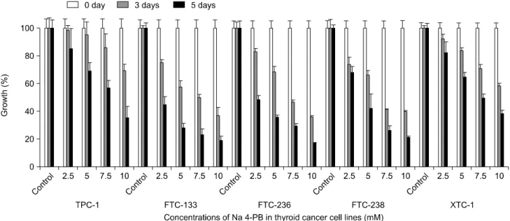
Fig. 1. Antiproliferative effects of Na-4-PB in five thyroid cancer cell lines. Na-4-PB inhibits cancer cell proliferation in all five thyroid
cancer cell lines in a dose and time dependent manner.증폭은 Takara Shuzo Co Ltd (Tokyo, Japan)의 Takara PCR thermal cycler personal을 이용하였다. 얻어진 PCR의 최후 산물을 가지고 ethium bromide (Sigma Chemical Company, Missouri, USA) 염색을 이용하여 2% agarose gel 전기 영동 을 시행하고 Biological Image Processing System (BiPS) 3.0 (BIOMEDICAL Co, Seoul, Korea)을 이용하여 gel image를 얻었다. 다른 양의 cDNA를 사용할 경우를 대비하여, GAPDH mRNA를 internal control로 사용하였다.
6) 통계 분석
통계 방법으로는 One-way ANOVA test와 student t-test를 사용하였고, P-value<0.05를 통계적으로 유용하다고 판단 하였다.
결 과
1) Na-4-PB의 세포증식 억제효과
Na-4-PB는 갑상선 유두상암, 소포암, 휘틀세포암 세포주 모두에서 처리 농도와 기간에 비례하여 유의하게 세포증식 을 억제하였다(Fig. 1). Na-4-PB의 세포증식 억제효과는 소 포암 세포주인 FTC-133, FTC-236, FTC-238에서 유두상암 세포주나 휘틀세포암 세포주에 비해 좀 더 현저하였다.
FTC-133 세포주의 경우, 각각 2.5, 5, 7.5, 10 mM의 농도로 3일간 처리하였을 경우, 대조군과 비교한 상대적 세포증식
억제는 각각 24.9±2.8% (mean±SD), 42.3±4.9%, 50.1±2.3%, 63.0±5.9%였고, 5일간 처리한 경우, 각각 55.4±5.8%, 72.1±
3.4%, 77.7±4.6%, 80.8±2.7%였다(P<0.05). TPC-1 세포주에 서는 3일간 처리한 경우에는 각각 17.5±2.2%, 20.4±5.6%, 28.2±6.3%, 42.3±3.1%였으며, 5일간 처리한 경우에는 각각 43.0±10.9%, 48.1±4.3%, 51.3±4.0%, 57.7±6.3%였다(P<0.05).
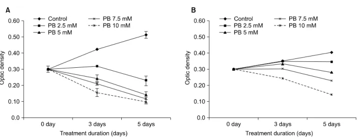
Na-4-PB 치료에 비교적 민감한 FTC-133 세포주에서는 모 든 치료 농도에서 세포 수의 실질적인 감소를 나타내었으 나, 상대적으로 덜 민감한 TPC-1 세포주에서는 7.5 mM 농 도 이상에서 5일 이상 치료했을 때에야 실질적인 세포 수의 감소를 보였다(Fig. 2).
2) Na-4-PB에 의한 세포 주기의 변화
갑상선암 세포주를 각각 0 (대조군), 2.5, 5 mM의 Na-4-PB 로 48시간 동안 처리한 후, 유세포 분석을 통해 각 세포 주 기의 분율을 측정하였다. FTC-133 세포주의 경우, 농도 증 가에 따라 G1 세포 주기 분율은 각각 68.1±1.5%, 66.1±0.8%, 68.5±0.7%, S 세포 주기 분율은 각각 9.9±1.5%, 7.1±0.8%, 5.1±0.7%, G2/M 세포 주기 분율은 각각 21.9±0.5%, 26.7±
1.0%, 26.4±2.2%였다(Fig. 3A). Na-4-PB 치료는 세포 주기 분율에 유의한 변화를 초래하지 않았지만, 세포사멸을 시 사하는 sub G1 세포 분율은 각각 7.1±2.2%, 16.8±1.3%, 34.3±2.2%로 치료 농도에 비례하여 유의하게 증가하였다(P
<0.05)(Fig. 3A). 이러한 소견은 TPC-1 세포주에서도 비
Fig. 2. Changes in optic density measured after Na-4-PB treatment in FTC-133 and TPC-1 cell lines. Treatment with Na-4-PB in different
concentrations induces cytotoxicity in FTC-133 cell line (A) whereas it induces cytotoxicity only in higher concentrations in TPC-1 cell line (B).Fig. 3. Changes in cell cycle populations after Na-4-PB treatment. Na-4-PB treatment increases sub G1 cell population in a dose dependent
manner. However it does not affect cell cycle significantly in FTC-133 (A) and TPC-1 (B) cell lines. a. control, b. 2.5mM of Na-4-PB, c. 5 mM of Na-4-PB.Fig. 4. Changes of PPARγ protein expression by treatment with
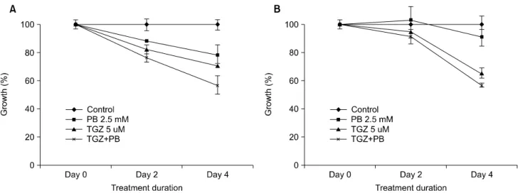
Na-4-PB and troglitazone. Na-4-PB increases PPARγ protein expression in FTC-133 and TPC-1 cell lines.Fig. 5. The antiproliferative effects of combination therapy with Na-4-PB and troglitazone. Combination therapy is not synergistic but
additive in antiproliferative effect in both FTC-133(A) and TPC-1(B) cell lines.Fig. 6. Changes in mRNA expressions of CD 97, sodium-iodide
symporter (NIS) and thyroglobulin (Tg) after Na-4-PB treatment. Na-4-PB treatment upregulates mRNA expre- ssions of NIS and Tg and downregulates mRNA expre- ssions of CD97 in FTC-133 and TPC-1 cell lines.슷하였다(Fig. 3B).
3) Na-4-PB와 PPARγ 촉진제의 병용 투여 효과
Na-4-PB 치료는 FTC-133, TPC-1 세포주에서 유의하게 PPARγ 단백질 발현을 증가시켰다. 그러나 성장 인자인 EGF, PPARγ 촉진제인 troglitazone은 PPARγ의 발현에 유 의한 영향을 미치지 않았다(Fig. 4).
Na-4-PB가 PPARγ 단백질 발현을 증가시킨다는 결과를 토대로, Na-4-PB와 PPARγ 촉진제인 troglitazone의 병용 치 료를 시도하였는데, 단독 치료의 효과를 합한 정도의 세포 증식 억제 효과가 관찰되었고, 그 이상의 상승작용(syner- gism)을 보이지는 않았다(Fig. 5).
4) Na-4-PB의 분화 유도 효과
FTC-133과 TPC-1 세포주 모두에서, Na-4-PB의 농도에 비
례하여 탈분화 표식자인 CD 97의 발현이 감소하였고, 갑상 선의 분화 표식자인 sodium-iodide symporter (NIS)와 thyro- globulin (Tg)의 발현이 증가되었다(Fig. 6).
고 찰
갑상선암의 암화과정은 분화 기능을 잃어가는 탈분화의 과정이라 할 수 있는데, 치료 후 잔존하거나 재발하는 분화 갑상선암의 약 30%에서 탈분화가 일어난다. 이런 탈분화의 결과로 생기는 분화가 나쁜 갑상선암 또는 역형성암의 경 우, 소포 세포의 분화된 기능에 의존하는 전통적인 치료에 반응하지 않게 되며, 항암 화학 요법이나 외부 방사선 조사 와 같은 이차 치료에도 반응성이 매우 낮다.(1) 재분화요법
(redifferentiation therapy)은 탈분화된 종양을 다시 분화된 종 양으로 역전시켜 종양 성장을 둔화시키고, 기존의 치료가 다시 유용해지도록 하는 것이다.(2,3) 재분화요법에 사용되 는 약제로는 retinoid를 비롯하여, phenylacetate, phenylbutyrate 등의 방향성 지방산(aromatic fatty acid), troglitazone, piogli- tazone 등의 PPARγ 촉진제, trichostatin A, suberoylanilide hydroxamic acid (SAHA) 등의 histone deacetylase 억제제, valproic acid 등이 있다.(2,7,8)
Phenylacetate는 retinoid와 더불어 중요한 재분화요법제 (redifferentiating agents)의 하나로 이미 오래 전부터 요소 회 로 대사 장애 환자에서 치료제로 사용되어 왔는데,(4) 최근 악성 신경아교종(glioma), 호르몬 치료에 반응하지 않는 전 립선암, 악성 흑색종, 신경 모세포종, 백혈병, 유방암, 대장 암, 폐암 등의 다양한 암종에서 세포 증식 억제와 분화 유도 효과가 보고되었다.(6,9) Kebebew 등(7)은 갑상선암 세포주 를 이용하여, phenylacetate가 세포 증식을 억제하고, NIS 유 전자 발현을 증가시켜, 방사성 요오드 흡착을 증가시킨다 고 보고하였다. 그러나 이런 효과를 보이는 치료 농도가 너 무 높고, 경구 투여에 어려움이 있어 직접 임상에 적용하지 는 못하였다. Phenylacetate의 전구 약물인 phenylbutyrate는 치료 효과가 다를 수 있는데, 최근 경구 투여가 가능한 Sodium-4-phenylbutyrate (Na-4-PB)가 FDA 공인을 받아 요소 회로 대사 장애 환자의 치료제로 사용되면서 새롭게 주목 받고 있다. Na-4-PB는 골수성 백혈병, 유방암, 폐암 및 간암 등의 세포주를 이용한 연구에서 세포증식을 억제하고, 세 포사멸을 촉진하며 재분화를 유도한다고 보고되었다.(10- 12) 그러나 갑상선암 세포에서의 Na-4-PB 투여 효과는 보고 가 거의 없다.(3)
저자들은 갑상선암 세포주를 이용하여 Na-4-PB가 임상 적으로 도달 가능한 2.5∼5 mM 농도에서 갑상선암의 세포 증식을 억제할 수 있음을 밝혔다. 이러한 세포증식 억제 효 과는 약물 투여 용량과 투여 기간에 비례하여 증가하였다.
Na-4-PB의 세포증식 억제효과는 소포암 세포주인 FTC-133, FTC-236, FTC-238에서 유두상암 세포주나 휘틀세포암 세 포주에 비해 좀 더 현저하였다. 소포암 세포주에서는 모든 치료 농도에서 실제적으로 세포 수(cell mass)의 감소를 나 타내었으나, 비교적 치료에 저항을 보이는 TPC-1에서는 치 료에도 불구하고 세포 수가 유지되는 소견을 보였다. 세포 증식의 억제는 세포주기 정지와 세포사멸로 설명될 수 있 는데, Kebebew 등(7)은, 갑상선 소포암 세포주에서 10 mM 농도의 phenylacetate로 24시간 치료한 경우 S 주기가 감소
하고 G0/G1 주기가 유의하게 증가함을 보고하였다. 또한 Greenberg 등(13)은 미분화 갑상선암 세포주에서 phenyl- acetate 치료로 G1 주기 및 G2/M 주기가 증가함을 보고하였 다. 그러나 Na-4-PB를 이용한 본 연구에서는, 세포주기의 변화에 특별한 유형을 찾기 어려웠지만, 세포사멸을 시사 하는 sub G1 세포분율은 치료 농도가 증가할수록 증가하였 다. 이런 사실을 볼 때, NA-4-PB의 세포증식 억제 효과는 세포 주기 정지(cell cycle arrest)보다는 세포사멸의 유도에 의한 것으로 추정된다. 이런 결과들은 여러 암종에서 보고 된 이전의 Na-4-PB에 대한 결과들과 유사했다.(10-12,14) Phenylacetate와 전구 약물인 phenylbutyrate는 기본적으로 같은 기전에 의해 작용하지만, phenylbutyrate가 세포사멸을 유도하는 능력이 더 뛰어난 것으로 보고된다. Phenylacetate 의 세포증식 억제 기전은 명확히 규명되지는 않았지만, 지 질대사 변화, peroxisome proliferator activator receptor-gamma (PPARγ)의 활성화, histone deacetylase 억제(HDAI), 단백 지질화(isoprenylation)의 억제, 글루타민 고갈 등이 관여할 것으로 추정하고 있다.(15,16) Na-4-PB에 의한 세포사멸은 bcl-2 단백의 발현, caspase 활성화, c-myc, k-ras, p53, p21 등 의 발현 변화, JNK pathway의 활성화 등을 통해 이루어지는 것으로 보고된다.(10,17) 본 연구에서는 갑상선암 세포주에 서의 세포사멸의 분자 기전을 밝히지 않았으나, 향후 추가 연구가 필요하겠다.
Phenylacetate가 세포증식을 억제하는 기전의 하나가 PPARγ의 활성화인데,(1,2) 본 연구에서 Na-4-PB가 FTC-133, TPC-1 세포주에서 PPARγ의 단백질 발현을 증가시킴을 확 인할 수 있었다. PPARγ는 중요한 재분화요법의 표적으로 서, 초기에 보고된 것과 달리 갑상선 소포암 외에도 갑상선 유두상암, 미분화 갑상선암 뿐 아니라 소포선종에서도 발 현이 보고된다. Ohta 등(18)은 유두상 갑상선 암 세포주를 이용하여 PPARγ 발현을 보이는 세포주에서 PPARγ 촉진제 인 troglitazone이 세포증식을 억제함을 보고하였고, Martelli 등(19)은 PPARγ 발현이 없는 갑상선 암 세포주에 wild- type PPARγ c-DNA 유전자 전달을 하면, PPARγ 촉진제의 세포증식 억제 효과가 증강됨을 확인하였다. 또한 Eigel- berger 등(20)은 두 가지 서로 다른 기전의 재분화요법 치료 제인 phenylacetate와 retinoid를 병용 투여하여 세포증식 억 제 효과가 증가함을 보고하였으나, 극적인 상승 효과 (synergism)를 보이는 데는 실패하였다. 저자들은 Na-4-PB 치료에 의해 증가된 PPARγ 단백질에 PPARγ 촉진제가 작용하면 세포증식 억제 효과가 증강될 것으로 기대하여
병용 투여를 시행하였다. 그러나 Na-4-PB와 troglitazone의 병용 처리 효과는 기대에 미치지 못했다. 단독 치료군에 비 교하여, 병용 투여군의 세포증식 억제 정도는 각각의 치료 효과의 합에 근사하여, 극적인 상승 효과를 나타내지는 않 았다. 최근 phenylbutyrate가 대장암, 비소세포성 폐암 등에 서 gemcitabine이나 5-FU의 세포증식 억제 효과를 증대시킨 다고 보고되는 바,(21,22) 향후 갑상선암에서도 Na-4-PB를 이용한 다양한 병용 투여의 결과를 확인할 필요가 있다. 그 러나 비록 병용 투여를 통해 치료 효과를 어느 정도 증가시 킬 수는 있겠지만, 부작용을 배가할 수 있는 가능성을 고려 하여 신중하게 득과 실을 따져봐야 하겠다.
갑상선암에서 재분화요법의 가장 중요한 목적은 소포 세 포의 분화 기능을 되살려 진단과 치료에 도움을 주는 것이 다. 이 때 가장 중요한 것은 TSH에 대한 반응도와 Tg의 생 성, sodium/iodide symporter (NIS)의 발현과, 요오드 흡착능 이다. Phenylacetate가 갑상선암 세포주의 분화 기능에 미치 는 영향은 다소 복잡한데, TSH에 대한 반응도를 감소시키 고, Tg 분비를 감소시키지만, sodium/iodide symporter (NIS) mRNA의 발현을 증가시켜, 방사성 요오드 흡착을 증가시킨 다고 보고된다.(3,7,20) Na-4-PB의 재분화 효과에 대한 보고 는 거의 없다. 저자들은 Na-4-PB가 갑상선암 세포주의 분화 기능에 미치는 영향을 CD 97, Tg, NIS의 유전자 발현을 통 해 알아보았다. CD 97은 EGF-TM7 subfamily의 하나로, 다 른 세포의 표면 수용체와 반응하여 세포간의 유착에 관여 한다.(23,24) CD 97은 갑상선 암의 공격성 및 림프절 전이 정도와 비례하여 발현이 증가하며,(25) 대표적인 재분화요 법 치료제인 retinoid 투여 시 CD 97을 발현하는 세포 수가 감소하여,(8) 갑상선암에서 탈분화의 표식자로 이용된다.
본 연구에서 Na-4-PB 치료는 FTC-133, TPC-1 세포주 모두 에서 CD 97 mRNA 발현을 감소시켜, 재분화의 간접적인 증거로 생각하였다. 갑상선 소포 세포의 대표적인 분화 기 능으로는 Tg를 생성하고, 요오드를 흡착하는 기능이다. Tg 는 소포기원 분화 갑상선암 수술 후 잔존암과 재발암의 유 무와 그 정도를 가늠하는데 매우 유용하다. NIS는 막 단백 질로 방사성 요오드를 흡수하는데 가장 중요한 기능을 하 는데, 전체 분화 갑상선암의 30∼90%에서 발현이 감소되 고, 탈분화가 진행될수록 발현 저하가 뚜렷해진다. 갑상선 암에서 NIS의 발현이 저하되는 것은 여러 가지로 설명되는 데, NIS 유전자의 메틸화, 아세틸화 등의 변화(epigenetic changes)로 인한 전사 과정의 실패도 주요한 원인의 하나이 다. NIS의 발현 정도는 임상적으로 방사성 요오드 흡수 정
도와 비례하므로 방사성 요오드 치료에 불응하는 갑상선암 을 재분화시켜 NIS 발현을 증가시킬 수 있다면 방사성 요 오드를 이용한 진단과 치료가 다시 효과적으로 될 수 있겠 다.(2,26,27) Na-4-PB는 histone deacetylase 억제제로 작용할 수 있는데, 본 연구에서 분화관련 유전자인 NIS와 Tg를 유 의하게 상승시켰다. trichostatin A와 suberoylanilide hydroxa- mic acid (SAHA) 등의 histone deacetylase 억제제 등이 NIS 발현을 현저하게 증가시키고, 방사성 요오드 흡착을 증가 시킨다고 보고되지만, 아직 임상 시험 단계로 당장 환자에 게 적용하기 어렵다는 점을 고려한다면 Na-4-PB가 NIS 발 현을 증가시키기 위한 좋은 선택이 될 수 있겠다. 그러나 본 연구에서는 Na-4-PB 치료에 의한 방사성 요오드 흡착능 의 변화는 살펴보지 못했다. 실제 Na-4-PB 치료가 방사성 요오드를 이용한 진단과 치료에 얼마나 도움이 될 수 있는 지 추가 연구가 필요하겠다.
우리는 본 연구를 통해 여러 종류의 갑상선암 세포주에 서, Na-4-PB의 세포증식 억제 효과를 확인하였고, 이는 세 포사멸과 PPARγ의 발현 증가와 관련되어 있었다. 또한 Na-4-PB의 투여는 탈분화 표식자인 CD 97의 발현을 감소 시키고, 분화 관련 유전자인 Tg, NIS의 발현을 증가시켰다.
이런 결과로 Na-4-PB는 전통적인 치료에 반응하지 않는 갑 상선암 환자에게 새로운 치료제의 하나가 될 수 있겠다. 현 재 치료에 반응하지 않는 고형암, 림프종, 낭성 섬유증 환자 등에서 Na-4-PB를 이용한 제1상 임상 시험이 진행되고 있 는 바,(28-30) 본 연구의 결과를 토대로 갑상선암에서도 Na-4- PB를 이용한 활발한 연구가 진행되어야 하겠다.
REFERENCES
1) Lea MA, Sura M, Desbordes C. Inhibition of cell proliferation by potential peroxisome proliferator-activated receptor (PPAR) gamma agonists and antagonists. Anticancer Res 2004;24:
2765-71.
2) Park JW, Clark OH. Redifferentiation therapy for thyroid cancer. Surg Clin North Am 2004;84:921-43.
3) Haugen BR. Redifferentiation therapy in advanced thyroid cancer. Curr Drug Targets Immune Endocr Metabol Disord 2004;4:175-80.
4) Brusilow SW, Danney M, Waber LJ, Batshaw M, Burton B, Levitsky L, et al. Treatment of episodic hyperammonemia in children with inborn errors of urea synthesis. N Engl J Med 1984;310:1630-4.
5) Samid D, Shack S, Sherman LT. Phenylacetate: a novel nontoxic inducer of tumor cell differentiation. Cancer Res
1992;52:1988-92.
6) Samid D, Shack S, Myers CE. Selective growth arrest and phenotypic reversion of prostate cancer cells in vitro by nontoxic pharmacological concentrations of phenylacetate. J Clin Invest 1993;91:2288-95.
7) Kebebew E, Wong MG, Siperstein AE, Duh QY, Clark OH.
Phenylacetate inhibits growth and vascular endothelial growth factor secretion in human thyroid carcinoma cells and modulates their differentiated function. J Clin Endocrinol Metab 1999;84:2840-7.
8) Schmutzler C, Kohrle J. Retinoic acid redifferentiation therapy for thyroid cancer. Thyroid 2000;10:393-406.
9) Samid D, Ram Z, Hudgins WR, Shack S, Liu L, Walbridge S, et al. Selective activity of phenylacetate against malignant gliomas: resemblance to fetal brain damage in phenylketo- nuria. Cancer Res 1994;54:891-5.
10) Zhang X, Wei L, Yang Y, Yu Q. Sodium 4-phenylbutyrate induces apoptosis of human lung carcinoma cells through activating JNK pathway. J Cell Biochem 2004;93:819-29.
11) Meng M, Jiang JM, Liu H, In CY, Zhu JR. Effects of sodium phenylbutyrate on differentiation and induction of the P21WAF1/CIP1 anti-oncogene in human liver carcinoma cell lines. Chin J Dig Dis 2005;6:189-92.
12) Carducci MA, Nelson JB, Chan-Tack KM, Ayyagari SR, Sweatt WH, Campbell PA, et al. Phenylbutyrate induces apoptosis in human prostate cancer and is more potent than phenylacetate. Clin Cancer Res 1996;2:379-87.
13) Greenberg VL, Williams JM, Cogswell JP, Mendenhall M, Zimmer SG. Histone deacetylase inhibitors promote apoptosis and differential cell cycle arrest in anaplastic thyroid cancer cells. Thyroid 2001;11:315-25.
14) Li XN, Parikh S, Shu Q, Jung HL, Chow CW, Perlaky L, et al. Phenylbutyrate and phenylacetate induce differentiation and inhibit proliferation of human medulloblastoma cells. Clin Cancer Res 2004;10:1150-9.
15) Appelskog IB, Ammerpohl O, Svechnikova IG, Lui WO, Almqvist PM, Ekstrom TJ. Histone deacetylase inhibitor 4-phenylbutyrate suppresses GAPDH mRNA expression in glioma cells. Int J Oncol 2004;24:1419-25.
16) Stepulak A, Stryjecka-Zimmer M, Kupisz K, Polberg K.
Histone deacetylase inhibitors as a new generation of anti- cancer agents. Postepy Hig Med Dosw (Online) 2005;59:
68-74.
17) Vilatoba M, Eckstein C, Bilbao G, Smyth CA, Jenkins S, Thompson JA, et al. Sodium 4-phenylbutyrate protects against liver ischemia reperfusion injury by inhibition of endoplasmic reticulum-stress mediated apoptosis. Surgery 2005;138:342-51.
18) Ohta K, Endo T, Haraguchi K, Hershman JM, Onaya T.
Ligands for peroxisome proliferator-activated receptor gamma inhibit growth and induce apoptosis of human papillary thyroid
carcinoma cells. J Clin Endocrinol Metab 2001;86:2170-7.
19) Martelli ML, Iuliano R, Le Pera I, Sama I, Monaco C, Cammarota S, et al. Inhibitory effects of peroxisome proliferator-activated receptor gamma on thyroid carcinoma cell growth. J Clin Endocrinol Metab 2002;87:4728-35.
20) Eigelberger MS, Wong MG, Duh QY, Clark OH. Phenylacetate enhances the antiproliferative effect of retinoic acid in follicular thyroid cancer. Surgery 2001;130:931-5.
21) Sung MW, Waxman S. Combination of cytotoxic-differen- tiation therapy with 5-fluorouracil and phenylbutyrate in patients with advanced colorectal cancer. Anticancer Res 2007;
27:995-1001.
22) Schniewind B, Heintz K, Kurdow R, Ammerpohl O, Trauzold A, Emme D, et al. Combination phenylbutyrate/gemcitabine therapy effectively inhibits in vitro and in vivo growth of NSCLC by intrinsic apoptotic pathways. J Carcinog 2006;5:25.
23) Aust G, Eichler W, Laue S, Lehmann I, Heldin NE, Lotz O, et al. CD 97: a dedifferentiation marker in human thyroid carcinomas. Cancer Res 1997;57:1798-806.
24) Hoang-Vu C, Bull K, Schwarz I, Krause G, Schmutzler C, Aust G, et al. Regulation of CD 97 protein in thyroid carci- noma. J Clin Endocrinol Metab 1999;84:1104-9.
25) Holting T, Siperstein AE, Clark OH, Duh QY. Epidermal growth factor (EGF)- and transforming growth factor alpha- stimulated invasion and growth of follicular thyroid cancer cells can be blocked by antagonism to the EGF receptor and tyrosine kinase in vitro. Eur J Endocrinol 1995;132:229-35.
26) Jeong H, Kim YR, Kim KN, Choe JG, Chung JK, Kim MK.
Effect of all-trans retinoic acid on sodium/iodide symporter expression, radioiodine uptake and gene expression profiles in a human anaplastic thyroid carcinoma cell line. Nucl Med Biol 2006;33:875-82.
27) Elisei R, Vivaldi A, Ciampi R, Faviana P, Basolo F, Santini F, et al. Treatment with drugs able to reduce iodine efflux significantly increases the intracellular retention time in thyroid cancer cells stably transfected with sodium iodide symporter complementary deoxyribonucleic acid. J Clin Endo- crinol Metab 2006;91:2389-95.
28) Carducci MA, Gilbert J, Bowling MK, Noe D, Eisenberger MA, Sinibaldi V, et al. A phase I clinical and pharmacological evaluation of sodium phenylbutyrate on an 120-h infusion schedule. Clin Cancer Res 2001;7:3047-55.
29) Gilbert J, Baker SD, Bowling MK, Grochow L, Figg WD, Zabelina Y, et al. A phase I dose escalation and bioavailability study of oral sodium phenylbutyrate in patients with refractory solid tumor malignancies. Clin Cancer Res 2001;7:2292-300.
30) Rubenstein RC, Zeitlin PL. A pilot clinical trial of oral sodium 4-phenylbutyrate (Buphenyl) in deltaF508-homozygous cystic fibrosis patients: partial restoration of nasal epithelial CFTR function. Am J Respir Crit Care Med 1998;157:484-90.