감귤 콤부차 발효액의 인체 방광암세포에 대한 성장억제와 Apoptosis에 미치는 영향
김청이․신승식․박성수 제주대학교 식품영양학과
Growth Inhibition and Induction of Apoptosis in Human Bladder Cancer Cells Induced by Fermented Citrus Kombucha
Chung-I Kim, Seung-Shick Shin, and Sung-Soo Park Department of Food Science & Nutrition, Jeju National University
ABSTRACT Kombucha is a slightly sour beverage fermented by symbiotic micro-organisms, including bacteria and yeasts. In this study, we examined the biological activities of citrus Kombucha (CK) produced by addition of citrus extract to original Kombucha (K). After fermentation for 10 days, radical scavenging activity examined by ABTS and DPPH assays increased by approximately 20% compared to that of K. Moreover, content of total phenolic compounds significantly increased by 60% compared to that of K. Cell proliferation assays utilizing MTT showed that CK treatment significantly inhibited growth of bladder cancer cells, T-24 and 5637, in a dose-dependent manner with IC50 values of 4 and 7 mg/mL, respectively. Annexin V staining showed that CK treatment led to apoptosis of cells in a dose-depend- ent manner. T-24 cells were more sensitive to CK treatment than 5637 cells, as 8 mg/mL of CK resulted in 97%
apoptosis of T-24 cells. Western blotting showed that CK treatment led to up-regulation of apoptotic proteins, including caspases-3, -8, -9, and PARP, in bladder cells not in K-treated cells. Taken together, these results demonstrate that CK may be developed as a functional beverage.
Key words: citrus, Kombucha, bladder cancer, apoptosis, caspase
Received 23 June 2016; Accepted 28 July 2016
Corresponding author: Sung-Soo Park, Department of Food Science
& Nutrition, Jeju National University, Jeju-si, Jeju-do 63243, Korea E-mail: [email protected], Phone: +82-64-754-3552
서 론
전 세계적으로 암 발병률이 꾸준히 증가하고 있으며 2013 년 통계청의 보고서에 따르면 한국인의 3대 사망 원인은 암, 뇌혈관 질환, 심장 질환이라 보고되고 있다. 또한, 세계 보건 기구 산하 국제 암연구소의 보고에 따르면 암 사망의 30%는 흡연, 30%는 식이요인, 그리고 18%는 만성 감염에 기인한 다고 알려져 있으며, 그밖에 직업, 유전, 음주, 생식요인 및 호르몬, 방사선, 환경오염 등도 각각 1~5%가량 기여하고 있는 것으로 알려져 있다(1). 그중 방광암은 우리나라에서 2012년 남성 기준 10대 암 중 아홉 번째로 높은 암으로 꼽혔 으며 방광암 치료를 위한 수술, 약물요법, 항암 치료의 부작 용으로 소화기계, 비뇨기계 감염, 혈액계, 감각계 합병증 및 골수 억제, 백혈구 감소 등의 다양한 부작용이 있다고 보고 되고 있다(2).
최근에는 항암, 항산화 등의 생리활성을 갖는 천연물에 대한 연구가 많이 진행되고 있으며 천연물에서 발견된 물질
들이 암세포의 자가 사멸을 유도하여 항암 효과를 나타낸다 는 연구 결과와 플라보노이드류와 폴리페놀류 같은 phy- tochemical이 암세포 증식 및 전이의 억제에 관여한다는 보 고들이 있다(3-5).
감귤은 정균 및 항균작용을 나타내는 플라보노이드 화합 물이 다량 함유되어 있으며, 알칼로이드 및 비타민 계열의 성분이 항산화 작용을 나타내며, 암 예방 효과, 지질과산화 물 형성 억제, 노화 지연, 항염증 효과, 모세혈관 보호, 항콜 레스테롤 활성이 있는 것으로 알려져 있다(6-12).
콤부차(Kombucha, K)는 국내에서 홍차버섯이라고 알려 진 발효음료로서 관절염 환자의 통증 완화, 고혈압 환자의 혈압 조절, 면역 T세포의 증가, 소화기 혹은 대사성 질환에 효과가 있다고 보고되었으며, 해독작용 및 항균 활성이 있는 usnic acid의 함량이 높고, glucuronic acid, vitamin B1, B2, B6와 같은 영양 성분들이 다량 함유되어 있음이 보고되 었다(13-16). 일반적으로 콤부차를 제조할 때에 홍차, 녹차, 우롱차 등을 사용하는데 그중 녹차는 홍차나 우롱차 같은 발효차에 비해 차 카테킨 함량이 높다고 보고되어 있다(17).
특히 차 카테킨은 식품이 변질되는 것을 방지하기 위한 산화 방지제로도 사용되며 체중, 허리둘레, 신체질량지수 등 혈중 지질 성분에 대한 저하 효과가 있다고 보고되어 있다(18).
이에 콤부차 발효 공정에 녹차 추출물과 감귤액을 첨가하여 각 성분의 생리적 활성에 대한 콤부차로의 이행 효과를 유도 하였으며, 감귤 콤부차(citrus Kombucha, CK) 발효액의 항 산화 능력과 총페놀 함량을 확인하였고, 방광암세포 증식 억제 및 apoptosis에 미치는 영향을 조사함으로써 천연물 소재 유래 발효음료의 건강 기능성 소재로서의 가치를 확인 하고자 본 연구를 진행하였다.
재료 및 방법
콤부차 배양
본 실험에서 사용한 콤부차는 Local Domestic Kombu- cha이며, 민간에서 분양하여 판매하는 것을 인터넷(www.
auction.co.kr)으로 구매하여 사용하였다.
콤부차는 녹차(동서녹차 티백, Seoul, Korea) 추출액 900 mL에 설탕 90 g을 교반한 후 전배양을 통해 얻은 배양액 100 mL와 pellicle 20 g을 넣어 전배양과 같은 조건인 30°C 에서 10일간 배양하였고, 감귤 콤부차는 콤부차 배양과 동 일한 과정에 전배양을 통해 얻은 배양액 100 mL에 녹차 추 출액 700 mL, pellicle 20 g, 감귤농축액(60 brix, (주)일해, Jeju, Korea) 200 mL에 acetic acid 500 μL를 첨가하여 콤부차와 동일한 조건에서 배양하였다.
시료처리
항산화 실험 및 페놀 함량 측정 시 콤부차(K)와 감귤 콤부 차(CK)를 원심분리 한 후 상등액을 사용하였고, 세포실험에 는 상등액을 주사기용 필터 유닛(pore size 0.45 μm, EMD Millipore Inc., Billerica, MA, USA)을 이용하여 여과한 후 RPMI-1640(Sigma-Aldrich Co., St. Louis, MO, USA)에 희석하여 사용하였다.
세포배양
실험에서 사용한 T-24와 5637 인체 방광암세포주는 한 국세포주은행(Seoul, Korea)에서 구입하여 RPMI-1640 (Sigma-Aldrich Co.) 배지에 10% FBS(Gibco Inc., San Francisco, CA, USA)와 1% Penicillin(Gibco Inc.)을 첨가 한 후 37°C, 5% CO2 조건에서 배양하였다.
DPPH 라디칼 소거능 측정
시료 1 mL에 에탄올로 용해한 0.2 mM DPPH(2,2-di- phenyl-1-picrylhydrazyl) 용액(Sigma-Aldrich Co.) 4 mL 를 첨가하여 혼합한 후 상온에서 20분간 반응하였다. 반응 시료는 ELISA microplate reader(Versa Max, Molecular Devices, LLC., Sunnyvale, CA, USA)를 사용하여 517 nm 에서 흡광도 값을 측정한 후 다음 식을 이용하여 결과 값을 나타내었다.
DPPH radical scavenging
activity (%) =
(
1- ExperimentControl)
×100ABTS 라디칼 소거능 측정
7.4 mM ABTS[2,2'-azino-bis(3-ethylbenzothiazo- line-6-sulphonic acid); Sigma-Aldrich Co.]와 potas- sium persulfate(Sigma-Aldrich Co.) 2.6 mM을 하루 동안 암소에 방치하여 ABTS 양이온을 형성시킨 후 이 용액을 735 mM에서 흡광도 값이 1.4~1.5가 되도록 증류수로 희석 하였다. 이후 시료 100 μL와 ABTS 용액 900 μL를 첨가하 여 30분간 방치하고, ELISA reader를 사용하여 735 nm에 서 측정한 후 다음 식을 이용하여 결과 값을 나타내었다.
ABTS radical scavenging
activity (%) =
(
1- ExperimentControl)
×100총폴리페놀 함량 측정
시료 1 mL와 10% Folin-Ciocalteu's(Sigma-Aldrich Co.) 용액 1 mL 및 2% sodium carbonate(Sigma-Aldrich Co.) 용액 1 mL를 각각 혼합하여 1시간 동안 방치 후 750 nm에서 흡광도를 측정하였다. 총페놀 함량 분석은 tannic acid(Sigma-Aldrich Co.)를 이용하여 작성한 표준곡선으 로 함량을 계산한 후 페놀 함량은 시료 중량당 μg/mL로 나 타내었다.
세포 증식 실험(MTT assay)
콤부차(K)와 감귤 콤부차(CK) 처리에 따른 세포증식 억 제 정도를 측정하기 위해 96-well plate에 방광암세포를 분 주하여 배양한 후 K와 CK의 농도를 달리하여 24시간 배양 하였다. 그 후 5 μg/mL MTT(thiazolyl blue tetrazolium bromide; Sigma Aldrich Co.) 시약을 10 μL씩 처리하여 1시간 반응시키고 배지를 제거한 후 DMSO를 100 μL씩 분 주하여 생성된 formazan을 모두 녹인 다음 560 nm에서 흡 광도를 측정하였다. 세포의 증식률은 시료의 흡광도를 대조 군(무처리군)의 흡광도에 대한 백분율로 나타내었다.
세포 형태 관찰 및 이동성 실험(scratch-wound assay) 방광암세포를 6-well plate에 각각 4×10⁵씩 분주하여 배 양한 후 K와 CK를 24시간 농도별로 처리하여 도립현미경 (CK40-CPG30, Olympus Optical Co., Tokyo, Japan)으 로 확인하였으며, 세포이동성은 세포의 수가 plate 면적의 약 90% 정도로 자랐을 때 팁 끝으로 긁어 스크래치를 내어 세포를 제거한 시점을 0으로 하고, 상층의 배지를 제거하고 K와 CK를 배지에 희석한 후 방광암세포에 처리하여 24시간 후 세포이동성의 변화를 도립현미경(Olympus Optical Co.) 으로 세포가 스크래치의 경계를 지나 자라는 정도를 관찰하 였다.
Annexin V 측정(flow cytometry)
방광암세포를 6-well에 배양하여 용기에 부착된 방광암 세포에 K와 CK를 24시간 동안 처리하였다. 이후 배지와 세
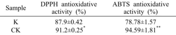
Table 1. Radical scavenging activity of Kombucha (K) and cit- rus Kombucha (CK)
Sample DPPH antioxidative
activity (%) ABTS antioxidative activity (%) K
CK
87.9±0.42 91.2±0.25*
78.78±1.57 94.59±1.81**
The antioxidant activities, based on the radical scavenging ca- pacity, of K and CK were assessed by DPPH and ABTS assays.
The results are expressed as mean±SD.
Statistically significant at *P<0.05 and **P<0.01 compared to K by Student's t-test.
Table 2. Contents of total phenolics in Kombucha (K) and citrus Kombucha (CK)
Sample Amount of total phenolics (μg/mL) K
CK
17.82±0.08 28.62±0.55**
The results are expressed mean±SD.
Statistically significant at **P<0.01 compared to K by Student's t-test.
포를 tube에 옮겨 1,000 rpm에서 2분간 원심분리 하였다.
분리된 세포를 1× cold PBS(Sigma-Aldrich Co.)로 세척 하여 다시 원심분리 한 후 상등액을 제거하였다. 1% Cold Binding Buffer(Sigma-Aldrich Co.) 200 μL로 1×10⁶의 세 포를 희석하여 여기에 5 μL PE Annexin V(Sigma-Aldrich Co.)를 넣고 약하게 vortex 한 후 25°C에서 15분간 방치한 다음, 1×Binding buffer 400 μL를 넣은 후 염색 정도를 flow cytometer(Moxi Flow Cytometry, ORPLO, Cam- bridge, MA, USA)로 측정하였다.
Apoptosis 관련 단백질 발현(western blot assay)의 검출 및 정량
방광암세포를 6-well plate에 1×10⁵씩 분주하여 배양한 후 K와 CK를 첨가하여 24시간 배양한 다음 배지를 제거하 고, 1× cold PBS로 1회 세척 후 40 μL RIPA lysis buffer (Bio-sesang Co., Seongnam, Korea)를 넣고 scraper로 세포 파쇄액를 모아 1.5 mL 튜브에 옮긴 후 20분간 얼음에 서 방치한 다음 원심분리를 통해 상등액을 취하였고, BCA protein assay kit(Pierce Inc., Appleton, WI, USA)을 이 용하여 단백질의 양을 정량하였다. 이후 12% SDS-PAGE gel에서 각 시료의 25 μg의 단백질을 취하여 3시간 동안 단백질을 분리하고 nitrocellulose membrane(Merck Mil- lipore, Billerica, MA, USA)에 2시간 transfer 한 후 5% 탈 지유로 blocking 하였다. 1차 항체는 4°C에서 18시간 반응 시켰고, 다음 날 2차 항체에 1시간 반응시킨 후 ECL+용액 (Pierce Inc.)과 반응시켜 단백질 ECL 화상분석기(Fusion- Fx7 Advance, Vilber Lourmat, Marne-la-Vallée, France) 시스템을 이용하여 측정하였다. Western band의 정량은 Fusion-Capt Advance Software 16.06(Vilber Lourmat) 을 사용하여 상대 강도를 측정하였다.
통계처리
본 실험은 독립적으로 3번 이상 반복실험을 시행하였다.
실험 결과는 통계분석용 프로그램 SPSS Version 18.0 Package Program(IBM, New York, NY, USA)을 이용하 여 각 실험군의 평균과 표준편차를 계산하고 t-test 분석 후, P<0.05 수준에서 처리군 간의 유의성을 검증하였다.
결과 및 고찰
DPPH와 ABTS 라디칼 소거 활성
DPPH assay는 빠르고 간단하게 유리 라디칼 소거능력을 측정하는 방법으로 항산화능을 측정하기 위해 널리 사용되 는 방법이다. DPPH는 짙은 보라색을 나타내는 organic ni- trogen radical로 에탄올 용액 상태에서 흡광도를 보이며 (19), ABTS assay는 양이온(ABTS・+)에 대한 항산화제의 소거능을 측정하는 방법으로 potassium persulfate와 반응 하여 녹색의 ABTS 라디칼을 형성하고 생성된 ABTS 라디
칼은 항산화력을 가진 물질로부터 전자를 받아 무색의 물질 로 환원된다(20).
이런 천연물의 항산화 활성의 활성 라디칼 소거능은 식품 중의 지방질 산화를 억제하거나 인체 내에서는 활성 라디칼 에 의한 노화를 억제시킬 수 있는 능력을 의미한다(21). 콤 부차 배양액(K)과 감귤액을 첨가한 감귤콤부차(CK)의 항산 화 활성은 Table 1에 나타낸 바와 같다.
1 mg/mL의 K와 CK에 대한 DPPH 라디칼 소거 활성은 각각 87.9%, 91.2%였으며, ABTS 라디칼 소거 활성은 각각 78.78%와 94.59%로 CK가 유리 라디칼에 대한 소거 활성 이 높은 것으로 나타났다. 이 결과는 콤부차의 발효에 의하 여 free-radical scavenging activity가 일반적으로 증가하 며 발효 시 첨가하는 재료에 따라 그 경향이 다르게 나타난 다는 Jayabalan 등(22)의 연구 결과와 일치하는 것으로 보 인다.
총폴리페놀 함량 측정
페놀 함량 측정법은 시료의 항산화능을 측정하는 방법은 아니지만 시료에 함유되어 있는 항산화능을 예측할 수 있으 므로 항산화 연구에 폭넓게 이용되는 방법이다(23). 폴리페 놀 화합물의 벤젠고리에 치환되어 있는 여러 개의 수산기가 유리 라디칼과의 환원 반응에 참여함으로써 항산화 활성이 나타나므로, 일반적으로 시료에 포함된 총폴리페놀 함량이 증가할수록 항산화 활성이 증가한다고 볼 수 있다(24). 발효 하기 전 측정한 총폴리페놀 함량보다 발효 후 총폴리페놀 함량이 전반적으로 약간 증가하는 결과를 나타내었으며 (data not shown), 발효 후 총폴리페놀 함량의 측정 결과는 Table 2에 나타낸 바와 같다.
K와 CK의 총페놀 함량이 μg당 tannic acid의 등량값으로 나타낼 때 K는 17.82 μg/mL, CK는 28.62 μg/mL로 감귤액
T-24
*
**
**
*** ***
*
**
**
***
***
0 20 40 60 80 100 120
0 1 2 4 6 8
Concentration (mg/mL)
Cell viability (% of control) .
k ck
K CK
5637
***
* **
* *
***
**
* *
*
0 20 40 60 80 100 120
0 1 2 4 6 8
Concentration (mg/mL)
Cell viability (% of control) .
K CK
Fig. 1. Effect of Kombucha (K) and Citrus Kombucha (CK) on the viability of bladder cancer cells. T-24 and 5637 cells were plated in 96-well plates at a 1×10⁴cell/well in RPMI 1640 supplemented with 10% FBS. The cells were treated with K or CK at concentrations as indicated for 24 h. Cellular survival rate was measured by MTT assay. The results are expressed as the mean±SD of three independent experiments. *P<0.05, **P<0.01, ***P<0.001; compared to the control by Student's two tailed t-test.
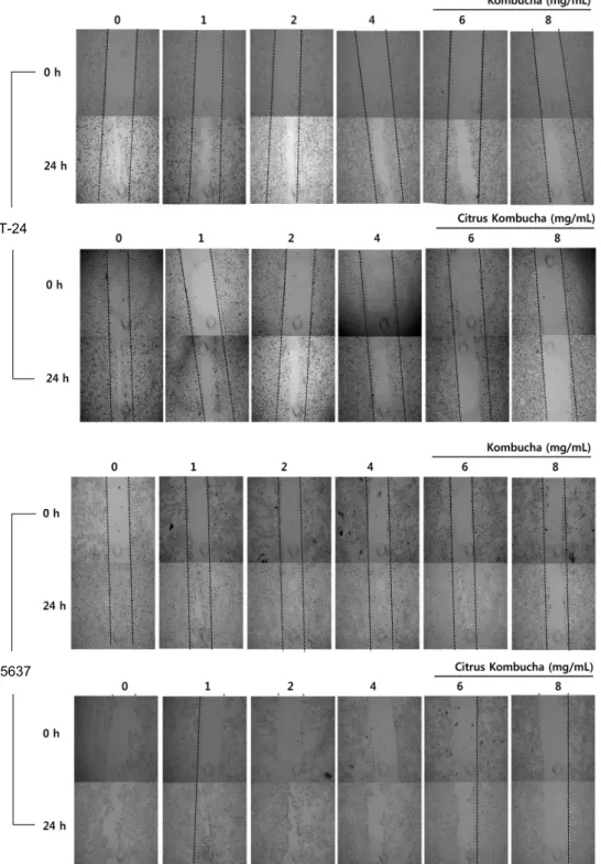
Fig. 2. Effect of Kombucha and citrus Kombucha on morphological change of bladder cancer cells. T-24 and 5637 cells were treated with Kombucha or citrus Kombucha for 24 h. The mor- phological changes were examined by optical microscopy with 100× (0.25 php) magnification.
을 첨가한 CK가 K보다 높은 페놀 함량을 보였다. 이상 라디 칼 소거능 측정과 총페놀 함량 측정 결과에 의하면 감귤액을 첨가한 CK가 K보다 항산화 능력이 높게 확인되었으며 총페 놀 함량도 CK가 K보다 높음을 확인하였다.
방광암세포(T-24와 5637)에 대한 증식 억제 효과 K 혹은 CK 자체의 세포 독성을 확인하기 위한 예비 실험 결과 유사 정상세포인 RAW 264.7 대식세포의 세포 생존율 은 8 mg/mL까지 대조군 대비 100% 이상이었다(data not shown). 그러므로 세포의 사멸에 큰 영향을 주지 않는 농도 인 0~8 mg/mL의 농도를 설정하여 방광암세포를 대상으로
하는 본 실험을 수행하였다. 방광암세포(T-24와 5637)에 K 와 CK를 0부터 8 mg/mL까지 처리하여 24시간 동안 배양한 후 세포 증식 억제 효과를 MTT assay를 통하여 측정하였 다. 방광암세포 T-24에 K를 처리한 결과 1, 2, 4, 6, 8 mg/
mL의 농도에서 생존율은 각각 82.57%, 69.01%, 50.12%, 33.47%, 26.06%로 나타났고, CK 처리군에서는 각 농도에 대해 81.96%, 72.77%, 50.36%, 19.49%, 13.44%의 생존 율을 나타내었다. K와 CK의 T-24 세포의 증식에 대한 저해 효과는 농도에 의존적인 저해로 나타났으며, K 처리군보다 CK 처리군의 성장이 더욱 저해되는 경향을 나타내었다(Fig.
1). 또한, 5637 방광암세포에 K를 처리한 결과는 1, 2, 4,
T-24
5637
Fig. 3. Scratch wound assays on bladder cancer cells. Capacity of wound recovery was assessed for bladder cancer cells, T-24 and 5637, treated with Kombucha or citrus Kombucha for 24 h. Cellular migration was examined by optical microscopy with 40×
(0.13 php) magnification.
6, 8 mg/mL 농도에서 생존율이 각각 84.22%, 84.71%, 85.63%, 80.63%, 68.22%였고, CK를 처리한 실험군에서 는 실험 농도에 대해 각각 82.5%, 74.24%, 76.03%, 65.71
%, 35.87%의 생존율을 보였다(Fig. 1). 이 결과에서도 T- 24에서와 마찬가지로 농도 의존적인 성장 억제 경향을 보였 으며, K 처리군보다 CK 처리군에서 방광암세포의 생존율이
크게 저하됨을 확인하였다. 특히 생존율에서 T-24는 5637 세포주에 비해 K 혹은 CK의 처리에 더 민감한 것으로 나타 났다.
방광암세포 형태 변화
방광암세포 T-24, 5637에 K와 CK를 24시간 동안 처리
T-24
* **
NS
***
**
* 0
20 40 60 80 100
0 4 6 8
Concentration (mg/mL)
% of apoptotic cells .
K CK
5637
* *
*
**
* **
0 20 40 60 80 100
0 4 6 8
Concentration (mg/mL)
% of apoptotic cells .
K CK
Fig. 4. Effect of Kombucha (K) and citrus Kombucha (CK) on apoptosis of bladder cancer cells. Apoptotic populations of T-24 and 5637 cells were measured by PE Annexin V kit after 24 h treatment of K or CK. Results were expressed as the mean±
SD of three independent experiments. *P<0.05, **P<0.01, ***P<
0.001; compared to the control by Student's two tailed t-test.
한 후 세포의 형태학적 변화를 도립현미경으로 관찰하였다 (Fig. 2). T-24 세포에 K를 8 mg/mL의 농도로 처리 시 생존 세포의 수가 감소한 것을 확인하였으나 형태학적인 변화는 보이지 않았다. T-24 세포에 CK를 처리하였을 때는 농도 6 mg/mL 이상에서 세포 형태의 변화를 관찰할 수 있었으 며, 농도가 증가할수록 부착능력이 상실되면서 세포의 외형 상 심한 변형이 관찰되었다. 그러나 5637 세포주에 K를 처 리 시 농도가 높아짐에 따라 세포수가 감소함을 확인할 수 있지만 세포의 형태에서는 큰 차이가 없었다. 5637 세포에 CK를 처리하였을 때는 8 mg/mL에서 약간의 형태상의 변화 를 관찰할 수 있었지만, T-24 세포에서의 변화와 비교할 때 그 변화가 크지 않았다.
방광암세포의 이동성에 미치는 영향(scratch-wound assay) 세포 이동(cell migration)은 동물의 성장과 생리활동에 있어서 매우 중요한 과정이며(25) 최근의 연구들에서는 세 포의 집단 이동이 다종의 종양전이에 기여한다는 보고가 있 다(26). 이에 T-24와 5637 방광암세포들을 K 혹은 CK에 24시간 노출시킨 후 scratch wound assay를 통해 세포들 의 이동 정도를 관찰하였다. 그 결과 방광암세포 T-24의 비교군에서는 24시간 후에 세포의 이동이 현저하게 증가하 였으나, K 혹은 CK 하에서는 농도가 높아짐에 따라 세포의 이동성이 점점 억제됨을 확인하였다(Fig. 3). 이 이동성의 저해는 K보다 CK에서 특히 높게 나타났다. 한편 방광암세 포 5637에서는 K를 8 mg/mL로 처리한 실험군에서만 세포 의 이동성 억제가 관찰되었다. CK 처리군에서는 농도 의존 적으로 세포의 이동이 저해되었지만 T-24와 비교할 때 그 수준은 낮았다.
Apoptotic cell 분석(Annexin V 측정)
T-24와 5637에 K 혹은 CK 처리 시 일어나는 성장 억제 및 세포의 형태학적 변화가 apoptosis에 의한 것인지를 확 인하기 위해 방광암세포를 K 혹은 CK로 24시간 처리한 후 PE Annexin V kit을 이용하여 세포사멸의 정도를 정량적으 로 확인하였다(Fig. 4). 방광암세포 T-24에 K를 처리하였 을 때 apoptosis의 유발 정도가 0, 4, 6, 8 mg/mL의 농도에 서 각각 4.47%, 4.07%, 6.4%, 11.1%로 나타났고, CK를 처리하였을 때는 각각 3.50%, 6.87%, 30.97%, 97.40%로 CK 8 mg/mL에서 apoptosis에 의한 세포사멸의 유발 정도 가 가장 높게 확인되었다. 또한, 방광암세포 5637에 K를 처리하였을 때 apoptosis에 의한 세포사멸의 비율은 각각 3.87%, 5.57%, 6.73%, 7.83%로 확인되었고, CK를 처리하 였을 때는 각각 3.87%, 6.27%, 7.3%, 15.73%로 나타났다.
Apoptosis 관련 단백질의 발현(western immunoblot assay) K와 CK의 처리에 의해 야기되는 apoptosis 관련 단백질 의 발현 정도를 살펴보기 위하여 western blot을 실시하였 다(Fig. 5). T-24와 5637에 K와 CK를 0부터 8 mg/mL까지
농도별로 처리한 후 단백질 시료를 제조하여 Bcl-2 관련 단백질들의 발현을 확인한 결과, T-24에서는 CK를 처리하 였을 때 anti-apoptotic 단백질인 Bcl-2의 발현이 6 mg/
mL 이상에서 현저히 감소하였고, pro-apoptotic 단백질인 Bax의 발현에는 차이가 없었다. 또한, cleaved caspase-9, cleaved caspase-8, cleaved caspase-3의 경우 CK의 농 도가 높아질수록 현저히 증가하였고, pro-caspase-9, pro- caspase-8, pro-caspase-3는 농도가 높아질수록 발현이 감소하였다. 특히 CK 처리 시 caspase의 활성화(cleaved- caspase)로 인한 PARP의 분절이 현저히 증가함을 관찰하 였다. 반면 T-24 세포주에 K를 처리하였을 때는 농도별 발 현의 차이를 보이지 않았으며, 5637 세포에서는 K와 CK 간의 변화가 없었으며, 농도별로도 별다른 발현의 차이를 보이지 않았다.
요 약
본 연구에서는 홍차버섯이라고 알려진 콤부차(Kombucha, K)에 플라보노이드 성분 및 각종 기능성 물질이 풍부한 감귤 액을 첨가하여 감귤의 생리활성 물질들이 콤부차로 이행되 는 효과를 기대하여 감귤 콤부차(citrus Kombucha, CK)를
A
T-24 (K)
0.00 0.50 1.00 1.50 2.00 2.50 3.00
0 4 6 8
μg/mL
Relative intensity .
cleaved caspase9/pro-caspase9 celaved caspase8/pro-caspase8 celaved caspase3/pro-caspase3
T-24 (CK)
0.00 1.00 2.00 3.00 4.00 5.00 6.00 7.00
0 4 6 8
μg/mL
Relative intensity .
cleaved caspase9/pro-caspase9 celaved caspase8/pro-caspase8 celaved caspase3/pro-caspase3
5637 (K)
0.0 2.0 4.0 6.0 8.0 10.0 12.0 14.0 16.0
0 4 6 8
μg/mL
Relative intensity .
cleaved caspase9/pro- caspase9
celaved caspase8/pro- caspase8
celaved caspase3/pro- caspase3
5637 (CK)
0.0 0.5 1.0 1.5 2.0 2.5 3.0 3.5 4.0 4.5
0 4 6 8
μg/mL
Relative intensity .
cleaved caspase9/pro- caspase9
celaved caspase8/pro- caspase8
celaved caspase3/pro- caspase3
B
Concentration (mg/mL) Concentration (mg/mL)
Concentration (mg/mL) Concentration (mg/mL)
Fig. 5. Expression of anti- or pro-apoptotic proteins in bladder cancer cells treated with Kombucha (K) or citrus Kombucha (CK).
(A) T-24 and 5637 cells were incubated in the absence or presence of K or CK for 24 h followed by separation of proteins by SDS-polyacrylamide gel electrophoresis. Numbers indicate relative intensities of western bands compared to 0. β-Actin was used as an internal control. (B) Relative intensity was measured by quantification of western immunoblot bands of caspases indicated.
배양한 후 항산화 능력 및 인체 방광암세포(T-24와 5637) 를 이용한 항암 효과를 확인하고 더 나아가 암의 증식을 억 제시킬 수 있는 천연소재 탐색을 목적으로 연구를 진행하였 다. 항산화 및 총페놀 함량 결과는 K보다 CK의 항산화 능력 과 페놀 함량이 높게 확인되었으며 방광암세포 T-24와 5637에 K 혹은 CK를 24시간 처리한 후 MTT assay를 통해 세포독성을 확인한 결과 농도 의존적으로 생존율이 감소하
였다. 특히 T-24 세포에서는 CK를 처리하였을 때 현저한 세포의 형태적 변화를 확인하였다. Western immunoblot을 통해 apoptosis 관련 단백질들의 발현을 확인하였는데 T-24에 CK 처리하였을 때 Bcl-2의 발현은 크게 감소하였 으며, pro-caspase-9, pro-caspase-8, pro-caspase-3 는 농도가 높아질수록 감소하였으며, cleaved caspase-9, cleaved caspase-8, cleaved caspase-3는 농도가 높아질
수록 증가하는 경향을 보였다. 또한, cleaved PARP가 증가 함을 확인할 수 있었다. 이상의 결과에서 일반 콤부차보다 감귤액을 첨가한 감귤 콤부차가 인체 방광암세포 T-24에 caspase에 의한 apoptosis가 유도됐음을 확인할 수 있었 다.
감사의 글
본 연구는 산업통산자원부에서 시행한 광역경제권 선도산 업 육성사업(R0001969) 및 교육인적자원부에서 시행한 지 역혁신창의인력양성사업(2014H1C1A1073112)의 지원으 로 수행된 연구 결과입니다.
REFERENCES
1. Cancer Statistics 2013. National Cancer Information Center.
http://www.cancer.go.kr/mbs/cancer/subview.jsp?id=cancer_
010101020000 (accessed Jun 2015).
2. Nargund VH, Tanabalan CK, Kabir MN. 2012. Management of non-muscle-invasive (superficial) bladder cancer. Semin Oncol 39: 559-572.
3. Frankfurt OS, Krishan A. 2003. Apoptosis-based drug screen- ing and detection of selective toxicity to cancer cells. Anti- cancer Drugs 14: 555-561.
4. Banerjee M, Singh P, Panda D. 2010. Curcumin suppresses the dynamic instability of microtubules, activates the mitotic checkpoint and induces apoptosis in MCF-7 cells. FEBS J 277: 3437-3448.
5. Dupuy J, Larrieu G, Sutra JF, Lespine A, Alvinerie M. 2003.
Enhancement of moxidectin bioavailability in lamb by a nat- ural flavonoid: quercetin. Vet Parasitol 112: 337-347.
6. Hwang YJ, Nam HK, Chang MJ, Noh GW, Kim SH. 2003.
Effect of Lentinus edodes and Pleurotus eryngii extracts on proliferation and apoptosis in human colon cancer cell lines.
J Korean Soc Food Sci Nutr 32: 217-222.
7. Cvetnić Z, Vladimir-Knezević S. 2004. Antimicrobial activ- ity of grapefruit seed and pulp ethanolic extract. Acta Pharm 54: 243-250.
8. Lee SJ, Moon SH, Kim T, Kim JY, Seo JS, Kim DS, Kim J, Kim YJ, Park YI. 2003. Anticancer and antioxidant activ- ities of Coriolus versicolor culture extracts cultivated in the citrus extracts. Kor J Microbiol Biotechnol 31: 362-367.
9. Guthrie N, Kurowska EM, Carrol KK. 1999. Use of citrus limonoids and flavonoids as well as tocotrienols for the treatment of cancer. PCT Patent WO1999015167 A2.
10. Reiss J. 1994. Influence of different sugars on the metabo- lism of the tea fungus. Z Lebensm-Unters Forsch 198: 258- 261.
11. Monforte MT, Trovato A, Kirjavainen S, Forestieri AM,
Galati EM, Lo Curto RB. 1995. Biological effects of hesper- idin, a Citrus flavonoid. (note Ⅱ): hypolipidemic activity on experimental hypercholesterolemia in rat. Farmaco 50:
595-599.
12. Okwu DE, Awurum AN, Okoronkwo JI. 2007. Phytochem- ical composition and in vitro antifungal activity screening of extracts from citrus plants against Fusarium oxysporum of okra plant (Hibiscus esculentus). Proceeding of 8th Afri- can Crop Science Society Conference. El-Minia, Egypt. p 1755-1758.
13. Steinkraus KH, Shapiro KB, Hotchkiss JH, Mortlock RP.
1996. Investigations into the antibiotic activity of tea fun- gus/kombucha beverage. Acta Biotechnol 16: 199-205.
14. Srinivasan R, Smolinske S, Greenbaum D. 1997. Probable gastrointestinal toxicity of Kombucha tea. J Gen Intern Med 12: 643-644.
15. Eric C, Jessica C. 2013. Kombucha: the amazing probiotic tea that cleans, heals, energizes, and detoxifies. 1st ed. Avery Publisher, New York, NY, USA. p 15-22.
16. Hesseltine CW. 1983. The future of fermented foods. Nutr Rev 41: 293-301.
17. Koo MWL, Cho CH. 2004. Pharmacological effects of green tea on the gastrointestinal system. Eur J Pharmacol 500:
177-185.
18. Hase T, Komine Y, Meguro S, Takeda Y, Takahashi H, Matsui Y, Inaoka S, Katsuragi Y, Tokimitsu I, Shimasaki H, Itakura H. 2001. Anti-obesity effects of tea catechins in humans. J Oleo Sci 50: 599-605.
19. Blois MS. 1958. Antioxidant determinations by the use of a stable free radical. Nature 181: 1199-1200.
20. Lemańska K, Szymusiak H, Tyrakowska B, Zieliński R, Soffers AE, Rietjens IM. 2001. The influence of pH on anti- oxidant properties and the mechanism of antioxidant action of hydroxyflavones. Free Radic Biol Med 31: 869-881.
21. Lee GD, Chang HG, Kim HK. 1997. Antioxidative and ni- trite-scavenging activities of edible mushrooms. Korean J Food Sci Technol 29: 432-436.
22. Jayabalan R, Subathradevi P, Marimuthu S, Sathishkumar M, Swaminathan K. 2008. Changes in free-radical scaveng- ing ability of kombucha tea during fermentation. Food Chem 109: 227-234.
23. Folin O, Denis W. 1912. On phosphotungstic-phosphomo- lybdic compounds as color reagents. J Biol Chem 12: 239- 243.
24. Park Y, Kim SH, Choi S, Han JK, Jung HK. 2008. Changes of antioxidant capacity, total phenolics, and vitamin C con- tents during Rubus coreanus fruit ripening. Food Sci Bio- technol 17: 251-256.
25. Halin C, Mora JR, Sumen C, von Andrian UH. 2005. In vivo imaging of lymphocyte trafficking. Annu Rev Cell Dev Biol 21: 581-603.
26. Rørth P. 2009. Collective cell migration. Annu Rev Cell Dev Biol 25: 407-429.