저작자표시-비영리-변경금지 2.0 대한민국 이용자는 아래의 조건을 따르는 경우에 한하여 자유롭게 l 이 저작물을 복제, 배포, 전송, 전시, 공연 및 방송할 수 있습니다. 다음과 같은 조건을 따라야 합니다: l 귀하는, 이 저작물의 재이용이나 배포의 경우, 이 저작물에 적용된 이용허락조건 을 명확하게 나타내어야 합니다. l 저작권자로부터 별도의 허가를 받으면 이러한 조건들은 적용되지 않습니다. 저작권법에 따른 이용자의 권리는 위의 내용에 의하여 영향을 받지 않습니다. 이것은 이용허락규약(Legal Code)을 이해하기 쉽게 요약한 것입니다. Disclaimer 저작자표시. 귀하는 원저작자를 표시하여야 합니다. 비영리. 귀하는 이 저작물을 영리 목적으로 이용할 수 없습니다. 변경금지. 귀하는 이 저작물을 개작, 변형 또는 가공할 수 없습니다.
Draw the anti-cancer effect via gene expression
profiles between well-differentiated and
undifferentiated thyroid cancer cell derived from
patient
Yong Sang Lee
Department of Medicine
The Graduate School, Yonsei University
[UCI]I804:11046-000000516024
[UCI]I804:11046-000000516024
Draw the anti-cancer effect via gene expression
profiles between well-differentiated and
undifferentiated thyroid cancer cell derived from
patient
Directed by Professor Hang-Seok Chang
The Doctoral Dissertation
submitted to the Department of Medicine,
the Graduate School of Yonsei University
in partial fulfillment of the requirements for the degree
of Doctor of Philosophy
Yong Sang Lee
This certifies that the Doctoral
Dissertation of Yong Sang Lee is approved.
---
Thesis Supervisor: Hang-Seok Chang
---
Thesis Committee Member #1: Eun Jig Lee
---
Thesis Committee Member #2: Soon Won Hong
---
Thesis Committee Member #3: Chang Geol Lee
---
Thesis Committee Member #4: Ki-Wook Chung
The Graduate School
Yonsei University
ACKNOWLEDGEMENTS
I very much appreciate my supervisor professor Hang-Seok Chang. I really respect him as
my mentor in both points of his right attitude for life and research.
During the period of this research for the doctoral thesis, he always emphasized the right
research even if it takes long time. He did not mind making time for discussing my
experimental results even though he was always busy for his work. I want to follow his love
for patients suffering cancer and pure research mind.
Professor Cheong Soo Park also helped my study very much. I am very grateful to them for
many insightful comments they gave. Their valuable comments have led our study to this point
well.
I also would like to express my thanks to my colleagues in our lab: Seok-Mo Kim, Bup-Woo
Kim, Hyeung Kyoo Kim, Ho Jin Chang, Soo Young Kim, Ki Cheong Park
helped me
immensely in experiments.
At last, I deeply appreciate my wife and son for supporting my long course of doctoral
dissertation.
<TABLE OF CONTENTS>
ABSTRACT --- 1
I.
INTRODUCTION --- 2
II.
MATERIALS AND METHODS
1. Patients/tissue specimens --- 16
2. Tumor cell isolation and primary culture --- 16
3. Cell culture ---16
4. Cell viability assay --- 17
5. Microarray experiment and data analysis --- 17
6. Immunofluorescence analysis and confocal imaging --- 18
7. Immunoblot analysis --- 18
8. Flow cytometry analysis of the cell cycle --- 19
9. Human thyroid cancer cell xenograft --- 19
10. Immunohistochemistry --- 20
11. Statistical analysis --- 20
III.
RESULTS --- 21
IV.
DISCUSSION --- 35
V.
CONCLUSION --- 37
REFERENCES --- 38
LIST OF FIGURES
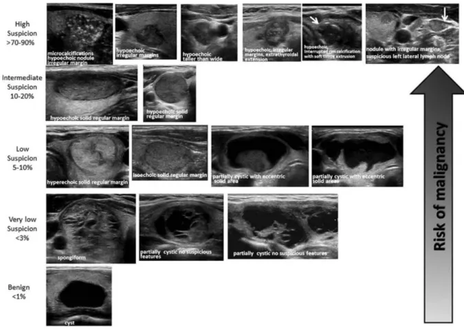
Figure 1. ATA nodule sonographic patterns and risk of malignancy --- 3
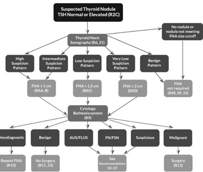
Figure 2. Algorithm for evaluation and management of patients with thyroid
nodules based on US pattern and FNA cytology --- 4
Figure 1. Scheme of step-wise dedifferentiation of follicular cell-derived thyroid
cancer --- 6
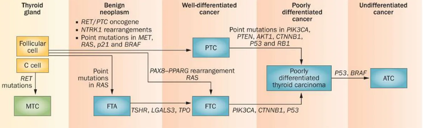
Figure 2. Classification of human thyroid carcinomas and subtype-specific
genetic alterations --- 8
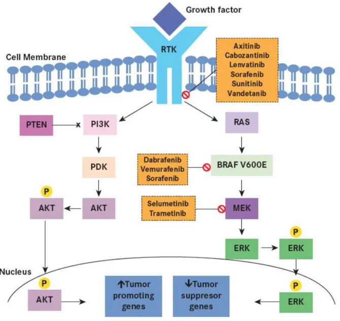
Figure 3. The PI3K-AKT pathway also plays a significant role in sporadic thyroid
tumorigenesis --- 9
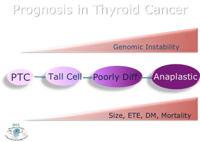
Figure 4. The prognosis system in current therapy of the thyroid cancer --- 12
Figure 7. Drug resistance was mediated by FGFR signaling pathway --- 13
Figure 8. FGFR signaling pathway mediated target gene expression --- 15
Figure 9. Gene expression profiles measured by microarrays. Hierarchical
clustering analysis in between differentiated-, poorly differentiated-and
dedifferentiated patient-tissue --- 21
Figure10. Gene expression profiles measured by microarrays. Hierarchical
clustering analysis in between differentiated-, poorly differentiated-and
dedifferentiated patient-derived thyroid cancer cell --- 22
Figure 11. Present study was to investigate the nuclear localization of β-catenin
in patient-derived-advanced thyroid cancer cells --- 23
Figure 12. Profiling of the FGFR and EMT signaling pathway between
patient-derived, advanced- and non-advanced thyroid cancer cells --- 24
Figure 13. Synergistic suppression of cancer cell proliferation by HNHA and
Lenvatinib was stronger than any other groups in patient –derived
thyroid cancer cells --- 26
Figure 14. The HNHA and Lenvatinib combination significantly induced
apoptosis and cell cycle arrest in patient–derived thyroid cancer cells
--- 28
Figure 15. More advanced cancer cells, cancer stem cells, were more resistant to
activation in GSP1, GSA1 and GSA2 --- 30
Figure 16.
β-catenin, EMT marker, plays a key role in the induction of EMT by
nuclear localization on advanced thyroid cancer cell --- 31
Figure 17. Tumor shrinkage was significantly induced by the combination
treatment of the HNHA and Lenvatinib in xenograft mode --- 32
Figure 18. Immunohistochemistry analysis of Bcl2, anti-apoptotic marker GSP1,
GSA1 and GSA2 cell xenograft tumors --- 33
LIST OF TABLES
Table 1. Cell line characteristics, viability after drug treatment of all thyroid
cancer cell lines --- 25
Table 2. IC50 (half maximal inhibitory concentration) determination using a cell
proliferation assay --- 25
Table 3. Flow cytometry analysis of the cell cycle of the GSP1, GSA1 and GSA2
--- 27
1
ABSTRACT
Draw the anti-cancer effect via gene expression profiles between well-differentiated and
undifferentiated thyroid cancer cell derived from patient
Yong Sang Lee
Department of Medicine
The Graduate School, Yonsei University
(Directed by Professor Hang-Seok Chang)
Thyroid gland presents a broad scope of tumors derived from follicular cells that range from well-differentiated cancer including papillary and follicular cancer (PTC and FTC), generally carrying a favorable prognosis, to the clinically aggressive, undifferentiated thyroid cancer (UTC). It is mostly recognized that UTC arise progress from a pre-existing well-differentiated cancer via a various procedure of genetic and epigenetic alterations that bring about clonal expansion and neoplastic progression. Mutations and epigenetic changes in UTC are not so clear. This presumes, of course, that UTC may derive from well-differentiated thyroid cancer (WDTC), it is look forward to some UTC would shelter genetic changes that are usual of PTC and FTC. It is the instance for several markers (BRAF, NRAS) that are existent in WDTC and UTC. The p53 genes is usually identified in less- and undifferentiated thyroid cancer, confirming a diagnosis of UTC. Especially, UTC are unusual but extremely aggressive malignancies with a greatly short survival that was acquired to multiple anticancer drug-resistant.
This study suggested that therapeutic approaches to undifferentiated thyroid cancer (UTC) via the gene expression profile.
Key words: papillary thyroid cancer (PTC), follicular thyroid cancer (FTC), well-differentiated thyroid cancer (WDTC), undifferentiated thyroid cancer (UTC), gene expression profile, drug resistance
2
Draw the anti-cancer effect via gene expression profiles between well-differentiated and
undifferentiated thyroid cancer cell derived from patient
Yong Sang Lee
Department of Medicine
The Graduate School, Yonsei University
(Directed by Professor Hang-Seok Chang)
I. INTRODUCTION
1. Thyroid cancer
Thyroid cancer is a cancer originating from follicular or parafollicular thyroid cells. These cells give rise to both well-differentiated cancers – papillary thyroid cancer (PTC) and follicular thyroid cancer (FTC) – and anaplastic thyroid cancer (ATC), whose anaplastic cells are poorly differentiated. The second cell type, the C or parafollicular cell, produces the hormone calcitonin and is the cell of origin for medullary thyroid cancer (MTC)1. The most effective management of aggressive thyroid
cancers is surgical removal of thyroid gland (thyroidectomy) followed by radioactive iodine ablation and TSH-suppression therapy. Chemotherapy or radiotherapy may also be used in cases of distant metastases or advanced cancer stage. Five-year survival rates are 98.1% in the United States [NIH, Cancer Statistical Summaries].
3
Figure 1. ATA nodule sonographic patterns and risk of malignancy (Thyroid. 2016 Jan 1; 26(1): 1– 133).
2. Signs and symptoms
Most often the first symptom of thyroid cancer is a nodule in the thyroid region of the neck2.
However, many adults have small nodules in their thyroids, but typically under 5% of these nodules are found to be cancerous3,4. Sometimes the first sign is an enlarged lymph node. Later symptoms that can
be present are pain in the anterior region of the neck and changes in voice due to an involvement of the recurrent laryngeal nerve5. Thyroid cancer is usually found in a euthyroid patient, but symptoms of
hyperthyroidism or hypothyroidism may be associated with a large or metastatic well-differentiated tumor. Thyroid nodules are of particular concern when they are found in those under the age of 20.
4
The presentation of benign nodules at this age is less likely, and thus the potential for malignancy is far greater3.
Figure 2. Algorithm for evaluation and management of patients with thyroid nodules based on US pattern and FNA cytology (Thyroid. 2016 Jan 1; 26(1): 1–133).
3. Causes
Thyroid cancers are thought to be related to a number of environmental and genetic predisposing factors, but significant uncertainty remains regarding their causes. Environmental
5
exposure to ionizing radiation from both natural background sources and artificial sources is suspected to play a significant role, and there are significant increased rates of thyroid cancer in those exposed to mantlefield radiation for lymphoma, and those exposed to iodine-131 following the Chernobyl6,
Fukushima, Kyshtym, and Wind scale7 nuclear disasters. Thyroiditis and other thyroid diseases also
predispose to thyroid cancer. Genetic causes include multiple endocrine neoplasia type 2 which markedly increases rates, particularly of the rarer medullary form of the disease8.
4. Diagnosis
After a thyroid nodule is found during a physical examination, a referral to an endocrinologist or a thyroidologist may occur. Most commonly an ultrasound is performed to confirm the presence of a nodule and assess the status of the whole gland. Measurement of thyroid stimulating hormone and anti-thyroid antibodies will help decide if there is a functional thyroid disease such as Hashimoto's thyroiditis present, a known cause of a benign nodular goiter9. Measurement of calcitonin is necessary
to exclude the presence of medullary thyroid cancer. Finally, to achieve a definitive diagnosis before deciding on treatment, a fine needle aspiration cytology test is usually performed and reported according to the Bethesda system. In adults without symptoms screening for thyroid cancer is not recommended10.
6
Figure 3. Scheme of step-wise dedifferentiation of follicular cell-derived thyroid cancer (Nikiforov, Y. E. & Nikiforova, M. N. (2011) Molecular genetics and diagnosis of thyroid cancer Nat. Rev. Endocrinol).
4.1 Classification
Thyroid cancers can be classified according to their histopathological characteristics11.
4.1.1 Papillary thyroid cancer (75% to 85% of cases) – often in young females – excellent prognosis. May occur in women with familial adenomatous polyposis and in patients with Cowden syndrome12.
4.1.2 Newly reclassified variant: noninvasive follicular thyroid neoplasm with papillary-like nuclear features is considered an indolent tumor of limited biologic potential.
7
4.1.3 Follicular thyroid cancer (10% to 20% of cases) – occasionally seen in patients with Cowden syndrome13.
4.1.4 Medullary thyroid cancer (5% to 8% of cases) – cancer of the parafollicular cells, often part of multiple endocrine neoplasia type 214.
4.1.5 Poorly differentiated thyroid cancer is aggressive pattern of thyroid cancer.
4.1.6 Anaplastic thyroid cancer (less than 5% of cases) is not responsive to treatment and can cause pressure symptoms15. The follicular and papillary types together can be classified as "differentiated
thyroid cancer"16. These types have a more favorable prognosis than the medullary and undifferentiated
types17. Papillary microcarcinoma is a subset of papillary thyroid cancer defined as measuring less than
or equal to 1 cm18. The highest incidence of papillary thyroid microcarcinoma in autopsy series was
reported by Harach et al. in 1985, who found 36 of 101 consecutive autopsies were found to have an incidental microcarcinoma19. Michael Pakdaman et al. report the highest incidence in a retrospective
surgical series at 49.9% of 860 cases20. Management strategies for incidental papillary microcarcinoma
on ultrasound (and confirmed on FNAB) range from total thyroidectomy with radioactive iodine ablation to observation alone. Harach et al. suggest using the term "occult papillary tumor" to avoid giving patients distress over having cancer19. It was Woolner et al. who first arbitrarily coined the term
8
Figure 4. Classification of human thyroid carcinomas and subtype-specific genetic alterations (Pallante, P. et al. (2013) Deregulation of microRNA expression in thyroid neoplasias Nat. Rev. Endocrinol).
5. Treatment
Thyroidectomy and dissection of central neck compartment is initial step in treatment of thyroid cancer in majority of cases. Thyroid-preserving operation may be applied in cases, when thyroid cancer exhibits low biological aggressiveness (e.g. well-differentiated cancer, no evidence of lymph node metastases, low MIB-1 index, no major genetic alterations like BRAF mutations, RET/PTC rearrangements, p53 mutations etc.) in patients younger than 45 years22. If the diagnosis of
well-differentiated thyroid cancer (e.g. papillary thyroid cancer) is established or suspected by FNA the surgery is indicated, whereas watchful waiting strategy is not recommended in any evidence-based guidelines22. Watchful waiting reduces overdiagnosis and overtreatment of thyroid cancer among old
patients. Radioactive Iodine-131 is used in patients with papillary or follicular thyroid cancer for ablation of residual thyroid tissue after surgery and for the treatment of thyroid cancer23. Patients with
medullary, anaplastic, and most Hurthle cell cancers do not benefit from this therapy. External irradiation may be used when the cancer is unresectable, when it recurs after resection, or to relieve pain from bone metastasis. Sorafenib and sunitinib, approved for other indications show promise for thyroid cancer and are being used for some patients who do not qualify for clinical trials24. Numerous
9
Figure 5. The PI3K-AKT pathway also plays a significant role in sporadic thyroid tumorigenesis. As with the MAPK pathway, an extracellular stimulus activates RTK at the cell membrane, and subsequently PI3K, ultimately leading to phosphorylation and activation of AKT.13 Activated AKT then enters the nucleus to upregulate tumor-promoting genes. Within the cytoplasm, activated AKT also activates other signaling molecules, including the mTOR pathway and phosphorylation of glycogen synthase kinase 3β. Common genetic alterations implicated in induction of the PI3K-AKT pathway include RAS and PTEN mutation or deletion, PI3KCA mutation or amplification, AKT1 mutation, and amplifications of the RTK genes (Thyroid Cancer: Molecular Pathogenesis, Tyrosine Kinase Inhibitors,
10
and Other New Therapies).
6. Prognosis
The prognosis of thyroid cancer is related to the type of cancer and the stage at the time of diagnosis. For the most common form of thyroid cancer, papillary, the overall prognosis is excellent. Indeed, the increased incidence of papillary thyroid carcinoma in recent years is likely related to increased and earlier diagnosis. One can look at the trend to earlier diagnosis in two ways. The first is that many of these cancers are small and not likely to develop into aggressive malignancies. A second perspective is that earlier diagnosis removes these cancers at a time when they are not likely to have spread beyond the thyroid gland, thereby improving the long-term outcome for the patient. There is no consensus at present on whether this trend toward earlier diagnosis is beneficial or unnecessary. The argument against early diagnosis and treatment is based on the logic that many small thyroid cancers (mostly papillary) will not grow or metastasize. This viewpoint holds the overwhelming majority of thyroid cancers are over diagnosed (that is, will never cause any symptoms, illness, or death for the patient, even if nothing is ever done about the cancer). Including these overdiagnosed cases skews the statistics by lumping clinically significant cases in with apparently harmless cancers. Thyroid cancer is incredibly common, with autopsy studies of people dying from other causes showing that more than one-third of older adults technically has thyroid cancer, which is causing them no harm. It is easy to detect nodules that might be cancerous, simply by feeling the throat, which contributes to the level of overdiagnosis. Benign (non-cancerous) nodules frequently co-exist with thyroid cancer; sometimes, it is a benign nodule that is discovered but surgery uncovers an incidental small thyroid cancer. Increasingly, small thyroid nodules are discovered as incidental findings on imaging (CT scan, MRI, ultrasound) performed for another purpose ; very few of these people with accidentally discovered, symptom-free thyroid cancers will ever have any symptoms, and treatment in such patients has the potential to cause harm to them, not to help them25. Thyroid cancer is three times more common in
11
women than in men, but according to European statistics, the overall relative 5-year survival rate for thyroid cancer is 85% for females and 74% for males. There is general agreement that stage I or II papillary, follicular or medullary cancer have a good prognosis, it is not possible when evaluating a small thyroid cancer to determine which ones will grow and metastasize and which will not. As a result, once a diagnosis of thyroid cancer has been established (most commonly by a fine needle aspiration), it is likely that a total thyroidectomy will be performed. This drive to earlier diagnosis has also manifested itself on the European continent by the use of serum calcitonin measurements in patients with goiter to identify patients with early abnormalities of the parafollicular or calcitonin-producing cells within the thyroid gland. As multiple studies have demonstrated, the finding of an elevated serum calcitonin is associated with the finding of a medullary thyroid carcinoma in as high as 20% of cases. In Europe where the threshold for thyroid surgery is lower than in the United States, an elaborate strategy that incorporates serum calcitonin measurements and stimulatory tests for calcitonin has been incorporated into the decision to perform a thyroidectomy; thyroid experts in the United States, looking at the same data sets have, for the most part, not incorporated calcitonin testing as a routine part of their evaluation, thereby eliminating a large number of thyroidectomies and the consequent morbidity. The European thyroid community has focused on prevention of metastasis from small medullary thyroid carcinomas; the North American thyroid community has focused more on prevention of complications associated with thyroidectomy. As demonstrated in the Table below, individuals with stage III and IV disease have a significant risk of dying from thyroid cancer. While many present with widely metastatic disease, an equal number evolve over years and decades from stage I or II disease. Physicians who manage thyroid cancer of any stage recognize that a small percentage of patients with low-risk thyroid cancer will progress to metastatic disease.
Fortunately for those with metastatic thyroid cancer, the last 5 years has brought about a renaissance in thyroid cancer treatment. The identification of some of the molecular or DNA abnormalities for thyroid cancer has led to the development of therapies that target these molecular defects. The first of these agents to negotiate the approval process is vandetanib, a tyrosine kinase
12
inhibitor that targets the RET proto-oncogene, 2 subtypes of the vascular endothelial growth factor receptor, and the epidermal growth factor receptor. More of these compounds are under investigation and are likely to make it through the approval process. For differentiated thyroid carcinoma, strategies are evolving to use selected types of targeted therapy to increase radioactive iodine uptake in papillary thyroid carcinomas that have lost the ability to concentrate iodide. This strategy would make it possible to use radioactive iodine therapy to treat "resistant" thyroid cancers. Other targeted therapies are being evaluated, making it possible that life will be extended over the next 5–10 years for those with stage III and IV thyroid cancer.
13
7. Targeted therapy for undifferentiated thyroid cancer
UTC are rare but highly aggressive malignancies with an extremely short survival. Poor prognosis is due to their unlimited growth, invasion, migration and resistance to common anticancer therapies. Advances in understanding the molecular alterations in thyroid carcinomas led to development of new therapeutic strategies such as kinase inhibitors [Resistance to Kinase Inhibitors in Poorly Differentiated and Anaplastic Thyroid Cancer: Preclinical In vitro Evidences].
8. Therapy of undifferentiated thyroid cancer
The advanced cancer subtype, include ATC, was that has poor prognosis due to its resistance to treatment and aggressive behavior. The total median survival is only a few months. Poorly differentiated cancers usually have an ability of the resistant to anti-cancer drug, and presently there is not propose the effective clinical guidelines for the therapy of ATC. One of the most well-known that drug resistance in cancer is the induction of epithelial-mesenchymal transition (EMT). Nevertheless, the mechanisms of EMT-mediated drug resistance dwell incompetently defined.
14
Epithelial-mesenchymal transition (EMT) is a physiological procedure depend on epithelial cells collapse cell-cell junctions and temporary or eternally transition on a condition that is more delegate of migratory cells. The morphological lead to EMT is a fundamental aspect of resistance to ErbB-targeting compounds a deficiency in knowledge of the molecular mechanisms of this incident has inhibited the progress of therapeutic approach of targeting this drug resistant state. Previous well-known research have proved that expression of fibroblast growth factor receptor 1 (FGFR1) is significantly induced while TGF-β mediated EMT and plays a crucial role of the metastatic cancer. These previous researchs are support to our hypothesis, at the same time lead to drug resistance of poorly differentiated cancer-cancer stem cell through EMT-mediated by FGFR signaling pathway. The molecules and mechanisms that are closely associated with the poor clinical results of the advanced thyroid cancer. Among of them, we concentrated to the epithelial-mesenchymal transition (EMT) and drug resistance on the cancer stem cell (CSC) properties that are caused by EMT as one of the potential bring about of the poor clinical result. Evidence of the lately research was proved that EMT of cancer cells not only causes induced metastasis, but also contributed drug resistance.
15
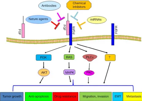
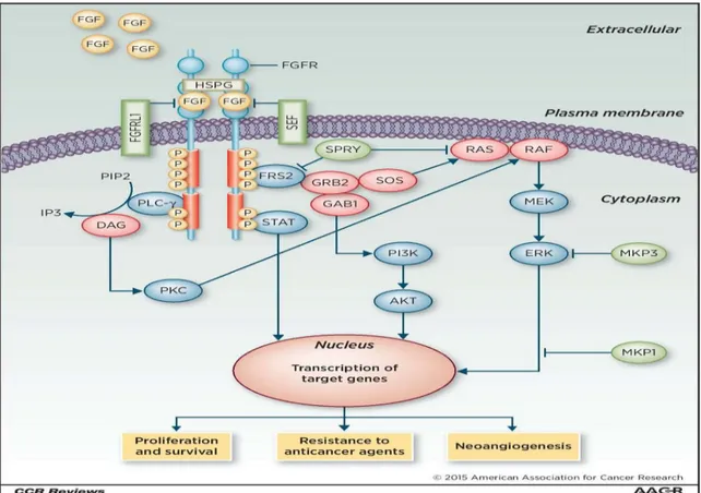
Figure 8. FGFR signaling pathway mediated target gene expression.
In this research, we based on gene expression profiles between PDTC and DTC, patient derived thyroid cancer cell, how inhibited to drug resistance via FGFR signaling and the epithelial-mesenchymal transition (EMT) develops in cancer in response to current treatments and how these problems are being addressed.
16
II. MATERIALS AND METHODS
Patients/tissue specimens
Fresh tumors were obtained from patients with biochemical and histologically proven PTC and ATC who were treated at the Thyroid Cancer Center, Gangnam Severance Hospital, Yonsei University College of Medicine, Seoul, Korea. Fresh tumors were acquired during surgical resection of thyroid cancer primary and metastatic sites. For goals of this research, we chose one patient with thyroid cancer. The research protocol was approved by Institutional Review Board of the Thyroid Cancer Center, Gangnam Severance Hospital, Yonsei University College of Medicine (IRB Protocol: 3-2016-0076).
Tumor cell isolation and primary culture
On the morrow of resection, tumors were kept into normal saline with antifungal and antibiotics, and moved to the laboratory. Normal tissue and fat were removed and the tissues were rinsed with 1X HBSS. Tumors were minced into tube with dissociation medium. Dissociation medium contained DMEM/F12 with 20% FBS supplemented with 1 mg/ml of collagenase type IV (Sigma, USA). Minced and suspended tumor cells were filtered through sterile 70-micron pores nylon cell strainers (BD Falcon) rinsed with 50 ml 1X HBSS and centrifuged at 1400 rpm for 5 min. Cells were resuspended with RPMI-1640 (Hyclone, South Logan, UT, USA) medium, with 10 % fetal bovine serum (Hyclone) and 2% penicillin/streptomycin solution (Gibco, Grand Island, NY, USA). Cell viability was decided by the trypan blue dye exclusion method used.
Cell culture
RPMI-17
1640 medium with 10% fetal bovine serum (Authentication by short tandem repeat profiling / karyotyping / isoenzyme analysis).
Cell viability assay
Cell proliferation was measured using the MTT assay. Cells were seeded in 96-well plates at 6 103 cells per well and incubated overnight to achieve 80% confluency. The indicated drugs were
added to achieve final concentrations of 0-100 μM. Cells were the incubated for the indicated times prior to determination of cell viability using the MTT reagent according to the manufacturer’s protocol, and measurement of absorbance at 490 nm. Viable cells were counted with trypan blue exclusion. Data were expressed as a percentage of the signal observed in vehicle-treated cells and shown as the mean ± standard error of the mean (SEM) of triplicate experiments.
Microarray experiment and data analysis
Gene expression data from the cancer cell lines were generated by hybridizing labeled RNAs to Human-6 v2. Expression BeadChips (Illumina). Total RNA was isolated from cells harvested after HNHA or Sorafenib or Lenvatinib alone or in combination with HNHA for 24 h, for the indicated time busing the mirVana miRNA Isolation Kit (Ambion Inc. AM1560) according to manufacturer’s protocol. Biotin-labeled cRNA was prepared using the Illumina Total Prep RNA Amplification Kit (Ambion Inc.). Total RNA (500 ng) was used for the synthesis of cDNA followed by amplification and biotin labeling. Biotinylated cRNA (1.5 μg) per sample was hybridized to Illumina Human-6 BeadChip v.2 microarray and signals were developed by Amersham fluorolink streptavidin-Cy3 (GE Healthcare Bio-Sciences). All statistical analysis was performed using R 2.3.0 and BRB Arraytools Version 3.5 (https://brb.nci.nih.gov/BRB-ArrayTools/) with quantile normalization26.
18
Immunofluorescence analysis and confocal imaging
Expression analysis of β-catenin was performed with immunofluorescent staining. Cells grown on glass-bottomed dishes glass bottom dishes (MatTek, Ashland, MA) were fixed with 4% formaldehyde solution (R&D systems, UK) for 10 min. and permeabilised with 0.5% TritonX-100 in PBS for 10 min. Slides were air-dried and washed with PBS, and incubated with anti-β-catenin (1:25, abcam) in 3% bovine serum albumin (BSA) in PBS. After washing with PBS, slides were incubated with Alexa 488 (1:200, Molecular Probes, Eugene, U.S.A.) Nucleus was stained with Hoechst 33342 (Life Technologies, Grand Island, NY, USA) to visualize nuclei. Images were observed under a confocal microscope (LSM Meta 700) and were analyzed with the Zeiss LSM Image Browser software, version 4.2.0121.
Immunoblot analysis
Cells were washed twice with cold phosphate-buffered saline and lysed on ice with protein extraction buffer (Pro-Prep, iNtRON Biotechnology, Korea) following the manufacturer’s protocol. Protein concentrations were determined by BCA assay (Pierce Biotechnology, Rockford, IL). Equal amounts of protein (20 μg) were separated in 8-10% SDS-polyacrylamide gels; the resolved proteins were then electro-transferred onto PVDF membranes (Millipore, Bedford, MA). The membranes were subsequently blocked with 5% nonfat milk in TBST for 1 h at room temperature and incubated with appropriate concentrations of primary antibodies for Ki-67 (Abcam), Cyclin D1 (Santa Cruz Biotechnology), CDK4 (Santa Cruz Biotechnology), p21 (Santa Cruz Biotechnology), p53(Santa Cruz Biotechnology), p-ERK 1/2 (Santa Cruz Biotechnology), ERK 1/2 (Santa Cruz Biotechnology), Apaf-1 (Abcam), p-NFκB (Santa Cruz Biotechnology), Bcl-2 (Santa Cruz Biotechnology), Caspase 3 (Santa Cruz Biotechnology), Vimentin (Abcam), E-cadherin (Abcam), Snail (Abcam), Zeb1 (Abcam) and β-actin (Santa Cruz Biotechnology) were overnight at 4°C. The membranes were then rinsed 3 ~ 5 times with TBS-T and probed with the corresponding secondary antibodies conjugated to HRP (Santa Cruz)
19
at room temperature for 1 h. After rinsing, blots were developed with ECL reagents (Pierce, Rockford, US) and exposed to Kodak X-OMAT AR Film (Eastman Kodak, Rochester, US) for 3 ~ 5 min.
Flow cytometry analysis of the cell cycle
Cells were treated with Sorafenib and Lenvatinib alone or in switching in RPMI-1640 medium containing 10% FBS for 40 h, harvested by trypsinization, and fixed with 70% ethanol. Cells were stained for total DNA, using PBS containing 40 μg/mL propidium iodide and 100 μg/mL RNase I for 30 min at 37°C. Cell cycle distribution was then analyzed in the FACSCalibur Flow Cytometer (BD Biosciences, San Jose, CA, USA). The proportions of cells in the sub-G0/G1, G0/G1, S, and G2/M phases were analyzed by FlowJo v8 software for MacOSX (Tree Star, Ashland, OR, USA). This experiment was repeated thrice, and the results were averaged.
Human thyroid cancer cell xenograft
The patient-derived PTC and ATC cells (3.5 106 cells / mouse) were cultured in vitro and
then injected subcutaneously into the upper left flank region of female BALB/c nude mice. After 11 days, tumor-bearing mice were grouped randomly (n = 10/group) and with 10 mg/kg Lenvatinib (given p.o.) and 40 mg/kg Sorafenib (given p.o.), Lenvatinib or Sorafenib once every 2 days for injections. Tumor size was measured every other day using calipers. Tumor volume was estimated using the following formula: L × S2/2 (where L, longest diameter; S, shortest diameter). Animals were maintained under specific pathogen-free (SPF) conditions. All experiments were approved by the Animal Experiment Committee of Yonsei University.
20
Immunohistochemistry
All tissues were fixed in 10% neutral-buffered formalin and embedded in paraffin wax by standard protocols. Tissue sections (5 μm) were dewaxed, and antigen retrieval was performed in citrate buffer (pH 6), using an electric pressure cooker set at 120°C for 5 min. Sections were incubated for 5 min in 3% hydrogen peroxide to quench endogenous tissue peroxidase. All tissue sections were counterstained with hematoxylin, dehydrated, and mounted.
Statistical analysis
Statistical analyses were performed using GraphPad Prism software (GraphPad Software Inc., La Jolla, CA, USA). Immunohistochemistry results were subjected to ANOVA followed by a Bonferroni post hoc test. Values are expressed as means ± SD. P values < 0.05 indicated statistical significance.
21
III. RESULTS
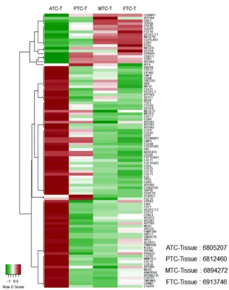
Figure 9. Gene expression profiles measured by microarrays. Hierarchical clustering analysis in between differentiated-, poorly differentiated-and dedifferentiated thyroid cancer of patient tissue.
We used expression microarrays to assess changes in gene expression of FGFR signalling cascades between differentiated- and poorly differentiated thyroid cancer of patient tissue. FGFR, EGFR and EMT markers were high expressed in poorly differentiated thyroid cancer.
22
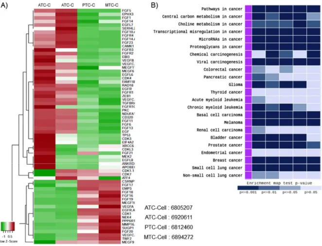
Figure 10. A, Gene expression profiles measured by microarrays. B, Gene expression pattern of the cancer subtype. Hierarchical clustering analysis in between differentiated-, poorly differentiated-and dedifferentiated patient-derived thyroid cancer cell.
The patient-derived ATC, PTC and MTC cells were isolation from the patient tissue. A, there is no significantly difference of gene expression compare microarrays of patient tissue. B, patient-derived ATC, PTC and MTC cells were confirmed by gene express pattern analysis of cancer subtype. The patient-derived ATC, PTC and MTC cells were showed coterminous gene express pattern compared with thyroid cancer FGFR, EGFR and EMT markers were high expressed in poorly differentiated thyroid cancer.
23
Figure 11. Present study was to investigate the nuclear localization of β-catenin in patient-derived-advanced thyroid cancer cells.
β-catenin, EMT marker, plays a key role in the induction of EMT, patient-derived advanced thyroid cancer cells were acquired target gene expression via EMT activation mediated β-catenin nuclear localization more than non-advanced thyroid cancer cells (Figure 8). β-catenin nuclear localization act synergistically to promote target gene expression related drug resistant.
24
Figure 12. Profiling of the FGFR and EMT signaling pathway between patient-derived, advanced- and non-advanced thyroid cancer cells.
FGFR signaling pathway was well known that an evolutionary conserved signaling cascade for contribute a few biological procedures, target gene expression, including for drug resistance. This signaling pathway was more activated by EMT induction mediated FGFR signaling pathway in advanced cancer cells for target gene expression (Figure 9).
25
Table 1. Cell line characteristics, viability after drug treatment of all thyroid cancer cell lines examined
GSP1 GSA1 GSA2
Age at diagnosis 31 74 56
Gender Female Female Male
Primary disease site Thyroid Thyroid Thyroid
Stage IVc IVc IVc
Primary pathology Papillary thyroid cancer Anaplastic thyroid cancer Anaplastic thyroid cancer Classification of
specimen used for culture
Fresh tumor Fresh tumor Fresh tumor Obtained from Hospital, Seoul, Korea Gangnam Severance Hospital, Seoul, Korea Gangnam Severance Hospital, Seoul, Korea Gangnam Severance
Table 2. IC50 (half maximal inhibitory concentration) determination using a cell proliferation assay. HNHA and Lenvatinib combination treatment is a lower IC50 than HNHA and Sorafenib combination or Sorafenib, Lenvatinib and HNHA alone. Each data point represents the mean of 3 independent MTS assays for IC50 performed in triplicate. SD, standard deviation.
Cell line Cell proliferation IC50 (µM)
Sorafenib Lenvatinib HNHA HNHA+S HNHA+L GSP1 9.42 ± 0.2 10.51 ± 0.1 5.15 ± 0.4 4.95 ± 0.5 3.84 ± 0.3* GSA1 23.51 ± 0.2 41.54 ± 0.5 20.05 ± 0.4 9.63 ± 0.1 6.49 ± 0.2* GSA2 21.11 ± 0.3 35.13 ± 0.2 18.33 ± 0.3 11.47 ± 0.5 7.32 ± 0.5*
26
Figure 13. Synergistic suppression of cancer cell proliferation by HNHA and Lenvatinib was stronger than any other groups in patient –derived thyroid cancer cells.
To investigate of the synergistic anticancer effects of Sorafenib or Lenvatinib with HNHA on patient-derived PTC and ATC, we assayed GSP1, GSA1 and GSA2 (Table 1, Information of established a PTC and ATC on Gangnam Severance hospital) cell proliferation in the presence and absence of these
27
compounds by MTT assay (Figure 11A, C and E). IC50 of the combination of HNHA and Lenvatib had
the lowest IC50 any other groups in GSP1, GSA1 and GSA2 (Table 2). Further characterization of the
synergistic effect of HNHA and Lenvatinib on GSP1, GSA1 and GSA2 cell viability showed that the combination reduced the viability of PTC and ATC cells to a greater extent than by either agent alone or combination of HNHA and Sorafenib. The combination of HNHA and Lenvatinib suppressed cell proliferation better than either agent used singly or combination of HNHA and Sorafenib (Figure 11A, C, and E); moreover, this effect was concentration-dependent (Figure 11 B, D, and F).
Table 3. Flow cytometry analysis of the cell cycle of the GSP1, GSA1 and GSA2. A) GSP1 Status Sub-G0G1 G0G1 S G2/M Control 2.4 ± 0.01 35.9 ± 0.03 34.8 ± 0.02 26.9 ± 0.02 Sorafenib only 18.4 ± 0.03 43.4 ± 0.04 22.6 ± 0.05 15.6 ± 0.02 Lenvatinib only 25.2 ± 0.02 45.5 ± 0.01 19.4 ± 0.02 9.9 ± 0.05 HNHA 33.4 ± 0.04 48.7 ± 0.01 11.4 ± 0.05 6.5 ± 0.05 HNHA + Sorafenib 61.4 ± 0.01 27.8 ± 0.06 6.8 ± 0.03 4.0 ± 0.03 HNHA + Lenvatinib 69.4 ± 0.02 23.7 ± 0.01 4.6 ± 0.01 2.3 ± 0.04 B) GSA1 Status Sub-G0G1 G0G1 S G2/M Control 1.3 ± 0.02 45.5 ± 0.01 30.2 ± 0.03 23.0 ± 0.03 Sorafenib only 14.2 ± 0.01 46.3 ± 0.02 20.0 ± 0.02 19.5 ± 0.03 Lenvatinib only 19.5 ± 0.04 49.5 ± 0.02 15.8 ± 0.05 15.2 ± 0.06 HNHA 26.8 ± 0.05 46.7 ± 0.02 15.9 ± 0.05 10.6 ± 0.01 HNHA + Sorafenib 51.8 ± 0.05 31.5 ± 0.04 11.5 ± 0.02 5.2 ± 0.06 HNHA + Lenvatinib 67.2 ± 0.05 21.3 ± 0.05 8.2 ± 0.04 3.3 ± 0.02
28 C) GSA2 Status Sub-G0G1 G0G1 S G2/M Control 0.7 ± 0.05 41.2 ± 0.02 41.9 ± 0.02 16.2 ± 0.04 Sorafenib only 4.5 ± 0.02 40.8 ± 0.01 39.7 ± 0.03 15.0 ± 0.01 Lenvatinib only 15.9 ± 0.02 48.4 ± 0.05 26.2 ± 0.03 9.5 ± 0.01 HNHA 23.4 ± 0.01 51.3 ± 0.03 20.5 ± 0.01 4.8 ± 0.04 HNHA + Sorafenib 45.7 ± 0.02 35.4 ± 0.01 14.8 ± 0.05 4.1 ± 0.02 HNHA + Lenvatinib 59.3 ± 0.02 30.7 ± 0.03 7.9 ± 0.02 2.1 ± 0.01
Figure 14. The HNHA and Lenvatinib combination significantly induced apoptosis and cell cycle arrest in patient–derived thyroid cancer cells
The combination treatment of HNHA and Lenvatinib showed the most significant induction of the sub-G0G1 population, showing the induction of cell death in GSP1, GSA1 and GSA2 cells (Table
29
3). The synergistic effect of HNHA and Lenvatinib most potently induced sub-G0G1 population, leading
to apoptosis, cell cycle arrest, and strong inhibition of GSP1, GSA1 and GSA2 cells viability.
Immunoblot analyses of protein levels in GSP1, GSA1 and GSA2 cell lines indicated that the HNHA and Lenvatinib combination induced most marked increases in the levels of p53 and p21-well-known arrestors of the cell cycle-and decreases in the levels of cyclin D1, CDK 4-positive regulators of the cell cycle-as compared with either agent alone or combination of HNHA and Sorafenib (Figure. 12A, B and C). It is a noteworthy fact that proliferation marker (Ki-67) and anti-apoptotic (phosphorylated NF-κB p65 and Bcl-2) markers were most suppressed to HNHA and Lenvatinib combination treated group compared with either agent alone or combination of HNHA and Sorafenib (Figure. 12A, B and C). Whereas the expression of apoptotic markers (Apaf- and cleaved-caspase 3) were most induced to HNHA and Lenvatinib combination treated group compared with either agent alone or combination of HNHA and Sorafenib (Figure. 12A, B and C).
These results suggest that HNHA and Lenvatinib combination was a potent agent for effectively suppressor on advanced cancer.
30
Figure 15. More advanced cancer cells, cancer stem cells, were more resistant to drug by EMT induction mediated FGFR signaling pathway activation in GSP1, GSA1 and GSA2.
We also investigated whether these combinations were restrained epithelial-mesenchymal transition (EMT) activation through prevention of fibroblast growth factor (FGFR) signal transduction. FGFR signaling pathway was well known that an evolutionary conserved signaling cascade for contribute a few biological procedures, target gene expression, including for drug resistance. HNHA and Lenvatinib combination was most suppressed FGFR signaling pathway (PKC, MEK and p-ERK1/2) which led to the inhibition of EMT (vimentin, E-cadherin, snail, and zeb1) (Fig. 13A, B and C) in GSP1, GSA1, and GSA2. On ligand binding, FGFR bring about a cascade of downstream signaling pathways, containing mitogen-activated protein kinase enzyme MEK and ERK (a component of the MAPK pathway) and PKC through PLCγ activation (Fig. 13A, B and C).
This evidence suggests that more advanced cancer cells, cancer stem cells, were more resistant to drug by EMT induction mediated FGFR signaling pathway activation, while synergistic effect of the
31
HNHA and Lenvatinib combination was an effective deterrent by suppression of EMT induction mediated inhibition of FGFR signalling pathway in GSP1, GSA1 and GSA2.
Figure 16. β-catenin, EMT marker, plays a key role in the induction of EMT by nuclear localization on advanced thyroid cancer cell.
32
β-catenin is a well-known multi-regulator and evolutionary conserved molecule that a crucial role in a tumorigenesis and correlates with poor prognosis. β-catenin nuclear localization act synergistically to promote target gene expression related drug resistant. Advanced thyroid cancer cells were acquired drug resistant via EMT activation mediated β-catenin nuclear localization. In GSA1 and 2, patient-derived anaplastic thyroid cancer 1 and 2, β-catenin nuclear localization is more increase than patient-derived papillary thyroid cancer 1, GSP1 (Fig. 14A, B and C, each control panel). β-catenin nuclear localization on GSA1 and 2 was significantly inhibited by FGFR inhibitor, Lenvatinib compare than non FGFR inhibitor, HNHA or Sorafenib (Fig. 14A, B and C).
This result implied many different things, one of them is disturbed EMT activation via inhibition of FGFR signaling pathway was contributed to drug resistant by Lenvatinib
Figure 17. Tumor shrinkage was significantly induced by the combination treatment of the HNHA and Lenvatinib in xenograft model.
33
Figure 18. Immunohistochemistry analysis of Bcl2, anti-apoptotic marker GSP1, GSA1 and GSA2 cell xenograft tumors.
34
To investigate the synergistic anti-cancer effect of combination treatment of the HNHA and Lenvatinib in vivo, we developed a mouse xenograft tumor model using GSP1 (patient-derived papillary thyroid cancer 1), GSA1 and GSA2 (patient-derived anaplastic thyroid cancer 1 and 2).
Each agent used alone or combination of HNHA and Sorafenib were not markedly suppressed GSP1, GSA1 and GSA2 cell xenograft tumors; however, combination treatment of the HNHA and Lenvatinib was exhibited a greater suppression of these tumors (Fig. 15A, D and G). Moreover, there is no proof of systemic toxicity or treatment-related death was found in any group. There was no significant effect on the body weight of mice treated with Sorafenib or Lenvatinib or HNHA (Fig. 15A, D and G). The HNHA and Lenvatinib combination treatment group indicated significantly smaller tumor volumes compared to each agent used alone or combination of HNHA and Sorafenib (Fig. 15C, F and I).
Anti-apoptotic activity is a fundamental factor in the evaluation of the biological conduct of tumorigenesis. Bcl-2 is the most important marker of anti-apoptosis. We identified this marker by immunohistochemistry analysis of GSP1, GSA1 and GSA2 cell xenograft tumors and found that the HNHA and Lenvatinib combination treatment group showed the strongest decrease in Bcl-2 (Fig. 7A, B and C).
When all the results were combined, the HNHA and Lenvatinib combination treatment has potent anti-cancer activity in the cancer stem cell, advanced cancer cell xenograft model.
35
IV. DISCUSSION
According to a recent study, EMT is an extremely connected cellular procedure that involved to cancer growth, including metastasis, therapeutic resistance and recurrent. Cancer stem cells (CSCs) delegate a portion of poorly differentiated cancer cells that prove stem cell-like features27. CSC has a
capability to self-renewal, metastases and accounts for therapeutic resistance28. Recent studies have
concentrated a link EMT as well as drug resistance on CSC29. A well-known study of the CSCs, CSCs
and EMT-type cells, which portion of molecular characteristics with CSCs, have been believed to performing crucial roles in drug resistance and cancer metastasis as proved on some human malignant cancer29. EMT is appropriate to gaining of stem cell-like properties and is adequate to provide
differentiated normal and cancer cells with stem cell properties. Furthermore, CSCs frequently show EMT properties. For a decade, there has been many studies on the relationship between EMT and drug resistance in CSCs. In this paper, we investigate to EMT-mediated drug resistance via FGFR signaling pathway of the patient-derived anaplastic thyroid cancer cell, cancer like cell. These cancer stem-like cells were highly induced marker of the EMT and FGFR signaling pathway. Some studies show that FGF2 and β3 integrin are part of an EMT signature that contribute to FGFR1-mediated drug resistance and metastatic progression30. One thing to look for is that drug resistance of the CSCs was
dependent to EMT mediated FGFR signaling pathway. Consequently, we focused to inhibition of FGFR signaling pathway by TKI (tyrosine kinase inhibitor) on the CSCs.
TKIs are suggested in Radioiodine (RAI)-refractory DTC patients with metastatic, rapidly progressive, symptomatic, and/or imminently threatening disease not otherwise amenable to local control with other approaches. Advantage of systemic therapeutics has been demonstrated in the form of improved progression-free survival in three randomized, double-blinded, placebo-controlled clinical trials: vandetanib, sorafenib and lenvatinib3,31,32. Sorafenib is known to inhibit RAF-1 a member of the
c-36
KIT33. Lenvatinib has a potent inhibitory effect on VEGFR-2, VEGFR-3, PDGFR/, KIT, RET and
other than sorafenib, FGFR 1-4. The most important difference of lenvatinib to other drugs are the ability to inhibit FGFR 1, representing an effective drug in those cases in which a resistance to VEGFR inhibitors is developed34-36. Although both lenvatinib and sorafenib show good results in phase III trials
and although they are the first line treatment in RAI refractory DTCs, most patients eventually stop responding to them and many are not able to continue medication because of its toxicity. In patients who have disease progression during initial kinase inhibitor therapy without prohibitive adverse effects, second-line kinase inhibitor therapy should be considered, whereas only lenvatinib may be used as second-line treatment3. There are several mechanisms for TKI resistance such as receptor
autophosphorylation, autophagy, hypoxia-inducing factor, epigenetic regulation and epithelial-mesenchymal transition37,38. Yet, there are several EMT inducing cytokines known such as TGF-, FGF,
HGF, insulin-like growth factor and IL-639,40.
EMT in thyroid cancer is known to be induced in more aggressive forms, with increased expression of ZEB1, which can promote drug resistance through EMT-dependent and EMT-independent mechanisms41-43. Studies showed that downregulation of ZEB1 expression could restore drug
sensitivity44,45. Sorafenib was able to inhibit EMT in hepatocellular carcinoma, to attenuate HGF
secretion in polarized macrophages and to decrease plasma HGF. Sorafenib abolished polarized-macrophage-induced activation of the HGF receptor Met46. Reversion of EMT resulted in overcoming
drug resistance in lung adenocarcinoma39.
This study proved that drug resistance of the CSC-like cancer cell- Sorafenib resistant-patient derived thyroid cancer cell- was inhibited by combination therapy of HNHA and Lenvatinib, through inhibition of EMT mediated FGFR signaling pathway. Synergistic effect of HNHA and Lenvatinib was more efficient than either agent used singly or combination of HNHA and Sorafenib. Which could induce markers of cell cycle arrest and apoptosis while reduce markers of anti-apoptosis, EMT and FGFR signaling pathway more efficient. Not only in cell culture studies, but also in in vivo studies of
37
xenograft model, significantly tumor shrinkage induced in HNHA and Lenvatinib combined treatment group. We propose that these effects might be due to reduced EMT-mediated drug resistance in CSCs model. HNHA and Lenvatinib combined treatment blocked FGFR signaling pathway. FGFR signaling pathway is a major signaling pathway for EMT and metastasis, and inhibition of this pathway showed significant reduction of EMT47.
The most important things of this study, the combination of targeted therapy with HNHA and Lenvatinib has possible new clinical approach of care in patients with CSCs, drug resistant properties.
V. CONCLUSION
These results propose that HNHA in combination with Lenvatinib has significant anti-cancer activity in preclinical models, potentially suggesting a new clinical approach for patients of advanced thyroid cancer type.
38
REFERENCES
1. Carling T, Udelsman R: Thyroid cancer. Annual review of medicine 2014, 65:125-37.
2. Bomeli SR, LeBeau SO, Ferris RL: Evaluation of a thyroid nodule. Otolaryngologic clinics of North America 2010, 43(2):229-38, vii.
3. Haugen BR, Alexander EK, Bible KC, Doherty GM, Mandel SJ, Nikiforov YE, Pacini F, Randolph GW, Sawka AM, Schlumberger M et al: 2015 American Thyroid Association Management Guidelines for Adult Patients with Thyroid Nodules and Differentiated Thyroid Cancer: The American Thyroid Association Guidelines Task Force on Thyroid Nodules and Differentiated Thyroid Cancer. Thyroid : official journal of the American Thyroid Association 2016, 26(1):1-133.
4. Popoveniuc G, Jonklaas J: Thyroid nodules. The Medical clinics of North America 2012, 96(2):329-49.
5. Maia FF, Zantut-Wittmann DE: Thyroid nodule management: clinical, ultrasound and cytopathological parameters for predicting malignancy. Clinics 2012, 67(8):945-54.
6. Neta G, Hatch M, Kitahara CM, Ostroumova E, Bolshova EV, Tereschenko VP, Tronko MD, Brenner AV: In utero exposure to iodine-131 from Chernobyl fallout and anthropometric characteristics in adolescence. Radiation research 2014, 181(3):293-301.
7. Bouville A, Linet MS, Hatch M, Mabuchi K, Simon SL: Guidelines for exposure assessment in health risk studies following a nuclear reactor accident. Environmental health perspectives 2014, 122(1):1-5.
8. Genetics of Endocrine and Neuroendocrine Neoplasias (PDQ(R)): Health Professional Version. In: PDQ Cancer Information Summaries. edn. Bethesda (MD); 2002.
9. Bennedbaek FN, Hegedus L: Management of the solitary thyroid nodule: results of a North American survey. The Journal of clinical endocrinology and metabolism 2000, 85(7):2493-8.
39
10. Force USPST, Bibbins-Domingo K, Grossman DC, Curry SJ, Barry MJ, Davidson KW, Doubeni CA, Epling JW, Jr., Kemper AR, Krist AH et al: Screening for Thyroid Cancer: US Preventive Services Task Force Recommendation Statement. Jama 2017, 317(18):1882-7. 11. Nguyen QT, Lee EJ, Huang MG, Park YI, Khullar A, Plodkowski RA: Diagnosis and treatment
of patients with thyroid cancer. American health & drug benefits 2015, 8(1):30-40.
12. Yu HM, Lee JM, Park KS, Park TS, Jin HY: Papillary Thyroid Carcinoma: Four Cases Required Caution during Long-Term Follow-Up. Endocrinology and metabolism 2013, 28(4):335-40. 13. Schneider DF, Chen H: New developments in the diagnosis and treatment of thyroid cancer.
CA: a cancer journal for clinicians 2013, 63(6):374-94.
14. Niafar M, Dabiri S, Bozorgi F, Niafar F, Gholami N: Metastatic medullary thyroid carcinoma: A case report. Journal of research in medical sciences : the official journal of Isfahan University of Medical Sciences 2011, 16(4):568-73.
15. Koussis H, Maruzzo M, Scola A, Ide EC, Fassina A, Marioni G, Lora O, Corti L, Karachontziti P, Jirillo A: A case of anaplastic thyroid cancer with long-term survival. Anticancer research 2010, 30(4):1273-8.
16. Nix P, Nicolaides A, Coatesworth AP: Thyroid cancer review 2: management of differentiated thyroid cancers. International journal of clinical practice 2005, 59(12):1459-63.
17. Nix PA, Nicolaides A, Coatesworth AP: Thyroid cancer review 3: management of medullary and undifferentiated thyroid cancer. International journal of clinical practice 2006, 60(1):80-4. 18. Shaha AR: TNM classification of thyroid carcinoma. World journal of surgery 2007,
31(5):879-87.
19. Harach HR, Franssila KO, Wasenius VM: Occult papillary carcinoma of the thyroid. A "normal" finding in Finland. A systematic autopsy study. Cancer 1985, 56(3):531-8.
20. Pakdaman MN, Rochon L, Gologan O, Tamilia M, Garfield N, Hier MP, Black MJ, Payne RJ: Incidence and histopathological behavior of papillary microcarcinomas: study of 429 cases. Otolaryngology--head and neck surgery : official journal of American Academy of
40
Otolaryngology-Head and Neck Surgery 2008, 139(5):718-22.
21. Woolner LB, Lemmon ML, Beahrs OH, Black BM, Keating FR, Jr.: Occult papillary carcinoma of the thyroid gland: a study of 140 cases observed in a 30-year period. The Journal of clinical endocrinology and metabolism 1960, 20:89-105.
22. American Thyroid Association Guidelines Taskforce on Thyroid N, Differentiated Thyroid C, Cooper DS, Doherty GM, Haugen BR, Kloos RT, Lee SL, Mandel SJ, Mazzaferri EL, McIver B et al: Revised American Thyroid Association management guidelines for patients with thyroid nodules and differentiated thyroid cancer. Thyroid : official journal of the American Thyroid Association 2009, 19(11):1167-214.
23. Perros P, Boelaert K, Colley S, Evans C, Evans RM, Gerrard Ba G, Gilbert J, Harrison B, Johnson SJ, Giles TE et al: Guidelines for the management of thyroid cancer. Clinical endocrinology 2014, 81 Suppl 1:1-122.
24. Sherman SI: Advances in chemotherapy of differentiated epithelial and medullary thyroid cancers. The Journal of clinical endocrinology and metabolism 2009, 94(5):1493-9.
25. Hofman MS: Thyroid nodules: time to stop over-reporting normal findings and update consensus guidelines. Bmj 2013, 347:f5742.
26. Lee E, Yang J, Ku M, Kim NH, Park Y, Park CB, Suh JS, Park ES, Yook JI, Mills GB et al: Metabolic stress induces a Wnt-dependent cancer stem cell-like state transition. Cell death & disease 2015, 6:e1805.
27. Liu X, Fan D: The epithelial-mesenchymal transition and cancer stem cells: functional and mechanistic links. Current pharmaceutical design 2015, 21(10):1279-91.
28. Borah A, Raveendran S, Rochani A, Maekawa T, Kumar DS: Targeting self-renewal pathways in cancer stem cells: clinical implications for cancer therapy. Oncogenesis 2015, 4:e177. 29. Sarkar FH, Li Y, Wang Z, Kong D: Pancreatic cancer stem cells and EMT in drug resistance
and metastasis. Minerva chirurgica 2009, 64(5):489-500.
41
Factor Receptor Inhibits Metastatic Breast Cancer. Molecular cancer therapeutics 2016, 15(9):2096-106.
31. Schlumberger M, Tahara M, Wirth LJ, Robinson B, Brose MS, Elisei R, Habra MA, Newbold K, Shah MH, Hoff AO et al: Lenvatinib versus placebo in radioiodine-refractory thyroid cancer. The New England journal of medicine 2015, 372(7):621-30.
32. Brose MS, Nutting CM, Jarzab B, Elisei R, Siena S, Bastholt L, de la Fouchardiere C, Pacini F, Paschke R, Shong YK et al: Sorafenib in radioactive iodine-refractory, locally advanced or metastatic differentiated thyroid cancer: a randomised, double-blind, phase 3 trial. Lancet 2014, 384(9940):319-28.
33. Wilhelm SM, Carter C, Tang L, Wilkie D, McNabola A, Rong H, Chen C, Zhang X, Vincent P, McHugh M et al: BAY 43-9006 exhibits broad spectrum oral antitumor activity and targets the RAF/MEK/ERK pathway and receptor tyrosine kinases involved in tumor progression and angiogenesis. Cancer research 2004, 64(19):7099-109.
34. Glen H, Mason S, Patel H, Macleod K, Brunton VG: E7080, a multi-targeted tyrosine kinase inhibitor suppresses tumor cell migration and invasion. BMC cancer 2011, 11:309.
35. Tohyama O, Matsui J, Kodama K, Hata-Sugi N, Kimura T, Okamoto K, Minoshima Y, Iwata M, Funahashi Y: Antitumor activity of lenvatinib (e7080): an angiogenesis inhibitor that targets multiple receptor tyrosine kinases in preclinical human thyroid cancer models. Journal of thyroid research 2014, 2014:638747.
36. Lorusso L, Pieruzzi L, Biagini A, Sabini E, Valerio L, Giani C, Passannanti P, Pontillo-Contillo B, Battaglia V, Mazzeo S et al: Lenvatinib and other tyrosine kinase inhibitors for the treatment of radioiodine refractory, advanced, and progressive thyroid cancer. OncoTargets and therapy 2016, 9:6467-77.
37. Zhu YJ, Zheng B, Wang HY, Chen L: New knowledge of the mechanisms of sorafenib resistance in liver cancer. Acta pharmacologica Sinica 2017, 38(5):614-22.
42
Khandjian EW, Mazroui R: Sorafenib, a multikinase inhibitor, induces formation of stress granules in hepatocarcinoma cells. Oncotarget 2015, 6(41):43927-43.
39. Kurimoto R, Iwasawa S, Ebata T, Ishiwata T, Sekine I, Tada Y, Tatsumi K, Koide S, Iwama A, Takiguchi Y: Drug resistance originating from a TGF-beta/FGF-2-driven epithelial-to-mesenchymal transition and its reversion in human lung adenocarcinoma cell lines harboring an EGFR mutation. International journal of oncology 2016, 48(5):1825-36.
40. Qi L, Song W, Li L, Cao L, Yu Y, Song C, Wang Y, Zhang F, Li Y, Zhang B et al: FGF4 induces epithelial-mesenchymal transition by inducing store-operated calcium entry in lung adenocarcinoma. Oncotarget 2016, 7(45):74015-30.
41. Montemayor-Garcia C, Hardin H, Guo Z, Larrain C, Buehler D, Asioli S, Chen H, Lloyd RV: The role of epithelial mesenchymal transition markers in thyroid carcinoma progression. Endocrine pathology 2013, 24(4):206-12.
42. Lan L, Luo Y, Cui D, Shi BY, Deng W, Huo LL, Chen HL, Zhang GY, Deng LL: Epithelial-mesenchymal transition triggers cancer stem cell generation in human thyroid cancer cells. International journal of oncology 2013, 43(1):113-20.
43. Zhang P, Sun Y, Ma L: ZEB1: at the crossroads of epithelial-mesenchymal transition, metastasis and therapy resistance. Cell cycle 2015, 14(4):481-7.
44. Meidhof S, Brabletz S, Lehmann W, Preca BT, Mock K, Ruh M, Schuler J, Berthold M, Weber A, Burk U et al: ZEB1-associated drug resistance in cancer cells is reversed by the class I HDAC inhibitor mocetinostat. EMBO molecular medicine 2015, 7(6):831-47.
45. Zhou G, Zhang F, Guo Y, Huang J, Xie Y, Yue S, Chen M, Jiang H, Li M: miR-200c enhances sensitivity of drug-resistant non-small cell lung cancer to gefitinib by suppression of PI3K/Akt signaling pathway and inhibites cell migration via targeting ZEB1. Biomedicine & pharmacotherapy = Biomedecine & pharmacotherapie 2017, 85:113-9.
46. Deng YR, Liu WB, Lian ZX, Li X, Hou X: Sorafenib inhibits macrophage-mediated epithelial-mesenchymal transition in hepatocellular carcinoma. Oncotarget 2016, 7(25):38292-305.
43
47. Ryu SH, Heo SH, Park EY, Choi KC, Ryu JW, Lee SH, Lee SW: Selumetinib Inhibits Melanoma Metastasis to Mouse Liver via Suppression of EMT-targeted Genes. Anticancer research 2017, 37(2):607-14.
44
ASTRACT (IN KOREAN)
분화갑상선암
및 미분화갑상선암 환자 유래 갑상선암 세포 사이에서
유전자 발현의 차이에 따른 치료 저항성 차이에 대한 연구
< 지도교수 장 항 석 >
연세대학교 대학원 의학과 이 용 상 갑상선은 일반적으로 좋은 예후를 보이는 분화갑상선암, 그리고 임상적으로 예후 가 매우 좋지 않으며 불완전하게 분화 된 미분화갑상선암에 이르기까지 난포 세포로부터 유래된 다양한 종양이 발생한다. 미분화갑상선암은 클론성 확장과 암으로의 진행을 일으키는 유전적 및 후생유전 학적 변화의 다양한 과정을 통해 기존의 잘 분화 된 암으로부터의 진행으로 인해 발생하 는 것으로 알려져 있다. 저분화갑상선암과 미분화갑상선안 간 유전 변이와 후성적 변화 는 아직 명확하게 밝혀진 바가 없는 실정이며, 다만 저분화갑상선암과 미분화갑상선암이 분화갑상선암에서 유래될 수 있다고 추정할 뿐이고, 미분화갑상선암이 유두갑상선암과 여포갑상선암의 일반적인 유전적 변화와는 차이가 있을 것이라고 생각한다. 분화갑상선암과 미분화갑상선암에 있어 BRAF, NRAS와 같은 유전 인자들의 변이45 를 근거로 삼는데 p53 은 일반적으로 저분화갑상선암이나 미분화갑상선암에서 많이 발현 되며 진단 마커로도 사용된다. 특히 미분화갑상선암은 예후가 매우 좋지 않아 생존율이 매우 낮은데 이러한 이유는 항암제에 대한 저항성을 가지기 때문에 표준항암치료법의 적 용을 할 수가 없는 실정이다. 본 연구는 분화갑상선암과 미분화갑상선암 간 유전적 발현 차이를 근거로 하여 항암제 저항성 관련 유전자 발현 차이를 이용한 항암제 조합을 발굴하고자 한다. 중심어: 유두갑상선암, 여포갑상선암, 분화갑상선암, 미분화암, 유전자 발현 프로파일, 약 물 저항성