Oriental Pharmacy and Experimental Medicine 2010 10(4), 254-262
Artemisia capillaris Thunb. inhibits cell growth and induces apoptosis
in human hepatic stellate cell line LX2
Young Il Kim
1†, Jang-Hoon Lee
2†, Seung-Won Park
3, In-Hwa Choi
4, Scott L. Friedman
5, Hong-Jung
Woo
2and Youngchul Kim
2,*
1
Medical Science Research Institute, Kyung Hee University Medical Center, Seoul 130-702, Korea;
2
Department of Internal Medicine, College of Oriental Medicine, Kyung Hee University, Seoul 130-702, Korea; 3Department of Agricultural Biology, National Academy of Agricultural Science, Suwon 441-701, Korea; 4Department of Oriental Ophthalmology, Otorhinolaryngology and Dermatology, East-West Neo Medical Center, Kyung Hee University, Seoul 134-090, Korea; 5Division of Liver Diseases, Mount Sinai School of Medicine, NY, NY USA
Received for publication September 15, 2010; accepted December 1, 2010
SUMMARY
Artemisia capillaris (A. capillaries) is known to play roles in many cellular events, such as cell proliferation, differentiation, and apoptosis. We investigated the antifibrogenic efficacy of A. capillaris in the immortalized human hepatic stellate cell line LX2. Cell proliferation was determined by the MTT assay. Cell cycle was analyzed by the flow cytometry. Apoptotic cells were measured using a cell death detection ELISA. Caspase activity was detected by a colorimetric assay. The mRNA level of Bcl-2 and Bax mRNA were measured by real-time PCR. MEK and ERK protein were detected by Western blot analysis. We provide evidence that A. capillaris induces cell cycle arrest, apoptosis, and potently inhibits the mitogen-activated protein kinase pathway. A. capillaris inhibited cell proliferation of LX2 cells in a dose- and time-dependent manner, increased the apoptosis fraction at cell cycle analysis with an accompanying DNA fragmentation, and resulted in a significant decrease in Bcl-2 mRNA levels and an increase in Bax expression. Exposure of LX2 cells to A. capillaris induced caspase-3 activation, but co-treatment of A. capillaris with the pan-caspase inhibitor Z-VAD-FMK, and the caspase-3 inhibitor Z-DEVE-FMK, blocked apoptosis. A. capillaris down-regulated Mcl-1 protein levels and inhibited phosphorylation of MEK/ERK, suggesting that it mediates cell death in LX2 cells through the down-regulation of Mcl-1 protein via a MEK/ERK-independent pathway.
Key words: Artemisia capillaris Thunb.; Apoptosis; Caspase activity; Immortalized human hepatic stellate cells LX2; MEK/ERK
*Correspondence:Youngchul Kim, Department of Internal Medicine, College of Oriental Medicine, Kyung Hee University, Seoul 130-702, Korea. Tel: +82-2-958-9236; fax: +82-2-958-9120; E-mail: [email protected]
†
Young Il Kim and Jang-Hoon Lee share 1st authorship equally.
INTRODUCTION
Liver fibrosis is a common response to chronic liver injury due to viral, metabolic, genetic, and cholestatic liver disease, and hepatic stellate cells (HSCs) are the primary cellular source of matrix components in chronic liver disease. HSCs therefore play a critical role in the development and maintenance of liver fibrosis (Friedman, 1993; Friedman, 2000). Cell adhesion, migration, proliferation, and apoptosis are central to many pathological processes involving tissue remodeling, including liver fibrosis, inflammation, angiogenesis, cancer growth, and metastasis. In liver fibrosis, through the binding of certain growth factors, cytokines, matrix metalloproteinases, and processing enzymes, activated HSCs produce an extracellular matrix that provides cells with positional information and a mechanical scaffold for adhesion and migration (Friedman, 2003). For example, fibroblast activation protein mediates enhancement of HSC adhesion and migration, and apoptosis facilitates tissue remodeling in chronic liver injury (Wang et al., 2005).
The pathogenesis of fibrosis may be modulated by the apoptotic susceptibility of transforming activated HSCs (Gong et al., 1998), which have an essential role in both fibrogenesis and fibrolysis (Gressner, 1998). Activation and proliferation of HSCs is central to fibrogenesis while apoptosis of HSCs is associated with the resolution of fibrosis (Iredale, 2001; Issa et al., 2001). Thus the inhibition of HSC apoptosis could serve as a target for antifibrotic strategies (Cales, 1998). Peptide mass fingerprinting of a purified, soluble mediator in conditioned medium from immortalized human hepatocytes demonstrated that gelsolin fragments play a role in the apoptosis of an immortalized human HSC line (LX2) via a caspase-dependent mechanism. These data indicate that a soluble mediator is involved in HSC growth regulation (Basu et al., 2006).
Artemisia capillaris Thunb. (A. capillaris)isa traditional Oriental medicinal herb that has been
used in many cultures for numerous indications, including liver ailments; in Korea, various liver diseases have been treated with A. capillaris. Studies in animal models showed that A. capillaris has hepatoprotective effects (Pan et al., 1998; Koo et al., 2002), and it has been used for the treatment of hypertension, respiratory disease, and chronic cervicitis (Hu et al., 2000). Pretreatment with A. capillaris was shown to inhibit ethanol-induced cytotoxicity, the secretion of interleukin-1α and tumor necrosis-α, and ethanol-induced apoptosis in a human liver carcinoma cell line (HepG2) (Koo et al., 2002).
We investigated the antifibrogenic potency and the mechanism of action of A. capillaris in LX2 cells. We provide evidence that a water extract of A. capillaris induces cell cycle arrest and apoptosis, and potently inhibits the mitogen-activated protein kinase (MAPK) pathway.
MATERIALS AND METHODS
Extraction of A. capillarisOne hundred grams of A. capillaris and 1 L distilled water were placed into a round-bottomed flask fitted with a condenser and a heated mantle, and boiled for 3 h. The resulting water extract was filtered through Whatman No. 1 filter paper and was reduced using a rotary evaporator (Buchi, Flawil, Switzerland). The concentrated extract was freeze-dried (EYELA, Tokyo, Japan) and stored at 4°C in a vacuum container until use. The final weight was 14.02 g (14.02% of natural product). Cell culture
Immortalized human HSCs (LX2) were used as previously described (Xu et al., 2005) and cultured in Dulbecco’s modified Eagle’s medium (Gibco, Grand Island, NY, U.S.A.) containing 1% fetal bovine serum, 100 units/ml penicillin, and 100 µg/ml streptomycin. Cells were incubated at 37°C in a humidified atmosphere of 5% CO2 in
MTT assay
Cell proliferation was measured with Cell Titer 96 Aqueous One Solution (Promega, Madison, WI, U.S.A.). Cells were seeded at 1 × 10⁴ cells/well in 96-well plates and incubated with several concentrations of A. capillaris (0 – 500 µg/ml) at 37°C for 24, 48, and 72 h. Cell viability was determined by a colorimetric assay with PMS/MTS solution. The absorbance was determined at 492 nm with background subtraction at 650 nm.
Cell cycle analysis
Cells were plated at a density of 5 × 10⁵ cells in 6-well plates and incubated with several concentrations of A. capillaris (0 – 500 µg/ml) for 72 h. After incubation, cells were harvested by trypsinization and washed with phosphate-buffered saline (PBS). Cell pellets were resuspended in 0.3 ml of PBS, fixed with 0.7 ml of cold 100% ethanol, and placed at 4°C. Cells were washed with PBS and resuspended with 0.5 ml PBS. The cell suspension was incubated with 10 µL of RNase A (10 mg/ml) at 37°C for 1 h and stained with 20 µL of propidium iodide (1 mg/ml). Flow cytometric analysis of DNA content was performed using a FACScalibur (Becton Dickinson Immunocytometry Systems, San Jose, CA, U.S.A.) and analyzed using Modfit cell cycle analysis software (Verity Software House, Topsham, ME, U.S.A.).
Apoptosis assay
For determining apoptosis in LX2 cells, the quantification of apoptotic cells was measured using the cell death detection ELISAplus (Roche Molecular Biochemicals, Mannheim, Germany). Cells were plated at a density of 1 × 10⁴ cells/well in 96-well plates. The cells were allowed to attach for 24 h, and then incubated with several concentrations of A. capillaris (0 – 500 µg/ml) for 72 h. After incubation, the wells were lysed with 0.2 ml (1 × 10⁴ cells) lysis buffer at room temperature for 30 min and the lysate was centrifuged at 200 × g for 10 min. Apoptotic cells in 20 µL cell lysates were
assayed for DNA fragments using the cell death ELISAplus according to the manufacturer’s protocol. DNA fragmentation was measured at 405 nm against an untreated control.
Measurement of caspase-3 protease activity The enzymatic activity of caspase proteases was measured using the caspase colorimetric protease assay sampler kit (BioVision, Mountain View, CA, U.S.A.). LX2 cells were plated at a density of 2 × 10⁶ cells/ml in 100-mm tissue culture dish and incubated with A. capillaris (500 µg/ml) for 0, 6, 24, and 48 h. Cells were harvested by trypsinization, added to 50 µL of lysis buffer, chilled on ice for 10 min, and centrifuged at 12,000 × g for 1 min. The supernatants were harvested and the protein concentration was determined by the BCA protein assay method (Pierce, Rockford, IL, U.S.A.). The activities of caspase-3-like proteases were measured by proteolytic cleavage of substrate (DEVD-pNA; caspase-3 substrate). This colorimetric substrate was added to cell lysates and incubated at 37°C for 2 h in the dark; color was measured with a microplate reader at 405 nm. Caspase-3 activities were determined by direct comparison with the level of the uninduced control.
Caspase inhibitor assay
Cells were seeded at a density of 1 × 10⁴ cells/well in 96-well plates. After preincubation with several concentrations of the pan-caspase inhibitor, Z-VAD-FMK, or a caspase-3-specific inhibitor, Z-DEVD-FMK (R&D Systems, Minneapolis, MN, U.S.A.), for 2 h, 500 µg/ml A. capillaris was added and cells were cultured for a further 72 h. Cell viability was determined by a colorimetric assay with PMS/ MTS solution. The absorbance was determined at 492 nm with background subtraction at 650 nm. RNA extraction and real-time PCR procedures Total RNA was purified from cultured cells using the RNA-Bee solution kit following the manufacturer’s protocol (Tel-Test, Friendswood, TX, U.S.A.).
First-strand cDNA synthesis was then made using 1 µg of RNA with a reverse transcriptase system (Promega). Reverse transcription was primed using random hexamers. The sequences for the gene-specific primers were Bcl-2, 5’-CTTTGAGTTCGG-TGGGGTCATGTG-3’ and 5’-TGACTTCACTTGT-GGCCCAGATAGG-3’ (275 bp); Bax, 5’-GCATC-GGGGACGAACTGG-3’ and 5’-GTCCCAAAGTA-GGAGAGGA-3’ (306 bp); and β-actin, 5’-CTTCTA-CAATGAGCTGCGTG-3’ and 5’-TCATGAGGTAG-TCAGTCAGG-3’ (305 bp). Real-time PCR was performed with the Chromo4 Detector real-time system (Bio-Rad, Hercules, CA, U.S.A.) using 2 µL of cDNA in a 20 µL reaction mixture of 10 µL SsoFast™ EvaGreen® supermix (Bio-Rad), 2 µL of primers, and 6 µL of PCR-grade water. The reaction was performed with a denaturation step at 95°C for 30 s, followed by 45 cycles at 95°C for 5 s, and 55°C to 58°Cfor 12 s. The crossing point of Bcl-2, Bax with β-actin was applied to the formula, 2-(target gene-β actin),
and the relative amounts were quantified. Immunoblot analysis
Cells were plated at 2 × 10⁶ cells in a 100-mm culture dish. The following day, cells were treated with A. capillaris in a dose-dependent manner for 72 h. After treatment, cells were washed with cold PBS and lysed using lysis buffer [20 mM Tris-HCl (pH 7.5), 150 mM NaCl, 1 mM Na2EDTA, 1 mM
EGTA, 1% Triton, 2.5 mM sodium pyrophosphate, 1 mM β-glycerophosphate, 1 mM Na3VO4, 1 µg/ml
leupeptin] containing 1 mM PMSF. The protein concentration was determined by means of the BCA protein assay according to the manufacturer’s protocol. Thirty micrograms of protein was fractionated on 12% SDS-PAGE and transferred by electrophoresis onto a nitrocellulose membrane. The membranes were blocked with 5% nonfat dry milk for 1 h at room temperature and incubated with anti-ERK1, anti-phospho-ERK1/2 (Thr 202), anti-MEK-1/2, anti-phospho-MEK-1/2 (Ser 218/ Ser 222), Mcl-1, and b-actin antibody (Santa Cruz Biotechnology, Santa Cruz, CA, U.S.A.) at a 1:250
dilution with Tris-buffered saline containing 0.05% Tween 20 (TBS-T) for 1 h. After washing with TBS-T for 1 h, the membranes were treated with horseradish peroxidase-conjugated secondary antibody diluted 1:2,500 with TBS-T for 1 h at room temperature. After washing the membranes with TBS-T for 1 h, proteins were detected using the Enhanced Chemiluminescence Kit (Santa Cruz Biotechnology). Statistical analysis
Values are expressed as the mean ± SD. Student’s t-test was used to evaluate differences between the control and A. capillaris-treated samples. The effect of the caspase inhibitors on cell viability was estimated by measuring the differences between the A. capillaris-, and the A. capillaris/caspase inhibitor-treated samples. *p < 0.05 and **p < 0.01 were considered statistically significant.
RESULTS
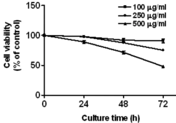
A. capillaris inhibits cell proliferation in LX2 Cells The effect of A. capillaris on cell proliferation was measured using the MTT assay. A. capillaris inhibited proliferation of LX2 cells in a dose- and time-dependent manner (Fig. 1).
Fig. 1. A. capillaris inhibited the proliferation of LX2 cells. LX2 cells were treated with various concentrations of A. capillaris (0 – 500 µg/ml) for 24, 48, and 72 h. Cell viability was determined by the MTT assay. The data represent the mean ± SD of triplicate samples. *p < 0.05 and **p < 0.01 compared to control.
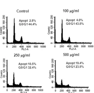
A. capillaris induces apoptosis during cell cycle progression of LX2 Cells
Following A. capillaris treatment for 72 h, flow cytometry showed a dose-dependent increase in the apoptosis phase and a decrease in the G1 phase of the cell cycle of LX2 cells (Fig. 2).
A. capillaris induces caspase-3 activation in LX2 Cells
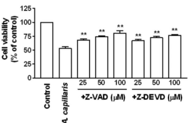
Induction of apoptosis by A. capillaris was evaluated using a cell death detection ELISA. LX2 cells were treated with different concentrations (50–500 µg/ml) of A. capillaris for 72 h. Apoptosis was increased in a dose- and time-dependent manner with A. capillaris (Fig. 3). Caspase-3 activity also increased in a dose- and time-dependent manner due to A. capillaris treatment (Fig. 4). Pretreatment of cells with the pan-caspase inhibitor Z-VAD-FMK and the caspase-3 inhibitor Z-DEVD-FMK increased A. capillaris-induced proliferation as measured by the MTT assay (Fig. 5).
A. capillaris causes changes in the expression of pro- and anti-apoptotic mRNA in LX2 Cells The levels of Bcl-2 and Bax mRNAs were examined Fig. 2. A. capillaris increased the percentage of LX2
cells in the apoptosis phase of the cell cycle. LX2 cells were cultured with various concentrations of A. capillaris (0 – 500 µg/ml) for 72 h. Cells were harvested, treated with RNAsin, and stained with propidium iodide. The DNA content was analyzed by flow cytometry. The percentages of cells in apoptosis and G0/G1 phases are indicated.
Fig. 3. A. capillaris induced apoptosis in LX2 cells. LX2 cells were cultured with various concentrations of A. capillaris (0 – 500 µg/ml) for 72 h. Apoptotic cells were measured using a cell death detection ELISA. The data represent the mean ± SD of three independent exper-iments. *p < 0.05 and **p < 0.01 compared to control.
Fig. 4. A. capillaris enhanced caspase-3 activation in LX2 cells. Cells were cultured with A. capillaris in a dose- and time-dependent manner. The enzymatic activity of caspase proteases was measured by a caspase colorimetric assay. The data represent the mean ± SD of three independent experiments. *p < 0.05 and **p < 0.01 compared to control.
by real-time PCR. The mRNA level of Bcl-2 decreased, while that of Bax increased in a time- and dose-dependent manner in LX2 cells treated with A. capillaris (Fig. 6).
A. capillaris inhibits the RAF/MEK/ERK signaling pathway in LX2 Cells
RAF kinases are best known as key regulators of the MEK/ERK cascade. Changes in the phos-phorylation levels of key proteins in the RAF/ MEK/ERK pathway in LX2 cells were determined by Western blot analysis to evaluate the effect of A. capillaris (Fig. 7). High concentrations (250 µg/ml, 500 µg/ml) of A. capillaris inhibited phos-phorylation of MEK and ERK, and decreased the production of Mcl-1 protein in LX2 cells (Fig. 7).
DISCUSSION
A. capillaris is known to play roles in many cellular events, such as cell proliferation, differentiation, and apoptosis. We found that A. capillaris inhibited cell proliferation in LX2 cells, in accordance with other studies reporting that five herbs did so in a dose-dependent manner (Chor et al., 2005). Our results showed that A. capillaris increased the apoptosis phase of the cell cycle and enhanced apoptotic cell death in LX2 cells. Taken together, these data suggest that A. capillaris not only inhibits Fig. 6. Effect of A. capillaris on the expression of
pro-and anti-apoptotic mRNAs in LX2 cells. LX2 cells were cultured either with different concentrations of A. capillaris for 72 h or with 500 µg/ml A. capillaris for different times. mRNA levels were measured by real-time PCR. The crossing point of Bcl-2, Bax with β-actin was applied to the formula, 2-(target gene-β actin)
, and relative amounts were quantified. The data represent the mean ± SD of three independent samples. *p < 0.05 and **p < 0.01 compared to control.
Fig. 7. A. capillaris inhibited the RAF/MEK/ERK signaling pathway and down-regulated Mcl-1 levels in LX2 cells. LX2 cells were cultured with different concentrations of A. capillaris for 72 h. The cells were lysed and 30 µg of soluble protein was separated by electrophoresis on a SDS-PAGE gel. Protein was detected by Western blot analysis.
Fig. 5. A. capillaris-induced apoptosis was restrained by caspase inhibitors in LX2 cells. LX2 cells were treated with A. capillaris (500 µg/ml) in the presence or absence of Z-VAD-FMK (25 – 100 µM) or Z-DEVD-FMK (25 – 100 µM) for 72 h. Cell viability was measured using the MTT assay. Values are the mean ± SD of three independent experiments. *p < 0.05 and **p < 0.01; A. capillaris-treated cells compared to A. capillaris and caspase inhibitor-treated cells.
LX2 cell growth and blocks cell cycle progression at the G1 phase, but also induces apoptosis. A previous study reported a decrease in the number of the HSC cell line T6 in the G1 phase and an accumulation of cells in the sub-G0/G1 phase after incubation with herbal extracts; Salvia miltiorrhiza demonstrated the strongest apoptotic effects of the five herbs used (Chor et al., 2005).
To investigate the molecular mechanism underlying the apoptosis of LX2 cells, we assessed caspase activity during A. capillaris-treatment. Caspases are central mediators in the process of apoptosis. We found that treatment of LX2 cells with A. capillaris increased intracellular caspase-3 activity. This finding was confirmed by experiments using the pan-caspase inhibitor Z-VAD-FMK and the caspase-3-specific inhibitor Z-DEVD-FMK, which enhanced A. capillaris-mediated cell proliferation. These results suggest that A. capillaris-induced apoptosis of LX2 cells is mediated by caspase activation.
We investigated the levels of pro- and anti-apoptotic mRNA in LX2 cells treated with A. capillaris and found enhanced expression of pro-apoptotic Bax, and reduced expression of anti-apoptotic Bcl-2. These results suggest that both decreased Bcl-2 expression and increased Bax expression are mediating the apoptotic activation of A. capillaris. In HSCs treated with inchin-ko-to (TJ-135), an increased expression of p53, a decreased expression of Bcl-2, and a decreased amount of phosphorylated Akt and Bad were observed. Hepatic stellate cell apoptosis is involved in the mechanism of spontaneous resolution of rat hepatic fibrosis; TJ-135 induced HSC apoptosis and experimental hepatic fibrosis in rats (Ikeda et al., 2006). Apoptosis induced by aloe emodin (a hydroxyanthraquinone present in aloe vera leaves) in rat hepatic stellate cells transformed by simian virus 40 (t-HSC/Cl-6) resulted in an increase in the expression of the Bax protein, while the expression of Bcl-2 was unchanged. Aloe emodin treatment caused
apparent DNA fragmentation and the activation of capase-3 and caspase-9. These data indicate an increased ratio of Bax/Bcl-2 expression may be associated with aloe emodin-induced apoptosis, and that caspase-3 and caspase-9 activation and the increase in Bax expression are closely linked (Lian et al., 2005).
We demonstrated that high concentrations of A. capillaris (250 µg/ml, 500 µg/ml) down-regulated the anti-apoptotic protein Mcl-1 and inhibited MEK and ERK phosphorylation in LX2 cells. This indicates that A. capillaris is able to inhibit RAF kinase and thus block the MEK/ERK signaling pathway in LX2 cells. These results suggest that A. capillaris down-regulates Mcl-1 protein levels through a MEK/ERK-independent pathway. The mechanism underlying A. capillaris-induced Mcl-1 down-regulation may be at the level of translation. Mcl-1, an anti-apoptotic member of the Bcl-2 family, has been shown to be an important factor for apoptosis resistance in hepatocellular carcinomas (Fleischer et al., 2006; Sieghart et al., 2006).
The MAPK pathway is a key signaling mechanism that regulates many cellular functions such as cell growth, transformation, and apoptosis (Rubinfeld and Seger, 2005). The MAPKs can mediate apoptotic signaling induced by antineoplastic agents (Fan and Chambers, 2001), and ERK-dependent activation of the Bcl-2 family can have a pro-apoptotic effect on HSCs (Saile et al., 2004). ERK, c-Jun NH1-terminal kinase, and p38 MAP kinase constitute three major subfamilies of MAP kinases that appear to mediate cellular responses, including proliferation, differentiation, and apoptosis (Davis, 1993). Induction of cell death can be mediated via activation of ERK1 or 2 (Stanciu et al., 2000; Wang et al., 2000).
In conclusion, we demonstrated that A. capillaris inhibits the growth of LX2 cells by caspase activation and induction of apoptosis. The study of the growth-inhibitory effect of A. capillaris in LX2 cells
will contribute to an increased understanding of the effects of natural herbs on liver diseases, and provide information about the involvement of critical intracellular signaling pathways. The inability of enforced activation of MEK/ERK to prevent Mcl-1 down-regulation suggests that A. capillaris down-regulates Mcl-1 protein levels through a MEK/ERK-independent pathway. These results suggest that A. capillaris is a candidate for further in vitro and in vivo study.
REFERENCES
Basu A, Saito K, Meyer K, Ray RB, Friedman SL, Chang YH, Ray R. (2006) Stellate cell apoptosis by a soluble mediator from immortalized human hepatocytes. Apoptosis 11, 1391-1400.
Cales P. (1998) Apoptosis and liver fibrosis: antifibrotic strategies. Biomedicine and Pharmacotherapy 52, 259-263.
Chor SY, Hui AY, To KF, Chan KK, Go YY, Chan HL, Leung WK, Sung JJ. (2005) Anti-proliferative and pro-apoptotic effects of herbal medicine on hepatic stellate cell. Journal of Ethnopharmacology 100, 180-186.
Davis RJ. (1993) The mitogen-activated protein kinase signal transduction pathway. Journal of Biological Chemistry 268, 14553-14556.
Fan M, Chambers TC. (2001) Role of mitogen-activated protein kinases in the response of tumor cells to chemotherapy. Drug Resistance Updates 4, 253-267. Fleischer B, Schulze-Bergkamen H, Schuchmann M,
Weber A, Biesterfeld S, Muller M, Krammer PH, Galle PR. (2006) Mcl-1 is an anti-apoptotic factor for human hepatocellular carcinoma. International Journal of Oncology 28, 25-32.
Friedman SL. (1993) The cellular basis of hepatic fibrosis. Mechanisms and treatment strategies. New England Journal of Medicine 328, 1828-1835.
Friedman SL. (2000) Molecular regulation of hepatic fibrosis, an integrated cellular response to tissue injury. Journal of Biological Chemistry 275, 2247-2250. Friedman SL. (2003) Liver fibrosis - from bench to
bedside. Journal of Hepatology 38, S38-S53.
Gong W, Pecci A, Roth S, Lahme B, Beato M, Gressner AM. (1998) Transformation-dependent susceptibility
of rat hepatic stellate cells to apoptosis induced by soluble Fas ligand. Hepatology 28, 492-502.
Gressner AM. (1998) The cell biology of liver fibrogenesis - an imbalance of proliferation, growth arrest and apoptosis of myofibroblasts. Cell and Tissue Research 292, 447-452.
Hu YQ, Tan RX, Chu MY, Zhou J. (2000) Apoptosis in human hepatoma cell line SMMC-7721 induced by water-soluble macromolecular components of Artemisia capillaris Thunberg. Japanese Journal of Cancer Research 91, 113-117.
Ikeda H, Nagashima K, Yanase M, Tomiya T, Arai M, Inoue Y, Tejima K, Nishikawa T, Watanabe N, Kitamura K, Isono T, Yahagi N, Noiri E, Inao M, Mochida S, Kume Y, Yatomi Y, Nakahara K, Omata M, Fujiwara K. (2006) The herbal medicine inchin-ko-to (TJ-135) induces apoptosis in cultured rat hepatic stellate cells. Life Sciences 78, 2226-2233.
Iredale JP. (2001) Hepatic stellate cell behavior during resolution of liver injury. Seminars in Liver Disease 21, 427-436.
Issa R, Williams E, Trim N, Kendall T, Arthur MJ, Reichen J, Benyon RC, Iredale JP. (2001) Apoptosis of hepatic stellate cells: involvement in resolution of biliary fibrosis and regulation by soluble growth factors. Gut 48, 548-557.
Koo HN, Hong SH, Jeong HJ, Lee EH, Kim NG, Choi SD, Ra KW, Kim KS, Kang BK, Kim JJ, Oh JG, Kim HM. (2002) Inhibitory effect of Artemisia capillaris on ethanol-induced cytokines (TNF-α, IL-1α) secretion in HepG2 cells. Immunopharmacology and Immunotoxicology 24, 441-453.
Lian LH, Park EJ, Piao HS, Zhao YZ, Sohn DH. (2005) Aloe emodin-induced apoptosis in t-HSC/Cl-6 cells involves a mitochondria-mediated pathway. Basic and Clinical Pharmacology and Toxicology 96, 495-502.
Pan J, Liu G, Liu H, Qiu Z, Chen L. (1998) Effects of Artemisia capillaris on blood glucose and lipid in mice. Zhong Yao Cai. 21, 408-511.
Rubinfeld H, Seger R. (2005) The ERK cascade: a prototype of MAPK signaling. Molecular Biotechnology 31, 151-174.
Saile B, DiRocco P, Dudas J, El-Armouche H, Sebb H, Eisenbach C, Neubauer K, Ramadori G. (2004) IGF-I induces DNA synthesis and apoptosis in rat liver hepatic stellate cells (HSC) but DNA synthesis and
proliferation in rat liver myofibroblasts (rMF). Laboratory Investigation. 84, 1037-1049.
Sieghart W, Losert D, Strommer S, Cejka D, Schmid K, Rasoul-Rockenschaub S, Bodingbauer M, Crevenna R, Monia BP, Peck-Radosavljevic M, Wacheck V. (2005) Mcl-1 overexpression in hepatocellular carcinoma: a potential target for antisense therapy. Journal of Hepatology 44, 151-157.
Stanciu M, Wang Y, Kentor R, Burke N, Watkins S, Kress G, Reynolds I, Klann E, Angiolieri MR, Johnson JW, DeFranco DB. (2000) Persistent activation of ERK contributes to glutamate-induced oxidative toxicity in a neuronal cell line and primary cortical neuron cultures. Journal of Biological Chemistry 275,
12200-12206.
Wang X, Martindale JL, Holbrook NJ. (2000) Requirement for ERK activation in cisplatin-induced apoptosis. Journal of Biological Chemistry 275, 39435-39443.
Wang XM, Yu DM, McCaughan GW, Gorrell MD. (2005) Fibroblast activation protein increases apoptosis, cell adhesion, and migration by the LX-2 human stellate cell line. Hepatology 42, 935-945.
Xu L, Hui AY, Albanis E, Arthur MJ, O’Byrne SM, Blaner WS, Mukherjee P, Friedman SL, Eng FJ. (2005) Human hepatic stellate cell lines, LX-1 and LX-2: new tools for analysis of hepatic fibrosis. Gut 54, 142-151.