45(4) : 288∼ 293 (2014)
288
Clitocybin A의 모유두 세포증식 효능
강정일1·김민경1·유은숙1·유익동2·강희경1*
1제주대학교 의학전문대학원 약리학교실, 2한국생명공학연구원 화학생물연구센터
Effect of Clitocybin A on the Proliferation of Dermal Papilla Cells
Jung-Il Kang1, Min-Kyoung Kim1, Eun-Sook Yoo1, Ick-Dong Yoo2, and Hee-Kyoung Kang1
*
1Department of Medicine, School of Medicine, Jeju National University, 102 Jejudaehakno, Jeju 690-756, Korea
2Chemical Biology Research Center, Korea Research Institute of Bioscience and Biotechnology, Daejeon 305-806, Korea
Abstract − The present study was conducted to evaluate the hair growth-promoting effect of Clitocybin A from mushroom Cli- tocybe aurantiaca with dermal papilla cells (DPCs), which play important roles in the regulation of hair cycle. Clitocybin A significantly increased the proliferation of immortalized rat vibrissa DPCs. Flow cytometry analysis revealed that Clitocybin A promoted cell-cycle progression through G0/G1 to S phase in immortalized rat vibrissa DPCs. In addition, Clitocybin A increased the level of cell cycle proteins such as cyclin D1, phospho-pRB, and phospho-CDK2. To elucidate the molecular mechanisms of Clitocybin A on the proliferation of DPCs, we examined the activation of wnt/β-catenin signaling which is known to regulate hair follicle development, differentiation and hair growth. Clitocybin A activated wnt/β-catenin signaling via the increase of phospho(ser552)-β-catenin, phospho(ser675)-β-catenin and phospho(ser9)-GSK3β. Furthermore, Clitocybin A markedly increased the activation of extracellular signal-regulated kinase (ERK). These results suggest that the Clitocybin A may induce hair growth by proliferation of DPCs via cell-cycle progression as well as the activation of Wnt/β-catenin signaling and ERK pathway.
Key words− Hair growth, RAF, Dermal papilla cells, Cell cycle, Wnt/β-catenin, ERK
탈모는 신체 또는 머리부위의 모발이 가늘어지며 감소하 는 증상을 말하며, 모낭의 축소화 및 성장기 모낭의 감소 등 의 특징을 수반하는 것으로 알려져 있다.1,2)탈모는 남성과 여성 모두에서 발생하며 미적 관심의 증대 및 탈모 인구의 증가로 치료제의 개발 및 그 기전에 대한 연구가 활발히 진 행되고 있지만, 탈모의 원인이 무엇인지는 정확히 알려져 있지 않다.1,3)현재 탈모방지 및 모발성장을 촉진하는 약물 로 미국식품의약국(Food and Drug Administration, FDA)의 승인을 받은 것으로서 finasteride와 minoxidil이 있다.
Finasteride는 type II 5α-reductase의 활성을 억제시키는 물 질로서 전립선 비대증 치료제로 개발되었으며,minoxidil은 고혈압 치료를 위한 혈관확장제로 개발되었으나, 두 약물 모 두 모발성장을 촉진함이 알려져 발모제로 이용되고 있다.4,5) Minoxidil의 작용기전은 명확히 밝혀지지 않았으나, Wnt/β- catenin 경로의 활성화6) 및 KATP 채널 개방효과7,8) 등이 발 모효과를 유도하는 것으로 생각되고 있다. Minoxidil은 또
한 모낭의 기저에 위치한 중배엽 유래의 모유두 세포의 apoptosis를 저해하여 모유두 세포를 증식함이 알려져 있다.9) 모발성장, 모발재생, 모낭 줄기세포의 활성화 및 hair germ cell의 증식 등의 과정에 Wnt/β-catenin 신호전달이 중요한 역할을 함이 알려져 있다.10-12) Wnt ligand가 수용체에 결합 하면 세포질 내의 β-catenin분해가 방지되고 세포핵으로 β- catenin의 이동이 증가하여 타겟 유전자의 발현을 조절하 게 된다. 특히, protein kinase A(PKA), Akt 및 glycogen synthase kinase-3β(GSK3β) 등의 활성화는 Wnt/β-catenin 신호전달 경로를 활성화시킴이 보고되어 있다.13-15)
모유두세포는 모낭내의 여러 종류의 상피세포들과 상호 작용을 하며 모낭의 형성, 모발의 재생 및 모발의 성장에 중요한 역할을 함이 알려져 있다.16) 특히 모유두세포의 성 장 증식 및 apoptosis 억제는 모발의 성장기를 유지하는데 필요함이 보고되어 있다.6,8,9) 세포의 증식은 G0/G1, S 및 G2/M phase로 구성되는 세포주기의 진행과 밀접하게 관련 되며, 이러한 세포주기에 관련된 단백질에는 cyclins, cyclin- dependent kinases(CDKs) 및 CDK 억제자 같은 단백질들이
*교신저자(E-mail): [email protected] (Tel): +82-64-754-3846
있다.17)이러한 세포주기 단백질 중 cyclin D1은 세포주기 중 G0/G에서 S phase로 진행될 때에 증가함이 알려져 있
고17-20), Wnt/β-catenin 신호전달 경로의 target gene의 하나
로 밝혀져 있다21). 한편, mitogen-activated protein kinases (MAPK) 중 extracellular signal-regulated kinases(ERK) 신 호전달 경로는 세포성장에서 필수적인 역할을 함이 잘 알 려져 있고, ERK의 활성화가 minoxidil에 의한 모유두세포 의 apoptosis 저해과정에 관여함을 보고되어 있다.9)
Clitocybe aurantiaca는 꾀꼬리큰버섯으로 불리는 국내 자 생버섯의 일종으로 9종의 Clitocybe속 균주 중 하나이다.22) Clitocybin A는 C. aurantiaca 배양액으로부터 추출 정제된 iso-indolinone계 단일 물질이다.23) Clitocybin A는 산소 라 디칼을 소거하는 항산화 작용, apoptosis 억제, 세포노화 억 제 및 세포의 이상증식 억제 등 다양한 효능을 나타냄이 보 고되어 있다.23-25) 육상식물 및 해조류에서 육모 효능 연구 는 활발히 진행되고 있으나, 버섯 및 그 유래 물질이 육모 및 탈모방지에 미치는 영향에 대한 연구는 아직까지 미미 한 실정이다.
본 연구에서는 육모효능에 중요한 역할을 하는 모유두 세 포를 이용하여 우리나라에 서식하는 국내자생버섯에서 유 래된 Clitocybin A의 육모 효능을 조사하여 이들을 탈모방 지제 및 탈모 치료제로 이용할 수 있는 근거를 마련하고자 하였다.
재료 및 방법
시료 − 실험재료인 Clitocybin A는 한국생명공학연구원 화 학생물연구센터에서 공급받아 사용하였다. 시료는 dimethyl sulfoxide(DMSO)로 녹여 실험에 사용하였으며, DMSO의 최종 농도는 0.2%를 초과하지 않도록 하였다.
모유두 세포의 배양 − 흰쥐 수염에서 분리된 모유두 세포 를 불멸화한 세포(Rat vibrissa immortalized dermal papilla cell)26)는 ㈜아모레퍼시픽 피부과학연구소로부터 제공받았 다. 모유두 세포를 100 units/ml penicillin-100 µg/ml strepto- mycin(Gibco Inc, NY, USA)과 10% heat-inactivated fetal bovine serum(FBS; Gibco Inc, NY, USA)이 함유된 DMEM (Hyclone Inc, UT, USA) 배지를 사용하여 37oC, 5% CO2 항온기에서 배양하였으며, 3일에 한 번씩 계대배양 하였다.
MTT assay − 모유두 세포의 증식은 3-(4,5-dimethylthia- zol-2-yl)-2,5-diphenyltetrazolium bromide(MTT) assay를 이 용하여 측정하였다.27) 모유두 세포(1.0×10⁴cells/ml)를 1%
FBS를 포함한 DMEM 배지에 혼탁하여 96 well plate에 넣 고 24시간 배양 후 Clitocybin A(0.001, 0.01, 0.1, 1, 10 및 50µM)를 처리하였다. 양성 대조군으로 minoxidil(Sigma, MO, USA)을 10 µM의 농도로 처리하였다. 4일 동안 배양 한 후 2 mg/ml 농도의 MTT(Sigma, MO, USA) 50 µl씩을
well에 첨가하고 4시간 동안 반응시켰다. 상층액은 제거하고 dimethyl sulfoxide(DMSO) 200µl을 가하여 침전물을 용해 시킨 후 microplate reader(Amersham Pharmacia Biotech, NY, USA)를 사용하여 540 nm에서 흡광도를 측정하였다. 각 시료군에 대한 평균 흡광도 값을 구하였으며, 대조군의 흡 광도 값과 비교하여 증식정도를 조사하였다.
세포주기 분석 − 모유두 세포는 1% FBS를 포함하는 DMEM 배지로 24시간 동안 배양한 후에 Clitocybin A (0.001, 0.01, 0.1µM) 또는 10 µM minoxidil을 처리한 다음 24시간 동안 배양하였다. 세포를 수확하고, PBS로 세척한 다음, -20oC에서 30분 동안 70% ethanol로 고정하였다.
RNase A(50µg/ml) 처리하고, propidium iodide(PI)로 염색 한 다음 FACScan flow cytometer(Becton-Dickinson, San Jose, CA, USA)로 세포주기를 분석하였다.
Western blot analysis − 모유두 세포를 1% FBS를 포함 하는 DMEM 배지로 24시간 동안 배양한 후에 Clitocybin A(0.001, 0.01, 0.1µM) 또는 10 µM minoxidil을 처리한 다 음 24시간 동안 배양하였다. 세포를 PBS로 2회 세척한 후 200µl의 lysis buffer [50 mM Tris-HCl(pH 7.5), 150 mM NaCl, 2 mM EDTA, 1 mM EGTA, 1 mM NaVO3, 10 mM NaF, 1 mM dithiothreitol(DTT), 1 mM phenylmethylsulfonyl- fluoride(PMSF), 25µg/ml aprotinin, 25 µg/ml leupeptin and 1% NP-40]를 첨가한 다음, 4oC에서 30분 동안 lysis 시 켰다. Cell lysate는 15,000 rpm에서 15분 동안 원심분리하 여 상층액을 얻었고 실험에 사용전까지 -20oC에 보관하였 다. 단백질 농도는 bovine serum albumin(BSA)를 표준물질 로 사용하여 Bradford method에 의하여 정량하였다.28) 20- 30µg의 lysate를 8-12% Sodium dodecyl sulfate polyacryl- amide gel electrophoresis(SDS-PAGE)로 변성 분리한 다음 polyvinylidene fluoride(PVDF) membranes(Bio-Rad, Hercules, CA, USA)으로 200 mA에서 2시간 동안 transfer하였다. 그 리고 membrane의 blocking은 5% nonfat dried milk가 함유 된 Tween-20-TBS(T-TBS)(50 mM Tris, pH 7.6, 150 mM NaCl, 0.1% Tween-20) 용액에서 2시간 동안 실시하였다.
Membrane은 여러 단백질의 발현을 조사하기 위해 Cyclin D1(1:1000), phospho-CDK2(1:1000), CDK2(1:1000), Cyclin E(1:2000), phospho(ser780)-pRB(1:1000), phospho-ERK1/2 (1:1000), phospho(ser552)-β-catenin(1:1000), phospho(ser675)- β-catenin(1:1000), β-catenin(1:2000), phospho(ser9)-GSK3β (1:1000), GSK3β(1:1000) and β-actin(1:5000)에 대한 primary antibody로 4oC에서 overnight 결합시켰다. Secondary anti- body는 Horse Radish Peroxidase(HRP)가 결합된 anti-rabbit IgG, anti-mouse IgG를 1:5000으로 희석하여 1시간 동안 상 온에서 반응을 진행하였다. 그 다음 T-TBS로 membane을 3 회 세척한 후, ECL 기질(Intron, Seoul, Korea)로 1분 동안 반응시킨 다음 X-ray film(AGFA, Mortsel, Belgium)에 감
광하였다. Band intensities는 NIH Image 소프트웨어를 이 용하여 정량하였다(http://rsb.info.nih.gov/ij/).
통계분석 − 모든 측정결과는 평균±표준편차로 나타내었으 며, 통계학적 유의성 검정은 student's t-test으로 검정하였으 며, p-value가 0.05 이하일 경우 유의성을 인정하였다. 통계 처리는 Sigma Stat software(Jandel Scientific Software, USA)를 사용하였다.
실험결과 및 고찰
Clitocybin A가 모유두 세포의 성장증식 효능이 있는지 불 멸화된 모유두 세포를 사용하여 조사하였다. 모유두 세포는 모낭의 기저에 위치하는 중배엽 유래 세포로써, 모기질 세 포(matrix cells)와의 상호작용, hair germ을 통한 stem cell 활성화 및 모발의 재생에 중요한 작용을 한다고 알려져 있 다.12,16,29,30) Clitocybin A을 0.001, 0.01, 0.1, 1, 10 및 50 µM 농도로 처리하였을 때, 대조군(100.0±7.1)%에 비하여 각각 116.7±11.3%(p<0.001), 115.3±11.7%(p<0.001), 112.6
±11.5%(p<0.01), 110.5±7.9%(p<0.01), 100.3±6.5% 및 80.2
±6.0% 정도 모유두 세포의 증식이 증가하였다. Clitocybin A(0.001 and 0.01µM)의 모유두 세포의 증식 효과는 양성 대조물질로 사용한 10 µM minoxidil의 115.7±9.9%(p<
0.001)와 비슷한 증식 증가 효과를 나타내었다(Fig. 1). 반면 50µM 이상의 농도에서 Clitocybin A는 세포성장을 억제함 을 확인하였다(Fig. 1). 이전 연구에서 Clitocybin A는 5 µM 이상의 농도에서 platelet-derived growth factor(PDGF) 에 의해 유도된 혈관평활근 세포의 증식을 억제함이 밝혀졌 다25). 즉, Clitocybin A는 세포성장을 억제하지 않는 낮은 농도에서 minoxidil 보다 높은 세포증식 효능을 나타냄을 확
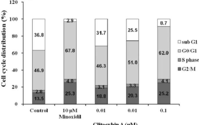
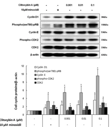
인할 수 있었다(Fig. 1). 이러한 결과를 통하여 Clitocybin A 가 모유두 세포의 증식을 통해 육모 효능을 나타낼 수 있음 을 확인하였다. G0/G1, S및 G2/M phase로의 cell cycle의 진행과정은 포유류 세포 증식의 주요과정이며,18-20) Clitocybin A가 cell cycle의 진행을 유도하는지 알아보기 위해 DNA에 결합하는 형광물질인 PI를 처리하여 유세포분석기로 조사 하였다. 그 결과, Clitocybin A 처리군에서 대조군에 비해 sub G1 phase 세포가 감소하였고 G1 phase 세포 및 G2/M phase 세포가 증가함을 확인할 수 있었다(Fig. 2). Cell cycle 의 진행은 cyclin E/CDK2 complex의 활성화, cyclin D1 증 가 및 pRB의 인산화 증가등의 단백질 발현을 동반함이 알 려져 있다.18-20) 이러한 cell cycle 진행과 관련된 단백질의 발 현이 Fig. 2의 결과와 일치하게 변화화하는지 알아보기 위 해, cyclin D1, cyclin E, phospho-CDK2, CDK2, phospho (ser780)-pRB의 발현을 조사하였다. Fig. 3에서 보는 바와 같이, 모유두 세포에 24시간 동안 Clitocybin A(0.001, 0.01 및 0.1 µM)를 처리하였을 때, cyclin D1, phospho-CDK2 및 phospho(ser780)-pRB의 발현이 유의하게 증가함을 확인할 수 있었다. 그러나 Clitocybin A 처리는 Cyclin E 및 CDK2 의 발현에는 영향을 미치지 않음을 관찰할 수 있었다.
Minoxidil를 처리하였을 때도 역시 Clitocybin A와 비슷한 결과를 확인할 수 있었다(Fig. 3). Clitocybin A가 모유두세 포의 성장 증식을 유도하는 분자적 기전을 밝히기 위해 Wnt/
β-catenin 신호 전달 경로에 관하여 조사하였다. Wnt/β- catenin 신호전달 경로는 모발성장, 세포증식 조절등의 과정 에서 중요한 역할을 하며,6,10,31) PKA, Akt 및 GSK3β 등의 다양한 인자에 의해 조절됨이 알려져 있다. Akt의 활성화 는 β-catenin의 인산화(ser552) 및 GSK3β의 인산화(ser9) 를 유도하며, PKA는 β-catenin의 인산화(ser552 및 ser675)
Fig. 1. The effect of Clitocybin A on the proliferation of DPCs.
Immortalized DPCs (1.0×10⁴ cells/ml) were plated in 96 well plates. Immortalized DPCs were treated with Clitocybin A (0.001, 0.01, 0.1, 1, 10 and 50µM). Stimulation with minox- idil served as a positive control. Cell proliferation was mea- sured using a MTT assay for 4 days. All experiments were per- formed in triplicate. Data are presented as the mean±the S.D.
*p<0.05, **p<0.01, ***p<0.001 vs. vehicle treated control.
Fig. 2. The effect of Clitocybin A on cell cycle progression in DPCs. Immortalized DPCs were stained with PI after 24 h exposure of Clitocybin A (0.001, 0.01 and 0.1 µM) or minox- idil (10 µM). The cell cycle was analyzed by flow cytometry.
The percentages of cells in each phase (subG1, G1, S and G2/
M) were quantified by CellQuest software.
를 유도하여 세포질 내에서 β-catenin의 분해를 저해하고 안정화를 증가시킨다.13,15,32) 그로 인해 β-catenin세포핵으로 의 이동이 촉진되고 타겟 유전자의 발현을 조절한다.13,15,32) 24시간 동안Clitocybin A를 처리한 경우, phospho(ser552)- β-catenin, phospho(ser675)-β-catenin, phospho(ser9)-GSK3β 의 레벨이 유의하게 증가하였다. 이런 결과로 Clitocybin A 가 β-catenin 신호전달 경로를 활성화함을 확인할 수 있었 다(Fig. 4). 또한 양성대조 물질인 minoxidil도 Kwack 등의 연구결과와 비슷하게 phospho(ser552)-β-catenin, phospho (ser675)-β-catenin, phospho(ser9)-GSK3β의 레벨을 증가시 키는 것으로 β-catenin 신호전달 경로를 활성화함을 확인하 였다.8) 한편, MAPK 경로 중 하나인 ERK는 성장인자들에 의해 활성화되어 세포 생존 및 성장을 조절하는 것으로 알 려져 있으며33), ERK의 활성화는 세포주기 단백질 중 하나 인 cyclin D1의 발현을 증가시킴이 보고되었다.34) 특히, minoxidil에 의한 ERK의 활성화는 모유두세포의 apoptosis 저해와 관련됨이 알려져 있다.9) Fig. 5에서 보이는 결과와 같이 모유두세포에 Clitocybin A를 처리하였을 때 phospho- ERK1/2의 레벨이 증가하는 것으로 ERK1/2의 신호전달을 활성화 시킴을 확인하였다. 위와 같은 연구결과로부터 Clitocybin A가 cyclin D1, phospho-CDK2, phospho-pRB Fig. 3. The effect of Clitocybin A on the level of cell cycle associated proteins in DPCs. Immortalized DPCs were treated with Clitocybin A (0.001, 0.01 and 0.1 µM) or minoxidil (10 µM) for 24 h. The effects of Clitocybin A on the levels of cyclin D1, cyclin E, phospho-CDK2, CDK2 and phospho-pRB were analyzed by immunoblotting.
Fig. 4. The effects of Clitocybin A on the level of Wnt/β- catenin signaling proteins in DPCs. Immortalized DPCs were treated with Clitocybin A (0.001, 0.01 and 0.1 µM) and minoxidil (10 µM) for 24 h. The effects of Clitocybin A on the levels of phospho(ser552)-β-catenin, phospho(ser675)-β- catenin, β-catenin, phospho(ser9)-GSK3β, GSK3β were ana- lyzed by immunoblotting.
Fig. 5. The effect of Clitocybin A on the level of phospho- ERK1/2 in DPCs. Immortalized DPCs were treated with Cli- tocybin A (0.001, 0.01 and 0.1 µM) and minoxidil (10 µM) for 24 h. The expression of phospho-ERK1/2 were measured by Western blotting.
같은 세포주기 조절 단백질의 레벨을 증가시킬 뿐만 아니 라 Wnt/β-catenin 신호전달 경로 및 ERK 신호전달 경로의 활성화를 유도하여 모유두 세포의 증식증가 효능을 나타냄 을 알 수 있다.
결 론
본 연구에서는 Clitocybin A이 모발의 성장에서 중요한 역할을 하는 모유두 세포에서 cell cycle proteins, Wnt/β- catenin 및 ERK 신호전달 경로의 활성화를 유도하여 모유 두세포의 성장증식을 촉진함을 밝혔다. 이와 같은 연구결과 는 Clitocybin A가 탈모치료 및 탈모예방에 이용될 수 있는 가능성을 가지고 있다는 근거를 제시하는 것이다.
사 사
이 논문은 2014학년도 제주대학교 학술진흥연구비 지원 사업에 의하여 연구되었음.
인용문헌
1. Price, V. H. (1999) Treatment of hair loss. N. Engl. J. Med.
341: 964-973.
2. Ellis, J. A., Sinclair, R. and Harrap, S. B. (2002) Androgenetic alopecia: pathogenesis and potential for therapy. Expert. Rev.
Mol. Med. 4: 1-11.
3. Cotsarelis, G. and Millar, S. E. (2001) Towards a molecular understanding of hair loss and its treatment. Trends Mol.
Med. 7: 293-301.
4. Kaufman, K. D. and Dawber, R. P. (1999) Finasteride, a Type 2 5alpha-reductase inhibitor, in the treatment of men with androgenetic alopecia. Expert. Opin. Investig. Drugs. 8: 403- 415.
5. Kaufman, K. D. (2002) Androgens and alopecia. Mol. Cell Endocrinol. 198: 89-95.
6. Kwack, M. H., Kang, B. M., Kim, M. K., Kim, J. C. and Sung, Y. K. (2011) Minoxidil activates beta-catenin pathway in human dermal papilla cells: A possible explanation for its anagen prolongation effect. J. Dermatol. Sci. 62: 154-159.
7. Hamaoka, H., Minakuchi, K., Miyoshi, H., Arase, S., Chen, C. H. and Nakaya, Y. (1997) Effect of K+ channel openers on K+ channel in cultured human dermal papilla cells. J. Med.
Invest. 44: 73-77.
8. Shorter, K., Farjo, N. P., Picksley, S. M. and Randall, V. A.
(2008) Human hair follicles contain two forms of ATP-sen- sitive potassium channels, only one of which is sensitive to minoxidil. FASEB J. 22: 1725-1736.
9. Han, J. H., Kwon, O. S., Chung, J. H., Cho, K. H., Eun, H.
C. and Kim, K. H. (2004) Effect of minoxodil on prolif-
eration and apoptosis in dermal papilla cells of human hair follicle. J. Dermatol. Sci. 34: 91-98.
10. Ouji, Y., Yoshikawa, M., Moriya, K. and Ishizaka, S. (2007) Effects of Wnt-10b on hair shaft growth in hair follicle cul- tures. Biochem. Biophys. Res. Commun. 359: 516-522.
11. Ito, M., Yang, Z., Andl, T., Cui, C., Kim, N., Millar, S. E. and Cotsarelis, G. (2007) Wnt-dependent de novo hair follicle regeneration in adult mouse skin after wounding. Nature 447:
316-320.
12. Greco, V., Chen, T., Rendl, M., Schober, M., Pasolli, H. A., Stokes, N., Dela Cruz-Racelis, J. and Fuchs, E. (2009) A two- step mechanism for stem cell activation during hair regen- eration. Cell Stem Cell 4: 155-169.
13. Hedgepeth, C. M., Conrad, L. J., Zhang, J., Huang, H. C., Lee, V. M. and Klein, P. S. (1997) Activation of the Wnt sig- naling pathway: a molecular mechanism for lithium action.
Dev. Biol. 185: 82-91.
14. Hino, S., Tanji, C., Nakayama, K. I., and Kikuchi, A. (2005) Phosphorylation of beta-catenin by cyclic AMP-dependent protein kinase stabilizes beta-catenin through inhibition of its ubiquitination. Mol. Cell. Biol. 25: 9063-9072.
15. Monick, M. M., Carter, A. B., Robeff, P. K., Flaherty, D. M., Peterson, M. W. and Hunninghake, G. W. (2001) Lipopoly- saccharide activates Akt in human alveolar macrophages resulting in nuclear accumulation and transcriptional activity of beta-catenin. J. Immunol. 166: 4713-4720.
16. Stenn, K. S. and Paus, R. (2001) Controls of hair follicle cycling. Physiol. Rev. 81: 449-494.
17. Johnson, D. G. and Walker, C. L. (1999) Cyclins and cell cycle checkpoints. Annu. Rev. Pharmacol. Toxicol. 39: 295- 312.
18. Sherr, C. J. (1996) Cancer cell cycles. Science 274: 1672- 1677.
19. Sherr, C. J. and Roberts, J. M. (1999) CDK inhibitors: Pos- itive and negative regulators of G1-phase progression. Genes Dev. 13: 1501-1512.
20. Prall, O. W., Sarcevic, B., Musgrove, E. A., Watts, C. K. and Sutherland, R. L. (1997) Estrogen-induced activation of Cdk4 and Cdk2 during G1-S phase progression is accompanied by increased cyclin D1 expression and decreased cyclin-depen- dent kinase inhibitor association with cyclin E-Cdk2. J. Biol.
Chem. 272: 10882-10894.
21. Tetsu, O. and McCormick, F. (1999) Beta-catenin regulates expression of cyclin D1 in colon carcinoma cells. Nature 398:
422-426.
22. Han, S. K., Cho, J. W., Cho, H. J., Kim, H. J. and Lee, Y. M.
(2013) A field guide to mushrooms. 2th Ed. Korea national arboretum, Hwang TS, pp. 386-393. Geobook, Seoul 23. Kim, Y. H., Cho, S. M., Hyun, J. W., Ryoo, I. J., Choo, S. J.,
Lee, S., Seok, S. J, Hwang, J. S., Sohn, E. D., Yun, B. S., Bae, K. H. and Yoo, I. D. (2008) A new oxidant, clitocybin A, from the culture broth of Clitocybe aurantiaca. J. Antibiot.
61: 573-576.
24. Moon, E. Y., Kim, Y. H., Ryoo, I. J. and Yoo, I. D. (2009) Clitocybins, novel isoindolinone free radical scavengers, from mushroom Clitocybe aurantiaca inhibit apoptotic cell death and cellular senescence. Biol. Pharm. Bull. 32: 1689- 1694.
25. Park, E. S., Yoo, K. D., Kang, S. I., Yoo, S. H., Won, H. H., Kim, Y. H., Yoo, I. D., Yoo, H. S., Hong, J. T. and Yun, Y.
P. (2012) Clitocybin A, a novel isoindolinone, from mush- room Clitocybe aurantiaca, inhibits cell proliferation through G1 phase arrest by regulating PI3K/Akt cascade in vescular smooth muscle cells. J. Pharmacological Sci. 118: 171-177.
26. Filsell, W., Little, J. C., Stones, A. J., Granger, S. P. and Bay- ley, S. A. (1994) Transfection of rat dermal papilla cells with a gene encoding a temperature-sensitive polyomavirus large T antigen generates cell lines a differentiated phenotype. J.
Cell Sci. 107: 1761-1772.
27. Carmichael, J., DeGraff, W. G., Gazdar, A. F., Minna, J. D.
and Mitchell, J. B. (1987) Evaluation of a tetrazolium-based semiautomated colorimetric assay: assessment of chemosen- sitivity testing. Cancer Res. 47: 936-942.
28. Bradford, M. M. (1976) A rapid and sensitive method for the quantitation of microgram quantities of protein utilizing the principle of protein-dye binding. Anal. Biochem. 72: 248-254.
29. Jahoda, C. A., Horne, K. A. and Oliver, R. F. (1984) Induc-
tion of hair growth by implantation of cultured dermal papilla cells. Nature 311: 560-562.
30. Horne, K. A., Jahoda, C. A. and Oliver, R. F. (1986) Whisker growth induced by implantation of cultured vibrissa dermal papilla cells in the adult rat. J. Embryol. Exp. Morphol. 97:
111-124.
31. Wangefjord, S., Brändstedt, J., Ericson Lindquist, K., Nodin, B., Jirström, K. and Eberhard, J. (2013) Associations of beta- catenin alterations and MSI screening status with expression of key cell cycle regulating proteins and survival from col- orectal cancer. Diagn. Pathol. 8: 10.
32. Brudvik, K. W., Paulsen, J. E., Aandahl, E. M., Roald, B. and Taskén, K. (2011) Protein kinase A antagonist inhibits β-cate- nin nuclear translocation, c-Myc and COX-2 expression and tumor promotion in Apc(Min/+) mice. Mol. Cancer. 10: 149.
33. Chuang, S. M., Wang, I. C. and Yang, J. L. (2000) Roles of JNK, p38 and ERK mitogen-activated protein kinases in the growth inhibition and apoptosis induced by cadmium. Car- cinogenesis 21: 1423-1432.
34. Lavoie, J. N., L'Allemain, G., Brunet, A., Muller, R. and Pouyssegur, J. (1996) Cyclin D1 expression is regulated pos- itively by the p42/p44MAPK and negatively by the p38/
HOGMAPK pathway. J. Biol. Chem. 271, 20608-20616.
(2014. 11. 28 접수; 2014. 12. 22 심사;
2014. 12. 24 게재확정)