Mitochondrial Respiration Is Required
for Activation of ERK1/2 and Caspase-3
in Human Eosinophils Stimulated With
Hydrogen Peroxide
YA Lee, MH Shin
Department of Environmental Medical Biology, Institute of Tropical Medicine, and Brain Korea 21 Project for
Medical Science, Yonsei University College of Medicine, Seoul, Korea
■ Abstract
Background: Eosinophils are important effector cells in the pathogenesis of allergic diseases such as bronchial asthma. Oxidative stress
in the form of cellular reactive oxygen species (ROS) has been implicated in the pathogenesis of several allergic diseases. Recently, it has become evident that mitochondrial-derived ROS are important transducers of apoptosis and intracellular signaling. In this study, we investigated the role of mitochondrial ROS in the activation of extracellular signal-regulated kinases (ERK) 1 and 2–mitogen-activated protein kinase (MAPK) and caspase-3 in human eosinophils stimulated with H2O2.
Methods: Human eosinophils were purifi ed using immunomagnetic negative selection and then stimulated with H2O2. H2O2-induced
eosinophil apoptosis was measured by staining cells with annexin V. Activation of ERK1/2 MAPK and caspases was assessed by Western blotting. Eosinophils were pretreated with rotenone, an inhibitor of the mitochondrial electron transport chain, before H2O2 was added.
Results: Treatment with 1 mM H2O2 induced externalization of phosphatidylserine (PS) and activation of caspases in eosinophils.
H2O2-triggered PS externalization and cleavage of caspase-3 were markedly inhibited by pretreatment with the mitochondrial ROS scavenger N-acetyl-L-cysteine. In addition, H2O2 strongly induced phosphorylation of ERK1/2, but not ERK5, in eosinophils. Hydrogen peroxide-triggered
activation of caspase-3 and ERK1/2 was attenuated by pretreatment with rotenone.
Conclusions: These results suggest that mitochondrial respiration is essential for activation of ERK1/2 and caspase-3 in human eosinophils
stimulated with H2O2.
Key words: Eosinophil. Hydrogen peroxide. Mitochondria. ERK1/2. Caspase-3.
■ Resumen
Antecedentes: Los eosinófi los son unas células efectoras importantes en la patogenia de las enfermedades alérgicas como el asma bronquial.
El estrés oxidativo en forma de especies reactivas de oxígeno (ERO) se ha implicado en la patogénesis de diversas enfermedades alérgicas. Recientemente, se ha hecho patente que las ERO derivadas de la mitocondria son importantes transductores de apoptosis y señalización celular. En este estudio, investigamos el papel de las ERO mitocondriales en la activación de las quinasas reguladoras de señal extracelular (ERK) 1 y 2-proteína quinasa activada por mitógenos (MAPK) y la caspasa-3 en eosinófi los humanos estimulados con H2O2.
Métodos: Se purifi caron eosinófi los humanos empleando selección negativa inmunomagnética y posteriormente estimulados con H2O2. Se midió la apoptosis de los eosinófi los inducida por H2O2 tiñendo las células con anexina V. La activación de ERK1/2 MAPK y caspases se evaluó mediante Western blot. Los eosinófi los se pretrataron con rotanona, un inhibidor de la cadena transportadora de electrones, antes de que el H2O2 se añadiera.
Resultados: El tratamiento con 1 mM de H2O2 indujo la externalización de fosfatidilserina (FS) y la activación de las caspasas en los eosinófi los. La externalización de la FS desencadenada por el H2O2 y la fragmentación de la caspasa-3 se inhibieron marcadamente
con el pretratamiento con el depurador de ERO mitocondriales NAC. Además, el H2O2 indujo marcadamente la fosforilación de ERK1/2, pero no de ERK5, en los eosinófi los. La activación desencadenada por peróxido de hidrógeno de la caspasa-3 y ERK1/2 se atenuó con el tratamiento con rotenona.
Conclusiones: Estos resultados sugieren que la respiración mitocondrial es esencial para la activación de ERK1/2 y caspasa-3 en eosinófi los
humanos estimulados con H2O2.
Introduction
Oxidative stress plays an important role in the pathogenesis of many infl ammatory diseases, including bronchial asthma [1]. In such diseases, activated infl ammatory cells respond with a burst of respiratory activity, which results in the production of large amounts of reactive oxygen species (ROS) such as H2O2 [2]. When released into infl amed tissues, these ROS may contribute to tissue injury. Indeed, it has been reported that a high level of H2O2 in exhaled breath condensate is positively associated with severity of asthma [3-5]. Therefore, the accumulation of high concentrations of ROS in infl amed tissues refl ects the underlying state of oxidative stress in asthmatic patients. Eosinophils are oxidant-sensitive cells and are regarded as key effectors in bronchial asthma [6]. Among ROS, H2O2 has emerged as a particularly important signaling molecule because of its ability to traverse the membrane, thereby gaining access to the interior of cells [7]. H2O2can cause cellular damage and oxidize protein thiol groups, thereby altering cellular functions and activating intracellular signaling molecules [8]. For example, in vitro experiments have revealed that exogenous H2O2 reverses interleukin (IL) 5-mediated survival and accelerates constitutive apoptosis of human eosinophils [9]. H2O2 can also stimulate eosinophil adhesion as an autocrine or paracrine mediator via the upregulation of ß2 integrin [6]. Many studies using various systems have also shown that exogenous H2O2 can activate
intracellular signaling molecules associated with cellular death. For example, H2O2-mediated apoptosis in mouse fi broblasts
or human neuroblastoma cells has been found to occur as a result of the activation of extracellular signal-regulated kinases (ERK) 1 and 2–mitogen-activated protein kinase (MAPK) [10,11]. H2O2-induced chondrocyte apoptosis also requires
caspase activation [12]. It is now generally accepted that mitochondria are both sensors and targets of ROS [13,14]. ROS produced by mitochondrial respiration have been shown to be closely associated with H2O2-mediated intracellular
signaling events [15,16]. Recent studies have also shown that exogenously added H2O2 can activate MAPKs such as ERK1/2
and ERK5 [10,17,18]. However, the proximal redox-sensitive targets required for H2O2-induced cell signaling are not well
understood in human eosinophils. The goal of this study, therefore, was to examine the role of mitochondrial respiration in the activation of caspases and ERK1/2 MAPK in human eosinophils stimulated with H2O2.
Materials and Methods
Reagents
We used the following reagents: pan-caspase inhibitor Z-VAD-FMK, MEK inhibitor PD98059, N-acetyl-L-cysteine (NAC), diphenyleneiodonium chloride (DPI), rotenone, and bongkrekic acid (EMD Biosciences, Madison, Wisconsin, USA); magnetic beads conjugated to anti-human CD16 mAb, phycoerythrin (PE)-labeled mouse immunoglobulin (Ig) G1, and PE-labeled annexin V (BD Pharmingen, San Diego, California, USA); 2’,7’-dichlorofl uorescein-diacetate (DCF-DA)
(Molecular Probes, Eugene, Oregon, USA); fetal calf serum (FCS) (Invitrogen, Carlsbad, California, USA) rabbit polyclonal Abs against caspase-3, caspase-9, phospho-ERK1/2 MAPK, and phospho-ERK5 (Cell Signaling Technology, Beverly, Massachusetts, USA); rabbit polyclonal Ab against ERK2 and ERK5 (Santa Cruz Biotechnology, Delaware, California, USA). Unless stated otherwise, all other reagents were obtained from Sigma-Aldrich (Saint Louis, Missouri, USA).
Isolation of Human Eosinophils
Human eosinophils were separated from the peripheral blood of healthy individuals by Percoll-gradient centrifugation and immunomagnetic negative selection using anti-human CD16 mAb conjugated with magnetic beads, as previously described by Shin et al [19]. The purity of eosinophils, as determined by Randolph staining, was consistently greater than 93%. The contaminating cells were neutrophils, and no mononuclear cells or basophils were observed.
Assay for Apoptosis
Eosinophil apoptosis was quantitated by examining the percentage of cells with annexin V binding on the cell surface. The phosphatidylserine (PS) externalization on the cells was evaluated by staining with a PE-conjugated annexin V. PE-conjugated mouse IgG1 was used as an isotype control. Flow cytometric analysis for the percentage of cells stained with annexin V was performed on at least 3 000 cells from each sample using a FACScan fl ow cytometer (BD Biosciences, San Jose, California, USA).
Eosinophil Stimulation and Preparation of Cell Lysates
Eosinophils (0.5 ⴒ106 cells/sample) were stimulated with
H2O2 at the concentrations and times indicated. Stimulation
with IL-5 (10 ng/mL) was used as a positive control. In some experiments, the cells were preincubated with specifi c inhibitors for 30 minutes before H2O2 was added. After stimulation, the reaction was stopped by a brief centrifugation. Cell pellets were lysed in 60 µL of lysis buffer containing 20 mM Tris-HCl, 60 mM ß-glycerophosphate, 10 mM EDTA, 10 mM MgCl2, 10 mM NaF, 2 mM DTT, 1 mM Na3VO4, 1 mM
APMSF, 1% NP-40, and 5 µg/mL leupeptin. After incubation on ice for 30 minutes, 20 µL of 4X sample loading buffer was added to the cell lysates, which were then boiled for 5 minutes. The samples were then centrifuged at 12 000g for 5 minutes to remove nuclear and cellular debris. The soluble supernatant fraction was then collected and stored at –20ºC or used immediately.
Sodium Dodecyl Sulfate-Polyacrylamide Gel Electrophoresis (SDS-PAGE) and Immunoblotting
Protein samples underwent 10% SDS-PAGE before being electrotransferred onto Immobilon P polyvinylidene fl uoride membranes (Millipore, Billerica, Massachusetts, USA). The membranes were blocked with 5% nonfat dry milk in Tris-buffered saline Tween at room temperature for 1 hour and then incubated with antibodies against phosphorylated proteins
(ERK1/2 and ERK5) and caspase-3 or caspase-9 at 4°C overnight. The membranes were subsequently incubated with horseradish peroxidase–conjugated anti-rabbit IgG at room temperature for 1 hour. Immunoreactivity was detected using LumiGLO (Cell Signaling Technology). Membranes were stripped using stripping buffer (100 mM 2-ME, 2% SDS, 62.5 mM Tris-HCl, pH 6.7) at 56°C for 30 minutes and reprobed with the corresponding antibodies against nonphosphorylated MAPK proteins.
Results
Stimulation of Human Eosinophils With H2O2 Causes Activation of ERK1/2, but not ERK5
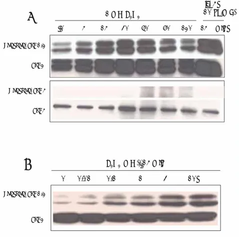
First, we investigated the effects of H2O2 on the activation of ERK1/2 and ERK5 in human eosinophils. As shown in Figures 1A and 1B, various concentrations of H2O2 (0, 0.01,
Phospho- ERK1/2 ERK2 1 m M H2O2 IL- 5 (10 ng/m L ) 0 5 15 30 60 90 120 15 min Phospho- ERK1/2 ERK2 H2O2 (m M), 15 min 0 0.01 0.1 1 3 10 P hospho- ERK5 ERK5
A
B
Figure 1. H2O2 induces phosphorylation of ERK1/2 in a concentration-dependent (A) and
time-dependent (B) manner. Eosinophils (5 ⴒ105/sample) were stimulated for 15 minutes with varying
concentrations (0.01-10 mM) of H2O2 before collection of cell lysates for immunoblotting with
phospho-specifi c ERK1/2 Ab. The membrane was reprobed with anti-ERK1/2 to control for protein
loading on the gel. In addition, eosinophils (5 ⴒ105/sample) were incubated with or without 1
mM H2O2 for specifi ed periods of time (1-120 min). Lysates from cells treated with IL-5 (10 ng/mL)
were used as a positive control. Each sample underwent 10% SDS-PAGE and blotting with anti-phospho-ERK1/2, anti-ERK, anti-phospho-ERK5, or anti-ERK5. The membrane was reprobed with anti-ERK1/2 or anti-ERK5 to control for protein loading on the gel. The fi gure is representative of 3 experiments showing similar results. ERK indicates extracellular signal-regulated kinases; IL, interleukin; SDS-PAGE, sodium dodecyl sulfate-polyacrylamide gel electrophoresis.
0.1, 1, 3, and 10 mM) induced phosphorylation of ERK1/2 in a time- and dose-dependent manner. The activity of ERK1/2 increased from 5 minutes after the addition of 1 mM H2O2, peaked at 30 minutes, and decreased thereafter. In contrast, 1 mM H2O2 failed to induce phosphorylation of ERK5 in eosinophils (Figure 1).
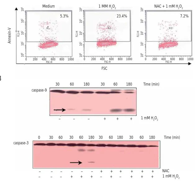
Treatment With Antioxidant NAC Inhibits H2O2-Induced PS Externalization and Activation of Caspases in Human Eosinophils
Stimulation with H2O2 has been known to accelerate constitutive apoptosis of human eosinophils. As shown in Figure 2A, 1 mM H2O2 induced translocation of PS to the outer surfaces of eosinophils, while pretreatment of the cells with the antioxidant NAC almost completely blocked H2O2-mediated PS externalization. In addition, activated cleaved forms of caspase-9 and caspase-3 were clearly detected in eosinophils
Medium 1 MM H2O2 NAC + 1 mM H2O2 Annexin-V 5.3% 23.4% 7.2% 10 4 10 3 10 2 10 1 10 0 FL1-H 0 200 400 600 800 1000 FSC-H 10 4 10 3 10 2 10 1 10 0 FL1-H 0 200 400 600 800 1000 FSC-H 10 4 10 3 10 2 10 1 10 0 FL1-H 0 200 400 600 800 1000 FSC-H R2 FSC – – – + + + 1 mM H2O2 0 30 60 30 60 180 30 60 180 30 60 180 – – – – – – + + + + + + – – – + + + – + – + + + NAC 1 mM H2O2 caspase-9
A
B
30 60 180 30 60 180 caspase-3 Time (min) Time (min)Figure 2. Antioxidant NAC treatment abolishes H2O2-induced apoptosis in human eosinophils. A, Effect of an antioxidant, NAC, on H2O2-induced PS
externalization in human eosinophils. Freshly isolated human eosinophils (5 ⴒ104/well) were preincubated with or without 1 mM NAC for 30 minutes at
37°C and stimulated with 1 mM H2O2 or medium alone for 2 hours at 37°C in a CO2 incubator. After treatment, cells were stained with FITC-conjugated
annexin V for fl ow cytometric measurement of the percentage of annexin V–positive cells on the cell surfaces. The fi gures are representative of 3 experiments
showing similar results. B, Effect of NAC on H2O2-induced caspase activation in human eosinophils. Eosinophils (5ⴒ105/sample) were pretreated with
or without 1 mM NAC for 30 minutes at 37°C and then incubated in the presence or absence of 1 mM H2O2 for 30-180 minutes at 37°C in a CO2
incubator. After incubation, whole cell lysates underwent 15% SDS-PAGE and blotting with anticaspase-9 or anticaspase-3. The fi gure is representative of 3 experiments showing similar results; FITC indicates fl uorescein isothiocyanate; NAC, N-acetyl-L-cysteine; PS, phosphatidylserine; SDS-PAGE, sodium dodecyl sulfate-polyacrylamide gel electrophoresis.
stimulated with H2O2 for 60 and 180 minutes, respectively (Figure 2B). In addition, pretreatment with NAC abrogated H2O2-induced cleavage of caspase-3 in eosinophils.
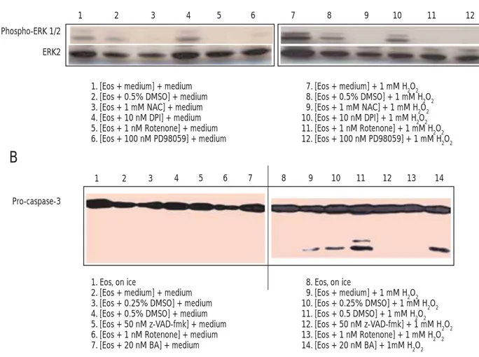
Inhibition of Mitochondrial Respiration Attenuates H2O2-Induced Activation of ERK1/2 and Caspase-3 in Human Eosinophils
To determine whether mitochondrial respiration plays a crucial role in activation of ERK1/2 and caspase-3 in eosinophils stimulated with 1 mM H2O2, cells were preincubated
with a distinct inhibitor of mitochondrial respiration before
exposure to 1 mM H2O2. As shown in Figure 3A, NAC or
rotenone completely abrogated the ability of H2O2 to activate ERK1/2 in eosinophils, as did PD98050, an ERK1/2 inhibitor. However, the control vehicle dimethyl sulfoxide and fl avin inhibitor diphenyliodonium did not eliminate H2O2-induced phosphorylation of ERK1/2. In addition, as shown in Figure 3B, inhibition of respiration at complex I with rotenone inhibited H2O2-induced cleavage of caspase-3 in eosinophils, as did direct inhibition of caspase activation with z-VAD-fmk.
A
B
Phospho-ERK 1/2
1 2 3 4 5 6 7 8 9 10 11 12
ERK2
1. [Eos + medium] + medium 2. [Eos + 0.5% DMSO] + medium 3. [Eos + 1 mM NAC] + medium 4. [Eos + 10 nM DPI] + medium 5. [Eos + 1 nM Rotenone] + medium 6. [Eos + 100 nM PD98059] + medium 7. [Eos + medium] + 1 mM H2O2 8. [Eos + 0.5% DMSO] + 1 mM H2O2 9. [Eos + 1 mM NAC] + 1 mM H2O2 10. [Eos + 10 nM DPI] + 1 mM H2O2 11. [Eos + 1 nM Rotenone] + 1 mM H2O2 12. [Eos + 100 nM PD98059] + 1 mM H2O2 1 2 3 4 5 6 7 8 9 10 11 12 13 14 Pro-caspase-3 1. Eos, on ice
2. [Eos + medium] + medium 3. [Eos + 0.25% DMSO] + medium 4. [Eos + 0.5% DMSO] + medium 5. [Eos + 50 nM z-VAD-fmk] + medium 6. [Eos + 1 nM Rotenone] + medium 7. [Eos + 20 nM BA] + medium
8. Eos, on ice 9. [Eos + medium] + 1 mM H2O2 10. [Eos + 0.25% DMSO] + 1 mM H2O2 11. [Eos + 0.5 DMSO] + 1 mM H2O2 12. [Eos + 50 nM z-VAD-fmk] + 1 mM H2O2 13. [Eos + 1 nM Rotenone] + 1 mM H2O2 14. [Eos + 20 nM BA] + 1mM H2O2
Figure 3. Effect of various pharmacologic inhibitors on the H2O2-induced activation of ERK1/2 (A) and cleavage of caspase-3 (B) in human eosinophils.
Human eosinophils were pretreated with 0.5% DMSO, 1 mM NAC, 10 µM DPI, 1 µM rotenone, or 100 µM PD98059 for 30 minutes at 37°C in a CO2
incubator. After preincubation, eosinophils were stimulated for 15 minutes with 1 mM H2O2. In addition, eosinophils (5 ⴒ 105/sample) pretreated for 30
minutes with 50 µM z-VAD-fmk (a pan-caspase inhibitor), 1 µM rotenone (a mitochondrial respiration inhibitor at complex I), 20 µM bongkrekic acid (a
mitochondrial membrane stabilizer), 0.25% DMSO (v/v), or 0.5% DMSO (v/v) were incubated for 2 hours in the absence or presence of 1 mM H2O2 at
37°C in a CO2 incubator. After incubation, whole cell lysates underwent 15% SDS-PAGE and blotting with antiphospho-ERK1/2, ERK1/2, or
anti-caspase-3. The fi gure is representative of 3 experiments showing similar results. Eos indicates eosinophils; ERK, extracellular signal-regulated kinases; DMSO, dimethyl sulfoxide; DPI, diphenyliodonium; NAC, N-acetyl-L-cysteine; SDS-PAGE, sodium dodecyl sulfate-polyacrylamide gel electrophoresis.
However, the mitochondrial membrane stabilizer, bongkrekic acid, did not show any inhibitory effect on H2O2-induced activation of caspase-3 in eosinophils, suggesting that non-PT pore-mediated release of cytochrome c is involved in H2O2 -induced activation of caspase-3.
Discussion
In this study, we found that mitochondrial respiration is required for activation of caspases and ERK1/2 in human eosinophils stimulated with H2O2. When eosinophils were treated with 1 mM H2O2 for 3 hours, the number of apoptotic cells was signifi cantly greater than the number of cells incubated
with medium alone. H2O2-induced eosinophil apoptosis
was effi ciently inhibited by pretreatment of cells with the mitochondrial ROS scavenger, NAC. In addition, H2O2-induced activation of caspase-9 and caspase-3 was dependent on both the time and dose of treatment, and NAC pretreatment clearly blocked H2O2-induced cleavage of caspase-3. Moreover, inhibition of mitochondrial respiration at complex I with rotenone inhibited H2O2-induced activation of caspase-3 in eosinophils, suggesting that H2O2-mediated caspase activation occurs downstream of mitochondrial injury. We also found that H2O2 stimulated eosinophils to induce phosphorylation of ERK1/2 but not ERK5. H2O2-mediated ERK1/2 activation was dramatically inhibited by pretreatment with NAC or rotenone. These results suggest that mitochondria-derived ROS play an important signaling role in H2O2-induced activation of caspase-3 and ERK1/2 in human eosinophils.
Although it has been reported that eosinophils contain low numbers of mitochondria that do not contribute signifi cantly to respiration, the numbers are suffi cient to induce apoptosis [20]. Our study suggests that mitochondrial respiration is important for caspase-3–mediated apoptosis in human eosinophils stimulated with H2O2. The mitochondrial electron transport chain contains redox centers that serve as the primary source of superoxide production. Although complex III is regarded as a possible site of O2- production, most of the O2- generated by intact mammalian mitochondria in vitro is produced at complex I [21]. This O2- production occurs primarily on the matrix side of the inner mitochondrial membrane. In addition to the respiratory chain, monoamine oxidase, a fl avoprotein localized on the outer mitochondrial membrane, is another important mitochondrial source of ROS [21]. In this study, monoamine oxidase did not appear to be a production site of ROS, since pretreatment with diphenyliodonium, an inhibitor of fl avoprotein, did not inhibit H2O2-triggered activation of ERK1/2 in eosinophils.
Although it is generally accepted that the ERK1/2 pathway delivers survival signals that counteract proapoptotic effects elicited by activation of JNK and p38, persistent activation of ERK1/2 by stimulation with H2O2 is closely linked to proapoptotic signaling [10]. ERK1/2 is known to be a downstream target for receptor tyrosine kinases and for Ras [22]. Another ERK1/2 activation pathway is dependent upon PI3-kinase [23]. In our study, the PI3-kinase inhibitor LY294002 signifi cantly reduced H2O2-induced phosphorylation of ERK1/2 in eosinophils (data not shown), suggesting an important role for PI3-kinase activation. We also found that a relatively high dose of H2O2 (1 mM) caused somewhat transient activation of ERK1/2, a result that is consistent with those of a previous study using the synthetic peptide WKYMVm [17]. In many studies with eosinophils, transient ERK1/2 activation has been closely linked to degranulation, migration, and adhesion [24-26]. In contrast to other reports that ERK5 is redox-sensitive [27], we found that ERK5 in eosinophils was not activated in response to H2O2.
Apoptosis is considered to be a noninfl ammatory mode of cell death because apoptotic cells are immediately and silently eliminated through phagocytosis [28]. In general, removal is thought to be accomplished through apoptosis followed by engulfment by macrophages. However, studies performed in vitro and in the airway lumen have shown that this current model of granulocyte apoptosis translates poorly to airway tissues in vivo [29]. In our study, apoptotic eosinophils were not detected, even during the resolution of airway infl ammation. In fact, even when signifi cant eosinophil apoptosis was induced in airway tissues in vivo, the number of phagocytizing cells engulfi ng apoptotic eosinophils was insuffi cient. For example, most of the apoptotic eosinophils developed in the Fas-treated airway tissue did not undergo phagocytosis, a process that leads to advanced signs of proinfl ammatory secondary necrosis [30]. Therefore, it has been suggested that accumulation of high concentrations of H2O2 in infl amed tissues causes mitochondrial ROS-dependent apoptosis in human eosinophils [31], thereby causing aggravation of eosinophil-mediated tissue infl ammation in patients with severe bronchial asthma.
In conclusion, we found evidence that mitochondria-derived ROS are required for apoptosis and activation of ERK1/2 and caspase-3 in human eosinophils stimulated with H2O2.
Acknowledgments
This study was funded by the Brain Korea 21 Project for Medical Science, Yonsei University College of Medicine.
References
1. Riedl MA, Nel AE. Importance of oxidative stress in the pathogenesis and treatment of asthma. Curr Opin Allergy Clin Immunol. 2008;8:49-56.
2. Barnes PJ. Reactive oxygen species and airway infl ammation. Free Radic Biol Med. 1990;9:235-43.
3. Emelyanov A, Fedoseev G, Abulimity A, Rudinski K, Fedoulov A, Karabanov A, Barnes PJ. Elevated concentrations of exhaled hydrogen peroxide in asthmatic patients. Chest. 2001;120:1136-9.
4. Fireman E, Shtark M, Priel IE, Shiner R, Mor R, Kivity S, Fireman Z. Hydrogen peroxide in exhaled breath condensate (EBC) vs eosinophil count in induced sputum (IS) in parenchymal vs airways lung diseases. Infl ammation. 2007;30:44-51.
5. Loukides S, Bouros D, Papatheodorou G, Panagou P, Siafakas NM. The relationships among hydrogen peroxide in expired breath condensate, airway infl ammation, and asthma severity. Chest. 2002;121:338-46.
6. Nagata M. Infl ammatory cells and oxygen radicals. Curr Drug Targets Infl amm Allergy. 2005;4:503-4.
7. Chen K, Thomas SR, Keaney JF Jr. Beyond LDL oxidation: ROS in vascular signal transduction. Free Radic Biol Med. 2003;35:117-32. 8. Droge W. Free radicals in the physiological control of cell
function. Physiol Rev. 2002;82:47-95.
9. Kankaanranta H, Giembycz M, Barnes PJ, Haddad EB,
Saarelainen S, Zhang X, Moilanen E, Lindsay MA. Hydrogen peroxide reverses IL-5 afforded eosinophil survival and promotes constitutive human eosinophil apoptosis. Int Arch Allergy Immunol. 2002;127:73-8.
10. Lee YJ, Cho HN, Soh JW, Jhon GJ, Cho CK, Chung HY, Bae S, Lee SJ, Lee YS. Oxidative stress-induced apoptosis is mediated by ERK1/2 phosphorylation. Exp Cell Res. 2003;291:251-66. 11. Ruffels J, Griffi n M, Dickenson JM. Activation of ERK1/2,
JNK and PKB by hydrogen peroxide in human SH-SY5Y neuroblastoma cells: role of ERK1/2 in H2O2-induced cell death. Europ J Pharmacol. 2004;483:163-73.
12. Lo MY, Kim HT. Chondrocyte apoptosis induced by hydrogen peroxide requires caspase activation but not mitochondrial pore transition. J Orthop Res. 2004;22:1120-5.
13. Simon HU, Haj-Yehia A, Levi-Schaffer F. Role of reactive oxygen species (ROS) in apoptosis induction. Apoptosis. 2000;5:415-8.
14. Andreyev AY, Kushnareva YE, Starkov AA. Mitochondrial metabolism of reactive oxygen species. Biochemistry (Moscow). 2005;70:246-64.
15. Chen K, Thomas SR, Albano A, Murphy MP, Keaney JF. Mitochondrial function is required for hydrogen peroxide-induced growth factor receptor transactivation and downstream signaling. J Biol Chem. 2004;279:35079-86.
16. Dumont A, Hehner SP, Hoffmann TG, Ueffi ng M, Droge W, Schmitz ML. Hydrogen peroxide-induced apoptosis is CD95-independent, requires the release of mitochondria-derived
reactive oxygen species and the activation of NF-B. Oncogens. 1999;18:747-57.
17. Abe JI, Kusuhara M, Ulevitchs RJ, Berk BC, Lee JW. Big mitogen-activated protein kinase 1 (BMK1) is a redox-sensitive kinase. J Biol Chem. 1996;271:16586-90.
18. Suzaki Y, Yoshizumi M, Kagami S, Koyama AH, Taketani Y, Souchi H, Tsuchiya K, Takeda E, Tamaki T. Hydrogen peroxide stimulated c-Src-mediated big mitogen activated protein kinase 1 (BMK1) and the MEF2C signaling pathway in PC12 cells. J Biol Chem. 2002;277:9614-21.
19. Shin MH, Lee YA, Bae YS, Kita H, Kim Y, Ryu SH. The synthetic chemoattractant peptide WKYMVm induces superoxide production by human eosinophils via the phosphoinositide 3-kinase-mediated activation of ERK1/2. Int Arch Allergy Immunol. 2005;137(S1):21-6.
20. Peachman KK, Lyles D, Bass DA. Mitochondria in eosinophils: Functional role in apoptosis but not respiration. PNAS. 2001;98:1717-22.
21. Orrenius S, Gogvadze V, Zhivotovsky B. Mitochondrial oxidative stress: Implications for cell death. Annu Rev Pharmacol Toxicol. 2007;47:143-83.
22. Lawrence MC, Jivan A, Shao C, Duan L, Goad D, Zaganjor E, Osborne J, McGlynn K, Stippe S, Earnest S, Chen W, Cobb MH. The roles of MAPKs in disease. Cell Res. 2008;18:436-42. 23. Moelling K, Schad K, Bosse M, Zimmermann S, Schweneker M.
Regulation of Raf-Akt cross talk. J Biol Chem. 2002;277:31099-106.
24. Adachi T, Choudhury BK, Stafford S, Sur S, Alam R. The differential role of extracellular signal-regulated kinases and p38 mitogen-activated protein kinase in eosinophil functions. J Immunol. 2000;165:2198-204.
25. Terakawa M, Tomimori Y, Goto M, Hayashi Y, Oikawa S, Fukuda Y. Eosinophil migration induced by mast cell chymase is mediated by extracellular signal-regulated kinase pathway. Biochem Biophys Res Commun. 2005;332:969-75.
26. Sano H, Zhu X, Sano A, Boetticher EE, Shioya T, Jacobs B, Munoz NM, Leff AR. Extracellular signal-regulated kinase 1/2-mediated
phosphorylation of cytosolic phospholipase A2 is essential for human eosinophil adhesion to fi bronectin. J Immunol. 2001;166:3515-21.
27. Suzaki Y, Yoshizumi M, Kagami S, Koyama AH, Taketani Y, Souchi H, Tsuchiya K, Takeda E, Tamaki T. Hydrogen peroxide stimulated c-Src-mediated big mitogen activated protein kinase 1 (BMK1) and the MEF2C signaling pathway in PC12 cells. J Biol Chem. 2002;277:9614-21.
28. Henson PM, Bratton DL, Fadok VA. The phosphatidylserine receptor: a crucial molecular switch? Nat Rev Mol Cell Biol. 2001;2:627-33.
29. Uller L, Persson CGA, Erjefalt JS. Resolution of airway disease: removal of infl ammatory cells through apoptosis, egression or both? Trend Pharmacol Sci. 2006;27:461-6.
30. Uller L, Rydell-Tormanen K, Persson CG, Erjefalt JS. Anti-Fas mAb-induced apoptosis and cytolysis of airway tissue eosinophils aggravates rather than resolves established infl ammation. Respir Res. 2005;6:90.
31. Nutku E, Hudson SA, Bochner BS. Mechanism of Siglec-8-induced human eosinophil apoptosis: role of caspases and mitochondrial injury. Biochem Biophys Res Commun. 2005;336:918-24.
Manuscript received August 12, 2008; accepted for publication October 13, 2008.
Myeong Heon Shin
Department of Environmental Medical Biology Yonsei University College of Medicine 134 Sinchon-dong, Seodaemun-gu Seoul 120-752, Korea