51 http://dx.doi.org/10.4196/kjpp.2013.17.1.51
ABBREVIATIONS: PMA, phorbol 12-myristate 13-acetate; LTP, long-term potentiation; PKC, protein kinase C; NMDA, N-methyl- D-aspartate.
Received October 28, 2012, Revised December 3, 2012, Accepted January 20, 2013
Corresponding to: Sun Seek Min, Department of Physiology and Biophysics, School of Medicine, Eulji University, 143-5, Youngdoo 2-dong, Jung-gu, Deajeon 301-746, Korea. (Tel) 82-42-259-1633, (Fax) 82-42-259-1639, (E-mail) [email protected]
*First two authors contributed to this work.
This is an Open Access article distributed under the terms of the Creative Commons Attribution Non-Commercial License (http://
creativecommons.org/licenses/by-nc/3.0) which permits unrestricted non-commercial use, distribution, and reproduction in any medium, provided the original work is properly cited.
Phorbol 12-Myristate 13-Acetate Enhances Long-Term Potentiation in the Hippocampus through Activation of Protein Kinase Cδ and ε
Eung Chang Kim1,*, Myeong Jong Lee2,*, Sang Yep Shin1, Geun Hee Seol3, Seung Ho Han1, Jaeyong Yee1, Chan Kim1, and Sun Seek Min1
1Department of Physiology and Biophysics, School of Medicine, Eulji University, Daejeon 301-746, 2Department of Anesthesiology and Pain Medicine, School of Medicine, Konkuk University, Chungju 380-701, 3Department of Basic Nursing Science, School of Nursing, Korea University, Seoul 136-703, Korea
Many intracellular proteins and signaling cascades contribute to the sensitivity of N-methyl-D- aspartate receptors (NMDARs). One such putative contributor is the serine/threonine kinase, protein kinase C (PKC). Activation of PKC by phorbol 12-myristate 13-acetate (PMA) causes activation of extracellular signal-regulated kinase (ERK) and promotes the formation of new spines in cultured hippocampal neurons. The purpose of this study was to examine which PKC isoforms are responsible for the PMA-induced augmentation of long-term potentiation (LTP) in the CA1 stratum radiatum of the hippocampus in vitro and verify that this facilitation requires NMDAR activation. W e found that PMA enhanced the induction of LTP by a single episode of theta-burst stimulation in a concentration- dependent manner without affecting to magnitude of baseline field excitatory postsynaptic potentials.
Facilitation of LTP by PMA (200 nM) was blocked by the nonspecific PKC inhibitor, Ro 31-8220 (10μM);
the selective PKCδ inhibitor, rottlerin (1μ M); and the PKCε inhibitor, TAT-εV1-2 peptide (500 nM).
Moreover, the NMDAR blocker DL-APV (50μ M) prevented enhancement of LTP by PMA. Our results suggest that PMA contributes to synaptic plasticity in the nervous system via activation of PKCδ and/or PKCε , and confirm that NMDAR activity is required for this effect.
Key Words: Hippocampus, Long-term potentiation (LTP), Phorbol 12-myristate 13-acetate, Protein kinase C (PKC), Synaptic plasticity
INTRODUCTION
Long-term potentiation (LTP) is a long-lasting, activ- ity-dependent enhancement of excitatory synaptic strength after application of a brief, high-frequency train of electrical stimulation [1,2]. Induction of LTP at pyramidal cell syn- apses in the CA1 region of the hippocampus requires the activity of α-amino-3-hydroxy-5-methyl-4-isoxazolepropio- nic acid receptors (AMPARs), metabotropic glutamate re- ceptors (mGluRs), and N-methyl-D-aspartate receptors (NMDARs) [3]. Calcium/calmodulin-dependent protein kin- ase II (CaMKII), protein kinase C (PKC), and cAMP-de- pendent protein kinase A−three major serine/threonine protein kinases−have also been implicated in NMDAR-de-
pendent LTP. In particular, PKC is involved in the in- duction of LTP in the hippocampus [4] and cerebellar Purkinje cells [5].
PKC plays an important role in transducing signals asso- ciated with a variety of cellular responses, including cell growth and differentiation, gene expression, hormone secre- tion, and membrane function. Ten PKC isoforms have been identified; these are divided into three classes based on se- quence homology, substrate preference, and activators [6,7].
Activation of the classical PKC isoforms, α, β (splice var- iants βI and βII) [8] and γ, is significantly enhanced by calcium, diacylglycerol, and phorbol esters. Activation of the novel PKC isoforms, δ, ε, η and θ, is also enhanced by diacylglycerol and phorbol esters, but not by calcium.
Activation of the atypical PKC isoforms, ξ and λ, is not influenced by calcium, diacylglycerol, or phorbol esters.
Multiple PKC isoforms are expressed throughout the cen- tral nervous system, including the hippocampus [9-11], and can modulate ligand-gated ion channel function [12].
Recent studies examining the role of specific PKC iso- forms in synaptic plasticity have identified protein kinase Mζ (PKMζ), an atypical isoform of PKC, as a candidate central player in maintaining late LTP. PKMζ maintains
late LTP by persistently modifying N-ethylmaleimide- sen- sitive factor (NSF)/mGluR2-dependent AMPAR trafficking to favor receptor insertion into postsynaptic sites [13,14].
PKCγ, like the other classical PKC isoforms, PKCα and β, is activated by a receptor-coupled mechanism that medi- ates breakdown of inositol phospholipid, generating inositol 1,4,5-trisphosphate and the PKC-activating lipid, diacylgly- cerol. Several studies have shown that normal neuronal function, including LTP and LTD, requires PKCγ; for ex- ample, LTP is altered in the hippocampus of PKCγ-defi- cient mice [11].
Phorbol 12-myristate 13-acetate (PMA; also known as 12-O-tetradecanoylphorbol 13-acetate) is a stable analog of the signaling membrane lipid diacylglycerol and is thus a specific activator of PKC. However, PMA, the most com- monly used phorbol ester, is a known tumor promoter that has been shown to act as a co-mitogen with other factors in a number of cell types, including fibroblasts [15]. PMA is also known to potentiate exocytosis and modulate vesicle fusion kinetics in neurons and endocrine cells [16]. PMA, acting through PKC, causes activation of extracellular sig- nal-regulated kinase (ERK) and formation of new spines in cultured hippocampal neurons [17]. Therefore, the ac- tions of PMA may reveal a role for PKC in NMDA re- ceptor-dependent LTP in the hippocampus and, ultimately, in learning and memory. However, the specific mechanism underlying the actions of PMA is not clear, largely owing to the diversity of PKC isoforms. Thus, the purpose of this study was to identify which PKC isoforms are responsible for PMA-induced augmentation of LTP in the hippocampus.
METHODS Experimental animals
Male, 3∼5-wk-old C57BL/6N mice (Samtako, South Korea) were used in this study. All animals were indivi- dually housed in a temperature-controlled room (22∼25oC) under a 12 h light/12 h dark cycle (lights on at 07:00 AM).
Food and water were available ad libitum. All experiments were approved by the institutional Animal Care and Use Committee of Eulji University (permit no. EUIACUC 11-12).
Drugs
The role of PKC activation in the induction of LTP was examined using PMA, Ro 31-8220 (a nonspecific PKC in- hibitor), rottlerin (a selective PKCδ inhibitor) and DL-APV (DL-2-amino-5-phosphonovaleric acid; NMRA receptor an- tagonist), obtained from Sigma Chemical Co. (St. Louis, MO, USA). The PKCε inhibitor, TAT-εV1-2 peptide, was from Anaspec (San Jose, CA, USA). All drugs were dis- solved in dimethylsulfoxide (DMSO) except for DL-APV, which was dissolved in normal saline. All drugs were stored at -20oC prior to use in experiments.
Preparation of hippocampal slices for extracellular recording
Mice were decapitated under deep enflurane anesthesia and brains were quickly removed and transferred to ice-cold dissection buffer containing sucrose (212.7 mM), KCl (2.6 mM), NaH2PO4 (1.23 mM), NaHCO3 (26 mM), dextrose (10
mM), MgCl2 (10 mM), and CaCl2 (0.5 mM). Horizontal brain sections (400μm in thickness), prepared using a vibratome (Campden Instruments, Loughborough, UK), were placed into dissection buffer that was continuously bubbled with 5% CO2/95% O2 (v/v). The slices were held at 35oC for 1 h in a chamber filled with continuously oxygenated artifi- cial cerebrospinal fluid (ACSF) with the following composi- tion: NaCl (124 mM), KCl (5 mM), NaH2PO4 (1.25 mM), NaHCO3 (26 mM), dextrose (10 mM), MgCl2 (1.5 mM), and CaCl2 (2.5 mM). The slices were then transferred to an open submersion-type recording chamber, maintained at 30oC, and perfused with oxygenated ACSF at a flow rate of 2 ml/min.
Electrophysiological recording
A bipolar stimulating electrode was inserted into the stratum radiatum to activate the Schaffer collaterals of CA1 hippocampal pyramidal neurons. A glass micropipette filled with ACSF was inserted into the CA1 pyramidal layer to record field excitatory post-synaptic potentials (fEPSPs).
fEPSPs in the CA1 layer were evoked by stimulating Schaffer collaterals with a 0.2 ms electrical pulse delivered through concentric bipolar stimulating electrodes (FHC;
Bowdoinham, ME, USA). The initial slope of extracellular fEPSPs was recorded in the CA1 stratum radiatum.
Baseline responses were obtained upon application of a 50%-maximal−intensity stimulus at 0.033 Hz. LTPs were induced using a conventional theta burst stimulation (TBS) protocol consisting of eight bursts, each composed of four 100-Hz pulses, administered at 200-ms intervals. The stim- ulus intensity during TBS was identical to that of the test pulse. All measurements are expressed as percentages of average values calculated 20 min prior to LTP induction.
Differences between groups were determined by comparing average LTP values 58∼60 min after LTP induction. To measure paired-pulse facilitation (PPF), we used inter- stimulation intervals (ISIs) of 25, 50, 100, 200, 400, 1,000, and 2,000 ms.
Statistics
Data were analyzed using SPSS version 10.0 software (SPSS Inc., Chicago, IL, USA). All values are given as means±SEMs. Statistical significance was assessed using Student’s t-test or analysis of variance (ANOVA) followed by Fisher’s PLSD (protected least-significant difference) post hoc test, as appropriate.
RESULTS PMA enhances the induction of LTP
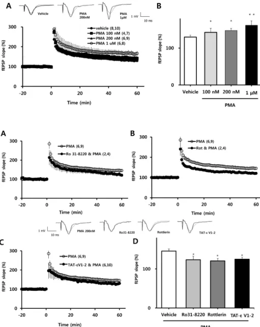
Because four episodes of TBS induce very robust LTP, effects of PMA on the magnitude of LTP might not be de- tectable under such conditions. Therefore, we measured the magnitude of LTP induced by TBS episode 1, 2, 3 and 4 and selected the episode that induced the lowest magnitude LTP. Next, we tested the effects of different concentrations of PMA on LTP 60 min after induction of LTP by one epi- sode of TBS. PMA was applied to the hippocampal slice at least 1 h prior to application of TBS. PMA did not affect the magnitude of baseline fEPSP values, but as shown in Fig. 1A, it exerted a concentration-dependent potentiation
Fig. 1. Effects of PMA on the induc- tion of LTP by one episode of TBS.
(A) PMA enhanced LTP induced by one episode of TBS in a concentrat- ion-dependent manner. fEPSP val- ues (as percentages of baseline) are plotted against time. (B) Averaged values from 58 to 60 min after TBS in the experiment reported on the left are represented as a bar graph.
All values are expressed as means±
SEMs (error bars). Values in paren- theses are numbers of animals and slices tested. *p<0.05, **p<0.01 com- pared to vehicle.
Fig. 2. Effects of PKCδ and PKCε inhibitors on facilitation of LTP by PMA. (A) The potentiating effect of PMA (200 nM) was blocked in the presence of Ro 31-8220 (10μM; a nonspecific PKC inhibitor), (B) rot- tlerin (1μM; a selective PKCδ in- hibitor), and (C) TAT-εV1-2 peptide (500 nM; a PKCε inhibitor). (D) Averaged values from 58 to 60 min after a single episode of TBS from the experiment reported in A, B and C, are represented in bar graph form.
All values are expressed as means±
SEMs (error bars). Values in paren- theses are numbers of animals and slices tested. *p<0.05 compared to vehicle. Rot, rottlerin; εV1-2, TAT- εV1-2.
of TBS-induced LTP. This is quantified in bar graph form in Fig. 1B, which shows stepwise percentage increases in average LTP values 58∼60 min after one episode of TBS (relative to baseline) with application of increasing concen- trations of PMA (100 nM: 142±4.38% ; 200 nM: 146±5.51%, p<0.05; 1μM: 164±11.14%, p<0.01) compared to vehicle (130±5.15%).
Inhibition of PKCδ or PKCε blocks the facilitation of LTP by PMA
We next determined which PKC isoform was involved in the facilitating effect of PMA (200 nM) on the induction of LTP. PMA facilitation of LTP was blocked in the presence of the nonspecific PKC inhibitor, Ro 31-8220 (10μM; Fig.
2A); the selective PKCδ inhibitor, rottlerin (1μM; Fig. 2B);
and the selective PKCε inhibitor, TAT-εV1-2 peptide (500 nM; Fig. 2C). These results are quantified in bar graph form in Fig. 1D, which shows the average LTP values 58∼60
min after TBS (as a percent of baseline) in slices pretreated with PMA plus 10μM Ro 31-8220 (123±9.1%), PMA plus 1μM rottlerin (121±6.0%), or PMA plus 500 nM TAT-ε V1-2 peptide (125±4.9%) compared to PMA alone (146±
5.5%). Collectively, these data indicate that PMA exerts its potentiating effect on TBS-induced LTP in the hippocampal CA1 region via activation of PKCδ or PKCε.
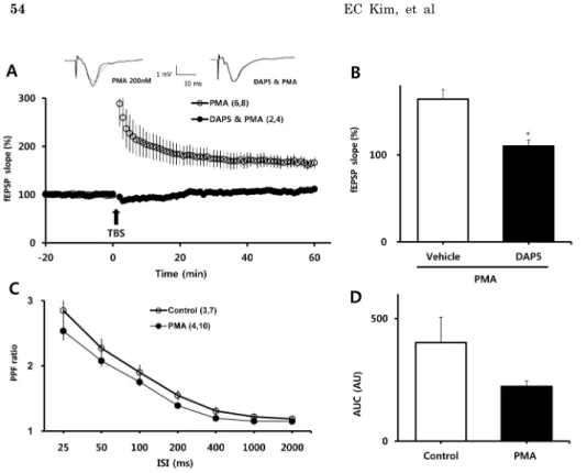
PMA-mediated facilitation of LTP induction requires NMDAR activity
To examine the involvement of NMDARs in PMA-induced enhancement of LTP, we measured LTP in the presence of DL-APV, an NMDAR blocker. As shown in Fig. 2A, B, DL-APV (50μM) eliminated the enhancing effect of PMA on LTP (110±7.13% vs. 164±11.14% of baseline for DL-APV and PMA, respectively; p<0.001). We then examined whether the facilitating effect of PMA was attributable to changes in basal synaptic transmission. PPF ratios, reflect-
Fig. 3. Involvement of NMDA re- ceptors in PMA-induced facilitation of LTP and PPF. (A) In the presence of DL-APV, PMA did not facilitate the induction of LTP by one episode of TBS. (B) Averaged values from 58 to 60 min after TBS in the experi- ment shown on the left are repre- sented as bar graphs. (C) PPF ratios were similar between PMA and con- trol groups. (D) Areas under the curves (AUC) of PPF ratio were calculated to compared the basal levels of synaptic transmission. All values are expressed as means±
SEMs (error bars). Values in paren- theses are numbers of animals and slices tested. *p<0.05 compared to vehicle. DAP5, DL-APV.
ing presynaptic function, were determined before and at least 30 min after treatment with PMA (Fig. 3C). PPF ra- tios were similar between the PMA-treated group and the vehicle-treated group, although there was tendency toward lower PPF ratios in slices treated with PMA.
DISCUSSION
In the present study, we showed that PMA dose-depend- ently facilitates the induction of hippocampal LTP and ex- erts this effect through PKCδ or PKCε. Moreover, this potentiating effect requires activation of NMDARs.
Because of its high sensitivity to intracellular concen- trations of calcium, PKC is thought to be a major coor- dinator of processes underlying activity-induced synaptic modifications. Consistent with this role, PKC activation is necessary for maintenance of LTP in cultured neurons [18]
and hippocampal slices [19,20]. In the present study, we have shown that activation of PKC also enhances the in- duction of LTP, an effect that may be attributable to NMDAR trafficking. Activation of PKC increases the NMDA channel opening rate and, importantly, delivers new NMDARs to the plasma membrane [21,22]. Recently, it has been shown that PKC promotes NMDA receptor trafficking and induces synaptic plasticity by indirectly triggering au- tophosphorylation of CaMKII, which subsequently becomes increasingly associated with NMDARs [23]. PKC also regu- lates NMDAR function by phosphorylating the glutamate receptor subunit NR1 and promoting export and slow trans- port of NMDARs from the endoplasmic reticulum to the plasma membrane over a period of hours [24,25] and by interacting with NMDAR-associated proteins and promot- ing the rapid insertion NMDAR channels in hippocampal neurons [22,26]. Alternatively, PKC activation may en- hance LTP due to synaptogenesis or phosphorylation or in- sertion of AMPARs into the postsynaptic membrane, there-
by altering the sensitivity of the membrane to glutamate [27,28]. In addition, the PKC activator, bryostatin-1, has been shown to increase the number of dendritic spines [29,30].
In CA1 pyramidal cells, PKC activation has also been shown to induce memory-specific enhancement of synaptic potentials elicited by Schaffer collateral stimulation [31].
Application of exogenous compounds known to activate PKC, such as oleic acid [32], arachidonic acid [33] or glucose [34], synergistically enhance the induction of LTP, convert- ing short-term potentiation to LTP [32,33]. Some PKC acti- vators, such as bryostatin-1, enhance memory-specific in- creases in the number of mushroom spines, which provide structural storage sites for long-term associative memory and sites for memory-specific synaptogenesis that involve PKC-regulated changes in spine shape as well as PKC-reg- ulated changes in pre- and postsynaptic ultrastructure [35].
Our demonstration that activation of PKC by PMA regu- lates the induction of LTP could support suggestions by pre- vious studies that PMA regulates a structural change in spines during memory retention [36-39].
We have shown that enhancement of LTP by PMA re- quires activation of PKCδ or PKCε. PMA has been pre- viously shown to modulate both the extent and fusion ki- netics of vesicle exocytosis and to facilitate exocytosis and vesicle fusion by activating PKCα in neuroendocrine PC12 cells [16]. Moreover, PMA potentiation of NMDA-induced currents in primary cultured cerebellar granule cells is mediated by protein kinase Cα [40]. Also, PMA stimulates neutrophil adhesion to endothelial cells in association with PKCδ translocation to the plasma membrane [41]. Con- trasts between literature reports and the results of the present study may be attributable to differences in cell types or species studied [42]. However, we did not evaluate the involvement of other PKC isoforms, perhaps also acti- vated by PMA, in the induction of LTP. Thus, we cannot exclude the possibility that such isoforms also facilitate
LTP induction by PMA.
In conclusion, our results suggest that PMA regulation of synaptic efficacy and synaptogenesis may be mediated by activation of PKCδ or PKCε via a mechanism that de- pends on NMDAR activity.
ACKNOWLEDGEMENTS
This study was supported by the Mid-career Researcher Program through an NRF grant funded by the MEST (2011-0027545). G.H. SEOL was partially supported by a Mid-Career Researcher Program (2012-007145) funded by an NRF grant from MEST.
REFERENCES
1. Bliss TV, Collingridge GL. A synaptic model of memory:
long-term potentiation in the hippocampus. Nature. 1993;361:
31-39.
2. Gustafsson B, Wigström H. Long-term potentiation in the hippocampal CA1 region: its induction and early temporal development. Prog Brain Res. 1990;83:223-232.
3. Izquierdo I, Cammarota M, Da Silva WC, Bevilaqua LR, Rossato JI, Bonini JS, Mello P, Benetti F, Costa JC, Medina JH. The evidence for hippocampal long-term potentiation as a basis of memory for simple tasks. An Acad Bras Cienc.
2008;80:115-127.
4. Collingridge GL, Isaac JT, Wang YT. Receptor trafficking and synaptic plasticity. Nat Rev Neurosci. 2004;5:952-962.
5. Carroll RC, Beattie EC, von Zastrow M, Malenka RC. Role of AMPA receptor endocytosis in synaptic plasticity. Nat Rev Neurosci. 2001;2:315-324.
6. Newton AC. Protein kinase C: structural and spatial regulation by phosphorylation, cofactors, and macromolecular interact- ions. Chem Rev. 2001;101:2353-2364.
7. Ohno S, Nishizuka Y. Protein kinase C isotypes and their specific functions: prologue. J Biochem. 2002;132:509-511.
8. Coussens L, Rhee L, Parker PJ, Ullrich A. Alternative splicing increases the diversity of the human protein kinase C family.
DNA. 1987;6:389-394.
9. Roisin MP, Barbin G. Differential expression of PKC isoforms in hippocampal neuronal cultures: modifications after basic FGF treatment. Neurochem Int. 1997;30:261-270.
10. Tanaka C, Saito N, Kose A, Ito A, Hosoda K, Sakaue M, Shuntoh H, Nishino N, Taniyama K. Possible roles of protein kinase C in neurotransmission. Adv Exp Med Biol. 1988;236:
277-285.
11. Saito N, Shirai Y. Protein kinase C gamma (PKC gamma):
function of neuron specific isotype. J Biochem. 2002;132:683- 12. Swope SL, Moss SJ, Raymond LA, Huganir RL. Regulation of 687.
ligand-gated ion channels by protein phosphorylation. Adv Second Messenger Phosphoprotein Res. 1999;33:49-78.
13. Yao Y, Kelly MT, Sajikumar S, Serrano P, Tian D, Bergold PJ, Frey JU, Sacktor TC. PKM zeta maintains late long-term potentiation by N-ethylmaleimide-sensitive factor/GluR2-de- pendent trafficking of postsynaptic AMPA receptors. J Neuro- sci. 2008;28:7820-7827.
14. Sacktor TC. PKMzeta, LTP maintenance, and the dynamic molecular biology of memory storage. Prog Brain Res. 2008;169:
27-40.
15. Rozengurt E. Early signals in the mitogenic response. Science.
1986;234:161-166.
16. Xue R, Zhao Y, Chen P. Involvement of PKC alpha in PMA-induced facilitation of exocytosis and vesicle fusion in PC12 cells. Biochem Biophys Res Commun. 2009;380:371-376.
17. Goldin M, Segal M. Protein kinase C and ERK involvement in dendritic spine plasticity in cultured rodent hippocampal
neurons. Eur J Neurosci. 2003;17:2529-2539.
18. Linden DJ, Routtenberg A. The role of protein kinase C in long-term potentiation: a testable model. Brain Res Brain Res Rev. 1989;14:279-296.
19. Angenstein F, Staak S. Receptor-mediated activation of protein kinase C in hippocampal long-term potentiation: facts, pro- blems and implications. Prog Neuropsychopharmacol Biol Psy- chiatry. 1997;21:427-454.
20. Van der Zee EA, Luiten PG, Disterhoft JF. Learning-induced alterations in hippocampal PKC-immunoreactivity: a review and hypothesis of its functional significance. Prog Neuropsy- chopharmacol Biol Psychiatry. 1997;21:531-572.
21. Lau CG, Takayasu Y, Rodenas-Ruano A, Paternain AV, Lerma J, Bennett MV, Zukin RS. SNAP-25 is a target of protein kinase C phosphorylation critical to NMDA receptor trafficking. J Neurosci. 2010;30:242-254.
22. Lan JY, Skeberdis VA, Jover T, Grooms SY, Lin Y, Araneda RC, Zheng X, Bennett MV, Zukin RS. Protein kinase C modulates NMDA receptor trafficking and gating. Nat Neuro- sci. 2001;4:382-390.
23. Yan JZ, Xu Z, Ren SQ, Hu B, Yao W, Wang SH, Liu SY, Lu W. Protein kinase C promotes N-methyl-D-aspartate (NMDA) receptor trafficking by indirectly triggering calcium/calmodu- lin-dependent protein kinase II (CaMKII) autophosphorylation.
J Biol Chem. 2011;286:25187-25200.
24. Scott DB, Blanpied TA, Ehlers MD. Coordinated PKA and PKC phosphorylation suppresses RXR-mediated ER retention and regulates the surface delivery of NMDA receptors. Neurophar- macology. 2003;45:755-767.
25. Scott DB, Blanpied TA, Swanson GT, Zhang C, Ehlers MD. An NMDA receptor ER retention signal regulated by phosphoryla- tion and alternative splicing. J Neurosci. 2001;21:3063-3072.
26. Moriguchi S, Shioda N, Han F, Yeh JZ, Narahashi T, Fukunaga K. Galantamine enhancement of long-term potentiation is mediated by calcium/calmodulin-dependent protein kinase II and protein kinase C activation. Hippocampus. 2009;19:844- 854.
27. Barria A, Muller D, Derkach V, Griffith LC, Soderling TR.
Regulatory phosphorylation of AMPA-type glutamate receptors by CaM-KII during long-term potentiation. Science. 1997;276:
2042-2045.
28. Giese KP, Fedorov NB, Filipkowski RK, Silva AJ. Autophos- phorylation at Thr286 of the alpha calcium-calmodulin kinase II in LTP and learning. Science. 1998;279:870-873.
29. Sun MK, Alkon DL. Dual effects of bryostatin-1 on spatial memory and depression. Eur J Pharmacol. 2005;512:43-51.
30. Sun MK, Hongpaisan J, Nelson TJ, Alkon DL. Poststroke neuronal rescue and synaptogenesis mediated in vivo by protein kinase C in adult brains. Proc Natl Acad Sci USA.
2008;105:13620-13625.
31. Olds JL, Alkon DL. Protein kinase C: a nexus in the bioche- mical events that underlie associative learning. Acta Neurobiol Exp (Wars). 1993;53:197-207.
32. Linden DJ, Sheu FS, Murakami K, Routtenberg A. Enhance- ment of long-term potentiation by cis-unsaturated fatty acid:
relation to protein kinase C and phospholipase A2. J Neurosci.
1987;7:3783-3792.
33. Luo Y, Vallano ML. Arachidonic acid, but not sodium nitro- prusside, stimulates presynaptic protein kinase C and phos- phorylation of GAP-43 in rat hippocampal slices and synapto- somes. J Neurochem. 1995;64:1808-1818.
34. Moriguchi S, Oomura Y, Shioda N, Han F, Hori N, Aou S, Fukunaga K. Ca2+/calmodulin-dependent protein kinase II and protein kinase C activities mediate extracellular glucose-re- gulated hippocampal synaptic efficacy. Mol Cell Neurosci. 2011;
46:101-107.
35. Hongpaisan J, Alkon DL. A structural basis for enhancement of long-term associative memory in single dendritic spines regulated by PKC. Proc Natl Acad Sci USA. 2007;104:19571- 19576.
36. Swannie HC, Kaye SB. Protein kinase C inhibitors. Curr Oncol Rep. 2002;4:37-46.
37. Bank B, DeWeer A, Kuzirian AM, Rasmussen H, Alkon DL.
Classical conditioning induces long-term translocation of pro- tein kinase C in rabbit hippocampal CA1 cells. Proc Natl Acad Sci USA. 1988;85:1988-1992.
38. Malenka RC, Madison DV, Nicoll RA. Potentiation of synaptic transmission in the hippocampus by phorbol esters. Nature.
1986;321:175-177.
39. Routtenberg A, Colley P, Linden D, Lovinger D, Murakami K, Sheu FS. Phorbol ester promotes growth of synaptic plasticity.
Brain Res. 1986;378:374-378.
40. Reneau JC, Reyland ME, Phillips J, Kindy C, Popp RL. Phorbol
12-myristate 13-acetate potentiation of N-methyl-D-aspartate- induced currents in primary cultured cerebellar granule cells is mediated by protein kinase C alpha. J Pharmacol Exp Ther.
2009;330:641-649.
41. Hendey B, Zhu CL, Greenstein S. Fas activation opposes PMA- stimulated changes in the localization of PKCdelta: a mecha- nism for reducing neutrophil adhesion to endothelial cells. J Leukoc Biol. 2002;71:863-870.
42. Hongpaisan J, Sun MK, Alkon DL. PKC ε activation prevents synaptic loss, Aβ elevation, and cognitive deficits in Alzhei- mer's disease transgenic mice. J Neurosci. 2011;31:630-643.