* Corresponding author: Jae-Sun Choi
Department of Biomedical Laboratory Science, Far East University, 76-32 Daehak-gil, Gamgok-myeon, Eumseong 27601, Korea
E-mail: [email protected]
* ORCID: https://orcid.org/0000-0002-1733-4088
ORIGINAL ARTICLE
Cisplatin Suppresses Proliferation of Ovarian Cancer Cells through Inhibition Akt and Modulation MAPK Pathways
Jae-Sun Choi
Department of Biomedical Laboratory Science, Far East University, Eumseong, Korea
Cisplatin의 난소암 세포 증식 억제에 관한 신호 전달 기전
최재선
극동대학교 임상병리학과
ARTICLE INFO ABSTRACT
Received January 7, 2020 Revised January 29, 2020 Accepted February 3, 2020
Cisplatin (CDDP) is a chemotherapy agent used for patients with ovarian cancers. CDDP activates multiple signaling pathways, which causes various cellular reactions according to the type of cancer cells. Therefore, it is difficult to clearly conclude its signaling pathways. The purpose of this study is to determine the role of the signal protein of Akt/ERK1/2 and MAPK by CDDP-induced apoptosis in ovarian cancer cells (SKOV3). As a result, the number of apoptosis increased according to the TUNEL assay, and flow cytometric analysis confirmed that the percentage of sub-G1 early apoptosis was 8.73% higher than the control. The PARP and caspase-3 activity that appeared in the process of apoptosis was increased and the Bcl-2 expression was decreased. It was verified that the Akt and ERK1/2 activity was decreased, and p38 and JNK activity increased in a time dependent fashion. In conclusion, these results demonstrate that cisplatin inhibits the proliferation of ovarian cancer cells by inhibiting Akt activity and induces apoptosis by modulating the MAPK signaling pathway.
However, a decrease in the ERK1/2 activity by CDDP was the opposite result to the result shown from the HeLa cells. For this reason, further research on signaling pathways is necessary. These results are expected to be useful for ovarian cancer treatment strategies targeting the MAPK pathway.
Copyright © 2020 The Korean Society for Clinical Laboratory Science. All rights reserved.
Key words Akt Apoptosis Cisplatin MAPK Ovarian cancer
서 론
난소암은 가장 치명적인 부인과 악성 종양으로 주로 상피성 난소암의 형태로 존재한다[1, 2]. 2018년 미국에서 난소암 발병 사례는 대략 22,240건 정도로, 이로 인한 암 사망 수치는 14,070건에 가까울 것으로 예상하였다. 이는 전체 암의 사망 원 인 중 5번째에 속하며 미국 여성의 암 사망률 5%를 차지한다[3].
Cisplatin (cis-[Pt(NH3)2 Cl2], CDDP)은 난소암 등의 고형 암 치료에 사용되는 가장 강력한 화학 요법제 중 하나이다.
Cisplatin을 포함하는 백금 기반의 항암제들은 실제 cisplatin 이 임상 실험에서 성공한 후 수많은 연구자들의 수고와 노력에 의하여 발견되고 발전하였다[4, 5]. 그러나 cisplatin의 광범위 한 항암 활성에도 불구하고 난소암 치료 과정 중 독성 부작용 및 종양 내성을 동반하여 재발율로 인한 치료에 한계를 갖는다. 이 에 따라 부작용을 줄이기 위해 cisplatin과 함께 사용할 수 있는 물질과의 병용요법이나 cisplatin이 가지는 다중 신호경로를 타겟으로 세포 사멸을 유도하는 새로운 치료 전략으로의 연구가 진행되고 있다. 세포 사멸(apoptosis)의 경로는 연속적인 진행
Korean Society for Clinical Laboratory Science
과정으로 초기 세포사멸의 활성화 단계인 핵 내의 신호전달, 전 이단계인 미토콘드리아에서의 단백질 조절 및 실행 그리고 실행 단계인 세포질에서의 단백질 분해 등으로 구조적 변화가 일어나 는 순서로 이어진다[6]. 세포 사멸을 일으키는 암세포의 성향은 화학 요법제와 반응하여 결정되는 중요한 요소이고 종종 암세포 는 세포 사멸을 조절하는 변이 유전자를 가진다[7, 8]. 예를 들어 pro-survival과 관련 있으며 PI3-K (phosphatidylinositol 3-kinase)에 의해 조절되는 Akt serine-threonine kinase는 사람에서 발생하는 암에 빈번하게 활성화된다[9]. 종양 억제 유 전자 MMAC1 (mutated in multiple advanced cancers 1) 으로 알려진 PTEN (phosphatase and tensin homolog)은 PI3-K/Akt 경로를 음성적으로 조절하여 몇몇 세포 사멸을 자 극하면서 세포 사멸에 반응하는 민감성을 증가시킨다[10]. 게다 가 PTEN은 성장인자에 의해 유도된 Shc (src homology domain C-terminal)의 인산화를 억제하고 MAPK (mitogen- activated protein kinases) 신호전달경로를 방해하기도 한다 [11]. MAPKs의 3가지 주요 그룹은 ERK (extracellular signal- regulated protein kinases), JUN (c-JUN N-terminal kinase/stress-activaed protein kinase)과 p38 subfamilies 로 포유동물 세포내 발견된다. 각각의 그룹은 다양한 세포 외부 의 자극과 스트레스를 통해 세포 내부의 경로가 변화될 수 있도 록 조절하는 단백질들을 가진다[12, 13]. Cisplatin은 세포의 종류 및 치료 용량에 따라 세포 반응을 조절하기 위하여 다중 신 호 경로를 활성화 시킨다[34]. 그러나 다양한 스트레스에 의해 세포 사멸을 유발하는 신호 경로 과정 중에 신호 단백질의 발현 변화로 단백질 역할을 규명하기에는 더 많은 연구가 필요한 실 정이다. 특히 cisplatin을 처리한 다양한 세포에서의 ERK1/2 활성 변화는 세포사멸 또는 세포 생존에 대한 논란을 가진다[14, 15]. 본 연구에서는 cisplatin으로 세포사멸을 유도한 SKOV3 세포에서 세포사멸 과정 중 나타나는 Akt와 MAPK 신호 단백질 의 작용을 규명하고자 하였다. 신호 단백질 발현 변화를 통해 이 들의 역할을 규명하기 위해서 cisplatin이 SKOV3 세포에 미치 는 세포사멸에 초점을 두고 시행하였다. 세포 사멸체 확인과 초 기 세포 사멸율 분석하고 caspase-3 및 Bcl-2의 발현을 고려하 여 Akt, ERK1/2, p38 그리고 JNK의 역할을 결정하였다.
재료 및 방법
1. 시약 및 항체
Cisplatin (cis-diamminedichloridoplatinum (II), CDDP) 은 Sigma- Aldrich (St. Louis, MO, USA)에서 구입하여 사용
하였다. p-AKT (ser 473), p-ERK1/2, p-JNK, p-P38, cleaved-PARP cleaved-caspase-3, Bcl-2와 -actin에 대 한 1차 항체는 모두 Cell Signaling Technology (Danvers, MA)에서 구입하여 1:1,000 또는 1:5,000으로 희석하여 사용 하였다.
2. 세포 배양
본 연구에 사용된 인간의 난소암 세포(SKOV3) (American Type Culture Collection, Rockville, MD, USA)는 서울 의 과대학에서 분양받아 RPMI-1640 배지(Gibco BRL, Gai- thersburg, MD, USA)에 2 mM L-glutamine (Gibco BRL, Gaithersburg, MD, USA)과 10% fetal bovine serum (HyClone, Thermo Scientific), 1% penicillin streptomycin (Invitrogen)을 첨가하여 37°C, 5% CO2 배양기에서 배양하였다.
3. 세포 생존율 측정
세포 생존율은 3-(4,5-dimetylthiazol-2-yl)-2, 5-diphenyl- tetrazolium (MTT) (Cell Titer 96Ⓡ AQueous Cell Proli- feration Assay kit; Promega Corporation, Madison, WI, USA) assay 방법에 따라 수행하였다. 동일한 수의 세포를 96 well plate에서 24시간 배양 후 다양한 농도의 cisplatin을 처 리하여 48시간 동안 다시 배양하였다. 배양액을 제거한 후 다시 100 L의 배양 액에 3-(4,5-dimetylthiazol-2-yl)-2, 5- diphenyl-tetrazolium (MTT) 용액(4 mg/mL) 20 L를 첨가 하여 37°C, 5% CO2 조건에서 4시간 동안 연속 배양하였다. 상 층액을 제거하고 DMSO 100 L/well 넣어 15분간 반응 후 microplate reader로 490 nm에서 흡광도를 측정하였다. 대 조군과 비교하여 백분율 수치를 계산하고 IC50 값을 결정하였 다. SKOV3 세포에 IC50에 해당하는 cisplatin을 처리하여 24 시간 또는 48시간 동안 배양하였다.
4. TUNEL assay를 이용한 세포사멸 확인
In Situ Cell Death Detection Kit (Roche, Penzberg, Germany)를 이용한 terminal deoxynucleotidyl transferase mediated digoxigenin-dUTP-biotin nick-end labeling (TUNEL) assay를 통해 SKOV3 세포의 세포 사멸체을 입증함 으로 세포 사멸을 확인하였다[16]. 세포는 4% 포름알데히드로 고정시키고 40분 동안 실온에서 배양하였다. 이어서 PBS에서 여러 번 세척하여 얼음 위에서 0.2% Triton X-100 용액에 5분 동안 투과시킨 후 50 L의 TUNEL reaction mixture 혼합물 을 chamber slide에 첨가하고 37°C, 5% CO2 암실 조건 하에
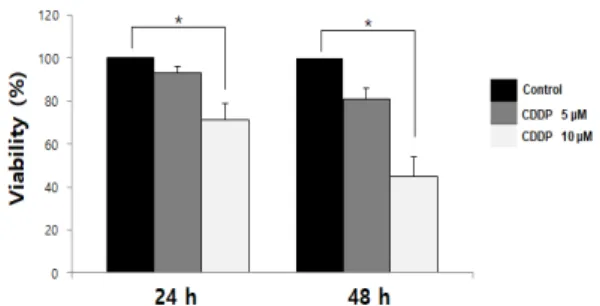
Figure 1. Effects of Cisplatin on viability in SKOV3 cells. Cells were seeded in 96-well plates and treated with 5 M and 10 M concentrations of cisplatin, and cell viability was determined with the MTT assay. The values are calculated relative to the control group (0 M cisplatin). The results are shown as mean±SD and are representative of three independent experiments. *P<0.05 versus the control group.
서 1시간 방치하였다. 마지막으로, 상층액을 제거하고 PBS로 5 분 동안 2번 세척한 후 confocal microscope Model LSM 510 (Carl Zeiss, Jen, Germany)으로 관찰하였다.
5. 세포주기 분석
Cisplatin 처리에 따른 SKOV3 세포의 초기 세포 사멸을 관 찰하기 위하여 제조사(Molecular Probes, Eugene, OR, USA)의 설명에 따라 Annexin V FITC로 염색하였다. 대략 1×10⁵개의 세포를 수확하여 pH 7.4 phosphate-buffered saline으로 세척 하였다. 세척한 세포는 100 L Annexin-V binding buffer (10 mM 4-(2-hydroxyethyl)-1-piperazi- neethanesulphonic acid, 140 mM NaCl과 2.5 mM CaCl2; pH 7.4)에 현탁시키고 5 L Annexin-V-FITC를 첨가하여 15 분 동안 실온에서 배양하고 2 mL Annexin-V binding buffer 를 첨가하여 5분간 원심분리 한 후 상층액을 제거하였다. 이후 배양 세포에 190 L Annexin-V binding buffer로 희석하여 세포가 최종 농도 1 g/mL PI (propidium iodide)가 되도록 염색한 후 Becton-Dickinson FACScan과 Cell Quest software (Becton-Dickinson, Mountain View, CA, USA)를 사용하 여 분석하였다. 세포주기 분석은 염색한 세포로부터 DNA양을 측정하여 결정하였다.
6. Western blot analysis
SKOV3 세포에 cisplatin을 처리한 후 단백질 저해제 칵테일 이 첨가된 radioimmuno- precipitation buffer (50 mM Tris; pH 8.0; Cell Signaling, USA)에 용해하여 상층액의 단 백질 양을 BCA Protein Assay kit (Thermo Fisher Scientific, Inc.)로 측정하였다. 단백질을 10% SDS-PAGE에 전기영동 한 후 nitrocellulose 막으로 전사하였다. 5% skim milk로 2시간 동안 실온에서 blocking 한 후 3회 세척하였다.
1차 항체 anti-cleaved caspase-3, anti-cleaved PARP, anti-Bcl-2, anti-phospho-Akt, anti-phospho-ERK, anti-phospho-p38, anti-phospho-JNK와 -actin은(all Cell Signaling Technology, Inc.) 4°C에서 24시간 반응시키 고 tris buffered saline with tween 20 (TBST)로 3번 세척 후, 2차 항체 horseradish peroxidase-conjugated anti-IgG (Invitrogen)로 실온에서 2시간 반응하였다. 세척 후 Super Signal West Pico Chemiluminescent Substrate (Pierce, IL, USA)을 이용하여 단백질 밴드를 확인하였다.
7. 통계학적 분석
본 연구에서의 모든 실험 결과는 평균(mean value)±표준 편차로 나타내었다. 두 그룹 간의 평균을 비교할 때는 Student’s t-test를 사용하여 비교하였고 통계적 유의성은 분산분석 (Analysis of Vatiance, ANOVA)을 실시하여 검증하였다.
결 과
1. Cisplatin이 SKOV3 세포에 미치는 세포 생존수와 초기 세포사멸
Cisplatin이 SKOV3 세포의 생존에 미치는 영향을 조사하기 위해 5 M과 10 M 농도로 0, 24 및 48시간 처리한 세포를 MTT assay 방법을 통해 세포 생존율을 결정하였다. SKOV3 세 포에 cisplatin을 48시간 동안 처리한 결과 50%의 세포 증식 억 제를 보이는 농도는 10 M로 나타났다(Figure 1). cisplatin에 의해 유도 관찰된 세포 사멸이 세포 sub-G1 주기에서 나타나는 초기 세포사멸과 관련이 있는지 조사하기 위해 유세포 분석을 수행하였다(Figure 2). SKOV3 세포에 10 M의 cisplatin을 48시간 처리한 결과, 처리하지 않은 대조군은 Annexcin-V- FITC positive 세포가 0.72%을 보였으나 cisplatin을 처리한 경우 9.46%로 증가하는 것을 확인하였다.
2. Cisplatin이 SKOV3 세포에 미치는 세포사멸과 caspase 활성
TUNEL assay는 세포 사멸의 신호전달경로[16] 과정 중에 나타나는 세포의 DNA 손상을 현미경으로 관찰할 수 있는 분석 방법이다. Cisplatin이 세포 사멸을 통해 항암 효과를 나타내는
Figure 2. Effects of Cisplatin on early apoptosis in SKOV3 cells.
Apoptosis of cells treated with 10 M cisplatin for 48 h. Cells in the sub-G1 phase were identified according to strong Annexin-V binding.
The data are representative of three independent experiments.
Figure 3. Cisplatin induces apoptotic cells in SKOV3 cells. Cells were treated with cisplatin and after treatment, the number of apoptotic cells was deter- mined with the TUNEL assay. Data are presented as the mean±SD and are representative of three independent experiments with similar results. *P< 0.05 versus the control group.
지의 여부를 평가하기 위해 48시간 동안 cisplatin 10 M로 처 리된 SKOV3의 사멸 세포 체를 TUNEL assay로 관찰하였고 세 포 사멸률을 24시간과 48시간에 확인하였다. Cisplatin으로 처리한 SKOV3 세포의 사멸 세포 수는 대조군과 비교하여 시간 이 증가할수록 유의하게 증가하였다(Figure 3).
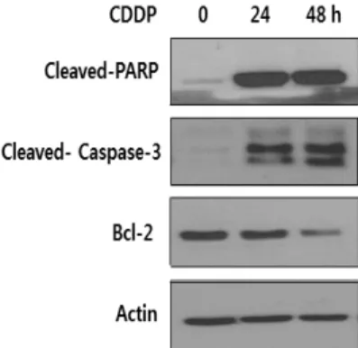
Caspase-3는 세포 사멸의 주요 단계에서 나타나는 단백질 이다. Cisplatin 처리한 SKOV3에서의 세포 사멸을 알아보기 위해 western blot으로 cleved PARP, cleved caspase-3 및 Bcl-2 단백질 수준을 측정하였다. Cleved PARP과 cleved caspase-3의 수준은 유의하게 증가하였으나 반대로 Bcl-2 수 준은 유의하게 감소하였다(Figure 4).
3. Cisplatin이 SKOV3 세포에 미치는 Akt와 MAPK 신호 단백질 발현
Cisplatin으로 세포 사멸을 유도한 신호전달 과정에는 어떠 한 신호 전달 단백질들의 활성이 변화하여 SKOV3 세포에 영향 을 미치는지 이들 활성 단백질에 대한 항체를 이용하여 western blot으로 확인하였다. Akt의 활성화는 세포 생존에 결정적인 역 할을 하는 단백질로 p-Akt와 p-ERK 단백질 발현은 24시간에 서 유의적으로 감소하였으나 p-Akt는 48시간에서 변화가 없었
다(Figure 5). MAPK은 세포에 주어진 스트레스를 통해 p38이 나 JNK 단백질들을 활성화하여 다양하게 세포의 신호를 조절하 여 세포 증식이나 세포 생존에 관여한다. Figure 5에 제시한 바 와 같이, p-p38과 p-JNK 단백질 발현은 24시간에서 유의하게 증가하였으나 48시간에서 p-38의 발현은 감소하였으며 p-JNK의 발현은 48시간에서 변화가 없었다.
고 찰
Cisplatin이 암세포에 미치는 중요한 기전은 세포 사멸을 통 해 나타난다. 세포 사멸의 중요한 항상성 기전은 세포 분열과 세 포 사멸의 균형을 유지하는 것이다[17, 18]. Cisplatin은 치료 과정 중 암세포에 내성이 보이는 부작용이 있음에도 불구하고 다양한 고형암에서 강력한 항암작용을 나타내므로 cisplatin과 함께 혼용하는 병용 요법 연구 또한 신호 전달에 초점을 두어 활 발히 진행 중이다. Cisplatin 등 백금을 기반으로 하는 신호전달 은 암세포의 종류에 따라, 치료 용량에 따라 다중 신호경로를 나 타내므로 신호 경로가 분명하지 않다. 따라서 본 연구에서는 cisplatin으로 세포 사멸을 유도한 SKOV3 세포에서 신호전달 과정 중 나타나는 Akt, ERK1/2, MAPK 신호전달 단백질의 변 화를 조사하였다. 세포사멸에 관련한 신호 전달 단백질들은 세 포 생존에 결정적인 역할을 하는 Akt 활성화 억제와 p38 활성화 조절이 세포사멸 유도와 관련되어 있음을 확인하였다.
세포 사멸은 내인성 경로와 외인성 경로로 구분한다. 내인성 경로에는 미토콘드리아를 매개로 여러 종류의 caspase (cystein protease의 일종)들이 순차적으로 절단되어 활성화되는 과정 을 거친다. Caspase 활성화는 세포 사멸 초기에 나타나는 과정 으로 세포의 자극을 통해 순차적으로 caspase 활성화를 초래한 다[19]. Caspase 활성화에 따라 세포막 bleb 형성, 세포 사멸체 (apoptotic body) 등의 형태학적 변화가 나타난다[20]. 이렇 게 세포 사멸 기전은 괴사(necrosis)와 다르게 세포의 독특한 형 태 및 caspase 계열의 효소 활성을 필요로 하는 생화학적 특징
Figure 4. Cisplatin induces apoptosis in SKOV3 cells. SKOV3 cells were treated with 10 M cisplatin for the indicated times, after which cells were lysed and analyzed for cleaved-PARP, cleaved-caspase-3, and Bcl-2 expression by western blotting using an anti-cleaved- PARP, anti-cleaved-caspase-3, and anti-Bcl-2 antibody Actin was used as an internal control. The western blots are representative of three independent experiments with similar results.
Figure 5. Effects of Cisplatin on phosphorylation of AKT and MAPK in SKOV3 cells. SKOV3 cells were treated with 10 M cisplatin for the indicated times, after which cells were lysed and Akt, ERK, p38 and JNK activation determined by western blotting with an anti- phosphor-Akt antibody, anti-phosphor-ERK antibody, anti-phosphor- p38 antibody and anti-phosphor-JNK antibody. Actin was used as an internal control. A representative image is shown from three independent experiments.
들이 유전자 활성에 의해 조절된다[21]. Figure 3에 제시한 결 과처럼 cisplatin 처리한 SKOV3 세포는 시간에 의존하여 TUNEL positive 세포 수가 증가하였고, Figure 2에 제시한 유 세포 분석에서는 48시간 동안 cisplatin 처리한 SKOV3의 초 기 세포 사멸 수가 대조군과 비교하여 9.45% 변화를 보였다.
Cisplatin에 의한 세포 사멸의 최종 실행 단백질인 caspase-3 의 절편은 처리 시간이 증가함에 따라 증가하였고 PARP의 절편 은 두드러지게 증가하는 결과를 보였다. 이는 caspase-3의 활 성화에 의해 PARP 절편의 잘림은 세포 사멸의 특징 중 하나이다 [22]. 또한 PARP의 활성은 손상된 DNA에 의해서뿐만 아니라 여러 다른 단백질들과의 상호작용을 통해서도 조절된다. 미토 콘드리아 막의 투과성도 세포 사멸과 관련이 있다. 세포 사멸과 관련이 있는 Bcl-2 family 단백질들은 기능에 따라 항 세포 사 멸 구성원과 초기 세포 사멸 구성원 분류한다. 그러나 Bcl-2의 항 세포 사멸(생존 촉진) 기능과 Bax의 사멸 촉진 기능이 길항을 일으켜 두 단백질 간의 발현 양을 조절하여 세포 생존과 사멸이 결정되는 것으로 알려져 있었으나 Bax을 비롯한 BH-3, Bcl-2 단백질들이 미토콘드리아로 표적화하는 단백질들의 균형을 결 정한다고 한다. 실험 결과에서 시간에 의존하여 항 세포 사멸 Bcl-2의 단백질의 발현은 감소하였다. 세포 사멸이 증가하고 있 음을 나타낸 것이다. 이는 cisplatin과 관련한 MCF-7 세포에 서 미토콘드리아 탈분극에 대하여[23] 보고한 바와 같이 SKOV3에 처리한 cisplatin이 세포 사멸을 유발하여 Bax의 단 백질들이 세포질에서 미토콘드리아로 전이 되어 Bcl-2 단백질 발현을 감소함을 시사한다. Hail 등[24]의 보고에 따르면 이러 한 세포 사멸의 유도 조절 기전은 미토콘드리아의 생체 에너지 상태에 따라 반응한 것이라 하였다. 정확하게 cisplatin이 어떻
게 미토콘드리아의 신호 기능을 변경 시킬 수 있는지는 알 수는 없으나 미토콘드리아의 조밀도가 cisplatin에 대한 세포의 민 감도를 결정하는 주요 열쇠가 될 수 있을 것으로 제안하기도 하 였다[25]. Cisplatin은 세포 사멸을 유도하는 MAPKs와 같은 신호전달경로를 활성화 시킨다[26]. 다른 연구에서는 MAPK의 활성화는 백혈병 세포의 죽음과 caspase의 활성화를 위해 필요 한 것으로 제안하였다[27]. 또한 MAPK 경로는 암세포가 사멸 하는 동안 몇몇 화학 요법제에 의해 유도된다고 보고하였다[28, 29]. MAPK은 serine/threonine kinase가 잘 보존된 계열로 다양한 세포 외 신호전달 경로를 조정한다. 그리고 ERK, JNK 및 p38 kinase 아형을 가지며 세포 생존, 증식, 종양 세포의 이 동에 중추적인 역할을 한다[30, 31]. 본 실험에서 cisplatin을 처리한 SKOV3 세포에서 신호 전달 단백질의 활성 변화는 다음 과 같이 나타났다. PI3K의 다음 단계에서 작용하는 Akt의 활성 은 24시간까지는 현저하게 감소하였으나 48시간에서는 24시 간에 나타난 활성과 동일하였다. Akt는 세포 생존에 결정적인 역할을 하는 serine/threonine kinase로[32, 33] 세포 증식을 억제하는 것으로 보여진다. Cisplatin과 관련한 ERK 활성 감소 는 내성을 갖는 변이 세포에서 나타날 수 있으며 ERK의 활성 증 가는 세포의 생존과 관련하여 활성을 유도한다. 또한 ERK의 상 위 경로인 RAS의 발현 증가는 cisplatin에 대한 민감성 증가와 관련이 있다고 하였다[14, 15]. 그러나 cisplatin은 세포에 따 라 치료 용량에 따라 다중 신호 경로를 활성화하여 다양한 반응 을 유도하기도 한다. 거기다가 ERK 활성화에 따른 세포 사멸과 세포 생존에 대한 논란은 여전히 존재한다[34]. HeLa 세포에서 cisplatin 농도와 시간이 증가함에 따라 ERK의 활성은 증가하
였고 세포 생존에 기여한다는 보고와는 달리[14] 본 실험은 10
M 농도로 SKOV3 세포에 24시간과 48시간까지 처리한 결과 ERK 활성은 시간이 증가함에 따라 감소하였다. 세포 사멸을 유 도한다는 결과이다. 더 많은 연구가 진행 되어야 하는 필요성이 강조되는 부분이다. p38의 활성은 cisplatin 처리시간이 24시 간까지는 현저하게 증가 하였으나 48시간에서는 다소 감소하였 다. JNK의 활성은 24시간까지는 증가하였으나 48시간에서는 24시간에 나타난 활성과 동일하였다. p38과 JNK는 DNA 손상 이나 다양한 스트레스에 의해 활성화되어 세포 사멸을 유도하는 것으로 잘 알려져 있다[34]. 따라서 cisplatin이 SKOV3 세포 에 스트레스 작용하면서 p38과 JNK의 활성을 24시간까지는 증가시켜 세포 사멸을 유도하였으나 48시간에서는 p38의 활성 을 감소하여 세포 사멸 조절자 역할을 하는 것으로 보여진다. 이 상의 종합적 결과들은 cisplatin이 SKOV3 세포에 Akt의 활성 을 감소하여 세포 증식을 억제하고 MAPK의 p38 활성을 조절 하여 세포 사멸을 유도하는 것으로 사료된다. MAPK 경로의 활 성화는 cisplatin과 같이 백금을 기반으로 하는 화합물들이 세 포 생존, 세포사멸, 세포주기 정지 등의 다양한 세포 반응을 유도 할 수 있다는 연구 결과와[35] 일치하는 결과를 나타낸 것이다.
실험에서 얻은 신호 전달 과정에서 발현한 신호 단백질들의 작 용과 스트레스에 반항하는 세포 사멸 시간을 고려하여 MAPK 의 활성 경로를 표적으로 하는 암 치료 전략에 유용한 자료가 될 수 있을 것으로 사료된다. 향후 다양한 세포 반응을 유도하는 백 금 기반 금속 화합물 화학요법제로 신호 전달 기작을 밝히기 위 해서는 지속적인 연구가 필요해 보인다.
요 약
Cisplatin (CDDP)은 난소암 치료에 사용되는 화학 요법제 로 암세포에 따라 그리고 치료 용량에 따라 다중 신호경로를 활 성화하여 세포 반응을 다르게 일으킬 수 있다. Cisplatin이 세포 에 작용하는 신호전달 기전은 분명하지 않아 더 많은 연구가 필 요해 보인다. 이에 본 연구는 cisplatin을 난소암 세포(SKOV3) 에 처리하여 세포사멸 유도 과정에서 나타나는 신호 단백질의 역할을 밝히고자 하였다. 결과는 cisplatin으로 처리한 난소암 세포에서 TUNEL assay와 유세포 분석을 통해 대조군과 비교 하여 세포 사멸수가 증가하였다. 세포 사멸 단계에서 나타나는 PARP 및 caspase-3 활성도 증가하였다. 그러나 Bcl-2의 발현 은 감소하였다. 신호 전달 경로에서 나타나는 단백질의 발현은 ERK1/2의 활성은 시간 의존적으로 감소하였으나 Akt 활성은 24시간에 감소하다 48시간에서의 활성은 일정하였다. p38과
p-JUN의 활성은 24시간에 증가하는 것으로 나타났으나 48시 간 에서 p38의 활성은 감소하였으며 p-JUN의 활성은 일정하 였다. 이상의 결과들을 토대로 결론은 cisplatin이 SKOV3 세 포에서 Akt 활성을 감소하여 세포 증식을 억제하고 MAPK의 p38 발현을 조절하여 세포사멸을 유도하는 것으로 판단된다.
향 후, 암치료 전략에 도움이 되는 cisplatin을 포함한 백금기반 화학요법제의 신호전달 기전을 밝히기 위한 더 많은 연구가 필 요할 것으로 생각된다. 본 실험을 통해 제시한 결과는 MAPK 신 호 경로를 타겟으로 하는 암 치료 전략에 유용하게 사용 될 수 있 기를 기대한다.
Acknowledgements: This work was supported by the 2019 Far East University Research (FEU 2019R37).
Conflict of interest: None
Author’s information (Position): Choi JS, Professor.
REFERENCES
1. Cho KR, Shih IeM. Ovarian cancer. Annu Rev Pathol. 2009;4:
287-313. https://doi.org/10.1146/annurev.pathol.4.110807.092246 2. Swisher EM, Taniguchi T, Karlan BY. Molecular scores to predict
ovarian cancer outcomes: a worthy goal, but not ready for prime time. J Natl Cancer Inst. 2012;104:642-645. https://doi.org/
10.1093/jnci/djs203
3. Siegel RL, Miller KD, Jemal A. Cancer statistics. CA Cancer J Clin.
2018;68:7-30. https://doi.org/10.3322/caac.21442
4. Frezza M, Hindo S, Chen D, Davenport A, Schmitt S, Tomco D, et al. Novel metals and metal complexes as platforms for cancer therapy. Curr Pharm Des. 2010;16:1813-1825. https://doi.org/
10.2174/138161210791209009
5. Chen D, Milacic V, Frezza M, Dou QP. Metal complexes, their cel- lular targets and potential for cancer therapy. Curr Pharm Des.
2009;15:777-791. https://doi.org/10.2174/138161209787582183 6. Goldstein P. Controlling cell death. Science. 1997;275:1081-
1082. https://doi.org/10.1126/science.275.5303.1081
7. Yuan ZQ, Sun M, Feldman RI, Wang G, Ma X, Jiang C, et al.
Frequent activation of AKT2 and induction of apoptosis by in- hibition of phosphoinositide-3-OH kinase/Akt pathway in hu- man ovarian cancer. Oncogene. 2000;19:2324-2330. https://do- i.org/10.1038/sj.onc.1203598
8. Lasky T, Silbergeld E. P53 mutations associated with breast, col- orectal, liver, lung, and ovarian cancers. Environ Health Perspect.
1996;104:1324-1331. https://doi.org/10.1289/ehp. 961041324 9. Kulik G, Weber MJ. Akt-dependent and -independent survival
signaling pathways utilized by insulin-like growth factor I. Mol Cell Biol. 1998;18:6711-6718. https://doi.org/10.1128/mcb. 18.
11.6711
10. Stambolic V, Suzuki A, de la Pompa JL, Brothers GM, Mirtsos C, Sasaki T, et al. Negative regulation of PKB/Akt-dependent cell survival by the tumor suppressor PTEN. Cell. 1998;95:29-39.
https://doi.org/10.1016/s0092-8674(00)81780-8
11. Gu J, Tamura M, Yamada KM. Tumor suppressor PTEN inhibits integrin-and growth factor-mediated mitogen-activated protein (MAP) kinase signaling pathways. J Cell Biol. 1998;143:1375- 1383. https://doi.org/10.1083/jcb.143.5.1375
12. Ip YT, Davis RJ. Signal transduction by the c-Jun N-terminal kinase (JNK)-from inflammation to development. Curr Opin Cell Biol. 1998;10:205-219. https://doi.org/10.1016/s0955- 0674(98)80143-9
13. Karin M. Mitogen-activated protein kinase cascades as regulators of stress responses. Ann N Y Acad Sci. 1998;30:139-146.
https://doi.org/10.1111/j.1749-6632.1998.tb08987.x
14. Wang X, Martindale JL, Holbrook NJ. Requirement for ERK acti- vation in cisplatin-induced apoptosis. J Biol Chem. 2000;275:
39435-39443. https://doi.org/10.1074/jbc.M004583200
15. Tang MK, Zhou HY, Yam JW, Wong AS. c-Met over expression contributes to the acquired apoptotic resistance of nonadherent ovarian cancer cells through a cross talk mediated by phosphati- dylinositol 3-kinase and extracellular signal-regulated kinase 1/2.
Neoplasia. 2010;12:128-138. https://doi.org/10.1593/neo.91438 16. Chen G, Gong M, Yan M, Zhang X. Sevoflurane induces endoplas-
mic reticulum stress mediated apoptosis in hippocampal neurons of aging rats. PLoS ONE. 2013;8:E57870. https://doi.org/10.
1371/journal.pone.0057870
17. Salvesen GS, Dixit VM. Caspases: intracellular signaling by proteolysis. Cell. 1997;91:443-446. https://doi.org/10.1016/
s0092-8674(00)80430-4
18. Fulda S, Debatin KM. Extrinsic versus intrinsic apoptosis path- ways in anticancer chemotherapy. Oncogene. 2006;25:4798- 4811. https://doi.org/10.1038/sj.onc.1209608
19. Salvesen GS, Abrams JM. Caspase activation-stepping on the gas or releasing the brakes? Lessons from humans and flies.
Oncogene. 2004;23:2774-2784. https://doi.org/10.1038/sj.onc.
1207522
20. Elmore S. Apoptosis: a review of programmed cell death.
Toxicol Pathol. 2007;35:495-516. https://doi.org/10.1080/
01926230701320337
21. Cohen TT. Apoptosis. Immunol. 1993;14:126-130. https://do- i.org/10.1016/0167-5699(93)90214-6
22. Kaufmann SH, Desnoyers S, Ottaviano Y, Davidson NE, Poirier GG. Specific proteolytic cleavage of poly (ADP-ribose) polymer- ase: an early marker of chemotherapy-induced apoptosis.
Cancer Res. 1993;53:3976-3985.
23. Muscella A, Calabriso N, Fanizzi FP, De Pascali SA, Urso L,
Ciccarese A, et al. Pt [(O,O’-acac)(gamma-acac)(DMS)], a new Pt compound exerting fast cytotoxicity in MCF-7 breast cancer cells via the mitochondrial apoptotic pathway. Br J Pharmacol.
2008;153:34-49. https://doi.org/10.1038/sj.bjp.0707576 24. Hail N Jr, Lotan R. Cancer chemoprevention and mitochondria:
targeting apoptosis in transformed cells via the disruption of mi- tochondrial bioenergetics/redox state. Mol Nutr Food Res.
2009;53:49-67. https://doi.org/10.1002/mnfr.200700527 25. Qian W, Nishikawa M, Haque AM, Hirose M, Mashimo M, Sato E,
et al. Mitochondrial density determines the cellular sensitivity to cisplatin-induced cell death. Am J Physiol Cell Physiol.
2005;289:C1466-1475. https://doi.org/10.1152/ajpcell.00265.2005 26. Siddik ZH. Cisplatin: mode of cytoxic action and molecular basis
of resistance. Oncogene. 2003;22:7265-7279. https://doi.org/
10.1038/sj.onc.1206933
27. Park SJ, Kim IS. The role of p38 MAPK activation in aur- anofin-induced apoptosis of human promyelocytic leukaemia HL-60 cells. Br J Pharmacol. 2005;146:506-513. https://doi.org/
10.1038/sj.bjp.0706360
28. Wagner EF, Nebreda AR. Signal integration by JNK and p38 MAPK pathways in cancer development. Nat Rev Cancer.
2009;9:537-549. https://doi.org/10.1038/nrc2694
29. Bulavin DV, Fornace AJ. p38 MAP kinase’s emerging role as a tu- mor suppressor. Adv Cancer Res. 2004;92:95-118. https://do- i.org/10.1016/S0065-230X(04)92005-2
30. Chang L, Karin M. Mammalian MAP kinase signalling cascades.
Nature. 2001;410:37–40. https://doi.org/10.1038/35065000 31. Johnson GL, Lapadat R. Mitogen-activated protein kinase path-
ways mediated by ERK, JNK, and p38 protein kinases. Science.
2002;298:1911-1912. https://doi.org/10.1126/science.1072682 32. Brazil DP, Hemmings BA. Ten years of protein kinase B signalling:
a hard Akt to follow. Trends Biochem Sci. 2001;26:657-664.
https://doi.org/10.1016/s0968-0004(01)01958-2
33. Kwon HW, Lee DH. The Inhibitory effects of cordycepin on phos- phoproteins including PI3K, Akt, and p38. Korean J Clin Lab Sci.
2017;49:99-107. https://doi.org/10.15324/kjcls
34. Achkar IW, Abdulrahman N, Al‑ulaiti H. Joseph JM, Uddin S, Mraiche F. Cisplatin based therapy: the role of the mitogen acti- vated protein kinase signaling pathway. J Transl Med. 2018;
16:96. https://doi.org/10.1186/s12967-018-1471-1
35. Smolle E, Taucher V, Pichler M, Petru E, Lax S, Haybaeck J.
Targeting signaling pathways in epithelial ovarian cancer. Int J Mol Sci. 2013;14:9536-9555. https://doi.org/10.3390/ijms14059536