甘松香 물추출물이 U937 백혈병 세포의 분화유도에 미치는 영향
김진국․주성민․전병제․양현모․전병훈*
원광대학교 한의과대학 병리학교실
Effect of Nardostachys chinensis on Induction of Differentiation in U937 Monomyelocytic Cells
Jin Kuk Kim, Sung Min Ju, Byung Jae Jeon, Hyun Mo Yang, Byung Hun Jeon*
Department of Pathology, College of Oriental Medicine, Wonkwang University
Nardostachyts chinensis (N. chinensis) belonging to the family Valerianaceae has been used to elicit stomachic and sedative effects. The MAPKs are serine/threonine kinases involved in the regulation of various cellular responses, such as cell proliferation, differentiation and apoptosis. The PKC also plays a key role in regulating the response of hematopoietic cells to both physiological and pathological inducers of proliferation and differentiation. This study investigated the signaling pathways on the U937 cell differentiation induced byN. chinensis.N. chinensis induced the differentiation of U937 cells, as shown by increased of differentiation surface antigen CD11b. Activation of ERK increased time-dependently in differentiation of U937 cells induced by N. chinensis, but activations of JNK and p38 were unaffected. Inhibitor of ERK (PD98059) significantly reduced CD11b expression induced byN. chinensis in U937 cells. In addition, N. chinensisincreased protein level of PKC βI and PKC βII isoforms, but the protein level of PKC α and PKC γwas constant. PKC inhibitors (GF 109203X and H-7) inhibited U937 cell differentiation and the ERK activation induced by N. chinensis. These results indicated that PKC and ERK may be involved in U937 cell differentiation induced by N. chinensis.
Key words : Nardostachyts chinensis (N. chinensis), Differentiation, U937, ERK, PKC
* 교신저자 : 전병훈, 익산시 신용동 344-2 원광대학교 한의과대학
․E-mail : [email protected], ․Tel : 063-856-6843
․접수 : 2011/01/06 ․수정 : 2010/01/20 ․채택 : 2010/02/11
서 론
급성 골수성 백혈병은 anthracycline과 cytarabine의 복합 항 암화학요법에 의해 약 65% 환자에서 완전 관해에 도달하지만 일 반적인 공고요법(consolidation therapy)으로는 약 25%에서만 지 속적인 관해 상태를 유지한다1). 이러한 불량한 예후를 향상시키 기 위해 공고요법의 cytarabine 용량을 증가시킨 결과 장기 생존 율이 약 40%까지 증가하였고, 고용량 항암 화학요법 및 동정 조 혈모세포 이식술을 시행할 경우 생존율은 60%까지 향상된다2).
그러나 궁극적으로 상당수의 환자는 재발하여 사망하게 된다. 이 러한 불량한 예후를 개선하기 위해 다양한 종류의 새로운 항 백 혈병 치료제의 개발이 필요한 상태이다.
마타리과에 속하는 甘松香의 뿌리와 뿌리줄기는 성미가 甘
溫하며 脾胃에 귀경하는 약물로서 理氣止痛하고 醒脾健胃하는 효능을 나타낸다. 임상에서는 신경성 위통 및 소화촉진과 항부정 맥 및 진정효과를 나타내는데 사용되고 있다3). 최근 보고된 연구 논문에 의하면 甘松香 물추출물이 급성 전골수구성 백혈병(acute premyelocytic leukemia)세포주인 HL-60 세포를 과립성 백혈구 분화를 유도하는 것으로 보고되었는데4), 甘松香 물추출물의 HL-60 세포의 분화 기전에 PKC와 MAPKs의 활성이 관여하는 것으로 밝혀졌다.
Mitogen-activated protein kinase (MAPKs)는 세포증식, 분 화, 아포토시스와 같은 다양한 세포반응들의 조절에 관여하는 serine/threonin kinases이다5-8). 또한 Protein kinase C (PKC)는 새로운 항암치료제 개발에 대한 표적단백질과 같이 동정되었다.
PKC는 세포증식과 분화에 대한 생리학적 병리학적 유도물질에 대한 조혈세포의 반응을 조절하는데 중요한 역할을 수행한다9,10). 또한 PKC isoform들 중 calcium-의존적 PKC들은 백혈병세포에 서 풍부하게 발현이되고 HL-60세포의 분화를 증대시키는 것으
로 보고되어 있다11).
따라서 본 연구에서는 급성 골수구성 백혈병 세포주인 HL-60 세포의 분화유도에 효능을 가진 甘松香 물추출물이 급성 골수단핵구성 백혈병(acute myelomonocytic leukemia) 세포주인 U937 세포의 분화 유도에 영향을 미치는지 조사하였고 이에 대 하여 유의한 결과를 얻었기에 보고하는 바이다.
재료 및 방법
1. 甘松香 물추출물 제조
甘松香 100 g은 증류수 1 L를 사용하여 100℃에서 3시간 추 출하였다. 추출물은 2,000 rpm에서 15분 원심분리하여 비수용성 성분을 제거하였다. 상층액은 부크너깔데기 위에 와트만 No. 4 여과지를 통하여 여과하였다. 여과물은 12시간 -20℃에 보관 후, 동결 건조하였다. 추출물의 수율(W/W)은 약 12.82%였다. 동결 건조된 추출물은 PBS (pH 7.4)에 20 mg/ml 농도로 용해하여 -2 0℃에 보관하였고, 실험에 사용하기 전에 희석하여 사용하였다.
2. 시약 및 항체
Anti-β-actin, all-trans-Retinoic acid (ATRA), protease inhibitor cocktail는 Sigma-Aldrich Chemical (St. Louis, MO, USA)사로부터 구입하였다. 2'-Amino-3'-methoxyflavone (PD98059), bisindolylmaleimideⅠ(GF 109203X), 1-(5-Isoquinolinesulfonyl)-2-methylpiperazine dihydrochloride (H-7)는 Calbiochem (La Jolla, CA, USA)사로부터 구입하였다.
RPE-conjugated anti-CD11b와FITC-conjugated anti-CD14 항체 들은 DAKO (Glostrup, Denmark)사로부터 구입하였다.
Anti-ERK, anti-phospho-ERK, anti-p38, anti-phospho-p38, anti-JNK, anti-phospho-JNK 항체들은 Cell Signaling Technology INC. (Beverly, MA, USA)사로부터 구입하였다.
Anti-PKC α, anti-PKC βⅠ, anti-PKC βⅡ, anti-PKC γ 항체들은 Santa Cruz Biotechnology, INC. (Santa Cruz, CA, USA)사로부 터 구입하였다.
3. 세포배양
U937 세포는American Type Culture Collection(ATCC, Rockville, MD, USA)로부터 분양 받았고, 10% fetal bovine serum(FBS, Invitrogen, Burlington, ON, Canada), 100 U/ml penicillin, 100 μg/ml streptomycin이 포함된 RPMI1640 배지 (Invitrogen, Burlington, ON, Canada)를 사용하여 37℃와 5%
CO2 상태에서 배양하였다.
4. 세포수 측정
세포수 측정은 Hemocytometer를 사용하여 측정하였다.
U937 세포는 1×10⁵cells/ml로 12 well 플레이트에 분주한 후, 甘 松香 물추출물을 0, 50, 100 μg/ml의 농도로 각각 1일 간격으로 5일 처리하였다. 처리 후에, 세포를 수확하여 PBS(pH7.4)에 현탁 하였고, 세포 현탁액 10 μl와 0.4% trypan blue 용액 10 μl를 혼합
하여 Hemocytometer에 적용하여 광학현미경으로 세포수를 측정 하였다. 세포수는 ml당 세포수(cells numbers/ml)로 계산하였다.
5. 분화도 검사
甘松香 물추출물을 처리한 세포는 수확하여 ice-cold PBS에 2번 세척한 후, 0.25% BSA/PBS 용액 100 μl에 suspension하였 다. 그 다음, RPE-conjugated anti-CD11b mAb 또는 FITC-conjugated anti-CD14 mAb를 10 μl첨가하여 4℃에서 30분 암실조건으로 배양하였다. 그 후, 세포는 0.25% BSA/PBS용액에 2번 세척하였고 1% formaldehyde/PBS용액 600 μl에 고정하였 다. 세포에 결합된 항체의 레벨은 fluorescence-activated cell sorting (FACS) Calibur (BD Biosciences, CA, USA)을 이용하여 측정하였다.
6. PKC 활성측정
PKC활성은 PKC Kinase Activity Assay kit (Non-Radioactive) (Stressgen, Victoria, Canada)를 이용하여 측 정하였다. 세포는 ice-cold PBS에 세척한 후, sample preparation lysis buffer (20 mM MOPS, 50 mM β-glycerolphosphate, 50 mM NaF, 1mM sodium vanadate, 5 mM EGTA, 2 mM EDTA, 1% NP-40, 1 mM dithiothreitol, 1% protease inhibitor cocktail) 로 얼음 위에서 5분 lysis하였고, 4℃에서13,000rpm으로 15분 원 심 분리하였다. 단백질농도는 Bradford assay를 이용하여 측정하 였다. 총 500 ng의 단백질을 kinase assay dilution buffer로 희석 하여PKC substrate peptide가 코팅된 96-well에 loading하였다.
PKC활성검사는 각 well에 10 μl의 ATP (diluted 1 mg/ml)용액 을 첨가하여30℃에서 1시간 배양하였다. 각 well은 antibody dilution buffer로 2번 세척한 후, 40 μl의 phosphospecific substrate 항체를 각 well에 첨가하여 1시간 배양하였다. 각 well 은 wash buffer로 10분4번 세척한 후, dilution buffer로 1:1000으 로 희석된 항-래빗 IgG HRP-conjugated 항체를 첨가하여 30분 RT에서 배양하였다. 각 well은 4번 세척한 후, 60 μl의 tetramethylbenzidine substrate를 첨가하여 30분 RT에서 배양하 였다, HRP반응은 20 μl의 acid stop solution을 첨가하여 반응을 정지시켰고, SpectraMAX 250 microplate spectrophotometer를 사용하여 450 ㎚에서 각 well의 흡광도를 측정하였다. PKC활성 퍼센트는 방정식[(mean OD of treated cells/mean OD of control cells) × 100]으로 계산하였다
7. Western blot analysis
세포는 얼음 냉장된 PBS(pH 7.4)로 세척하고 1% protease inhibitor 혼합액이 포함된 얼음 냉장 용해 완충액(50 mM Tris-HCl, pH7.4, 150 mM NaCl, 1% NP-40, 0.5% sodium deoxycholate, 0.1% SDS, 1 mM sodium vanadate)으로 부유 시 킨 후 얼음 위에서 30분 용해하였다. 세포 용해액은 4℃에서 14,000 rpm으로 20분 원심분리 하였고, 단백질 농도는 Bradford 검사법을 이용하여 측정하였다. 총 30 μg의 단백질 샘플을 SDS-PAGE 젤로 분리하였고 40V에서 3 시간 nitrocellulose 막
위로 단백질을 전이시켰다. 단백질이 전이된 막은 5% BSA가 포 함된Tris-buffered saline+Tween-20(20 mM Tris-HCl, pH7.6, 150 mM NaCl, 0.05% Tween-20)으로 차단시킨 후, 1차 항체를 결합시켰다. 면역 활성은 peroxidase가 붙어있는 anti-rabbit 또 는 anti-mouse 2차 항체를 사용하여 SuperSignal West Pico Chemiluminescent (Pierce, Rockford, IL, USA)에 의해 탐지하 였다.
8. 통계처리
모든 데이터의 결과는 마이크로소프트 오피스 엑셀(EXCEL) 2003 프로그램을 통하여 통계처리 하여 mean±standard deviation(SD)로 기록하였다.
결 과
1. 세포 중식억제에 대한 효과
U937 세포의 분화에 따른 甘松香 물추출물에 대한 세포 증 식억제의 효과를 조사하기 위해 甘松香 물추출물을 50 μg/ml과 100 μg/ml 농도로 1, 2, 3, 4, 5일간 처리하였다. 세포수는 trypan blue 염색을 통한 heomocytometer를 이용하여 측정하였다. 甘松 香 물추출물을 1~5일간 처리한 결과 처리 농도에 비례하여 세포 수를 감소시켰고, 5일간 처리한 甘松香 물추출물50 μg/ml과 100 μg/ml 농도에서 대조군과 비교하여 각각 약 20%와 50% 세포수 를 감소시켰다(Fig. 1).
Fig. 1. Effects ofN. chinensis on proliferation of U937 cells.The cells were incubated with various concentrations ofN. chinensis for 1, 2, 3, 4 or 5 days.
Cell numbers were counted using hemocytometer. Value are means ± SD, N = 3. ● Control; ▲: 50 μg/ml N. chinensis ■: 100 μg/ml N. chinensis
2. 분화유도 효과
甘松香 물추출물의 증식 억제효과가 U937세포의 분화에 영 향을 미치는지 조사하기 위해 분화표지자인 CD11b와 CD14의 발현을 분석하였다. CD11b (FITC-conjugated) 발현은 과립성 백 혈구 분화에서 발현하고, 반면에 CD14 (RPE-conjugated)는 단핵 백혈구/대식세포 분화에서만 발현한다. U937세포에 50 μg/ml과 100 μg/ml 농도의 甘松香 물추출물을 5일간 처리한 후, 세포표 면표지자는 면역 염색하여 유세포 분석기를 통하여 분석하였다.
甘松香 물추출물을 50 μg/ml농도에서 CD11b-양성(+) 세포가 약 간 증가하였지만, 100 μg/ml 농도에서 CD11b-양성(+) 세포가 대
조군과 비교하여 뚜렷하게 증가를 보였다(Fig. 2). 그러나 CD14- 양성(+) 세포는 대조군과 비교하여 아무런 차이가 없었다.
A
B
Fig. 2. Effect ofN. chinensis on differentiation of U937 cells.The cells were treated with 50 μg/ml and 100 μg/mlN. chinensis for 5 day. The cells were assessed by FACS analysis using RPE-conjugated anti-CD11b mAb (A) or FITC-conjugated anti-CD14 mAb (B).
3. MAPKs 활성에 대한 효과
MAPKs 신호전달경로는 분화조절에서 중요한 역할을 수행 한다. 甘松香 물추출물에 의해 유도된 U937세포의 분화에서 MAPKs의 관련을 조사하기 위해 甘松香 물추출물 100 μg/ml 농 도로 1~5일 간 처리 후, ERK, JNK, p38의 활성을 분석하였다. 甘 松香 물추출물은 ERK활성을 시간 의존적으로 증가하였지만, JNK활성과 p38활성에는 아무런 영향을 미치지 못하였다(Fig. 3).
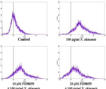
4. 세포분화유도에 대한 ERK 활성억제 효과
甘松香 물추출물에 의한 ERK 활성이 U937세포의 분화와의 관련성을 조사하기 위해 ERK 활성 억제효과를 조사하였다.
U937세포는 PD98059 (ERK inhibitor) 1시간 전 처리한 후, 甘松 香 물추출물을 처리하였다. 甘松香 물추출물에 의해 증가된 CD11b의 발현은 10 μM ERK inhibitor의해 약간 감소하였지만, 20 μM ERK inhibitor에서 뚜렷하게 감소하였다(Fig. 4).
Fig. 3. Effects of N. chinensis on activation of ERK, JNK and p38 in U937 cells.The cells were treated withN. chinensis (100 μg/ml) for 1, 2, 3, 4 and 5 days. Whole cell lysates were subjected to SDS-PAGE followed by Western blot analysis with an anti-ERK, anti-p-ERK, anti-p38, anti-p-p38, anti-JNK and anti-p-JNK antibodies.
Fig. 4. Effect of inhibition for ERK on U937 cell differentiation induced by N. chinensis.The cells were preincubated with each inhibitor (ERK inhibitor, PD98059) for 1 hr, and then treated with 100 μg/ml N. chinensis for 5 day. The cellular differentiation was assessed by FACS analysis using RPE-conjugated anti-CD11b mAb.
5. PKC 발현과 활성에 대한 효과
PKC의 활성은 세포의 분화에 대해서 필수적이다. 甘松香 물 추출물에 의해 유도된 U937세포의 분화에서 PKC의 관련성을 조 사하기 위해, U937세포는 100 μg/ml 농도의 甘松香을 1~5일간 처리한 후, PKC의 활성과 발현을 조사하였다. 甘松香 물추출물 은 시간의존적으로 PKC 활성을 증가시켰다 (Fig. 5A.). 甘松香 물추출물에 의한 PKC 활성 증가에 따른 PKC 발현을 조사하기 위해, Western blot을 통하여 PKC isoform들의 발현량을 분석하
였다. 甘松香 물추출물은 PKC βⅠ과 PKC βⅡ의 발현을 상당히 증가시켰지만, PKC α와 PKC γ의 발현에는 아무런 영향을 미치 지 못하였다 (Fig 5B).
A
B
Fig. 5. Effect ofN. chinensis on PKC activity and protein levels of PKC isoforms in U937 cells. The cells were treated with 100 μg/ml N.
chinensis for the indicated times (0-5 days). (A) PKC activity was measured by using a PKC Kinase Activity Assay kit (Non-Radioactive). PKC activity represents the percentage of PKC activity of each treated group relative to the untreated control group. Values are mean ±SD, N = 3. (B) The conventional PKC isoforms were determined by Western blot analysis with an anti-PKC α, anti-PKC βⅠ anti-PKC βⅡand anti-PKC γ antibodies.
Fig. 6. Effect of inhibition for PKC on U937 cell differentiation induced by N. chinensis. The cells were treated with PKC inhibitors (GF 109203X and H-7) for 1 h, followed by incubation withN. chinensis. for 5 day. The cellular differentiation was assessed by FACS analysis using RPE-conjugated anti-CD11b mAb.
6. 세포분화유도에 대한 PKC 활성억제 효과
甘松香 물추출물에 의한 PKC 발현과 활성이 U937세포분화 에 영향을 주었는지를 조사하기 위해, U937세포는 PKC inhibitor (GF 109293X, H-7)를 1시간 전 처리한 후,甘松香 물추 출물을 처리하였다. PKC inhibitor들은 甘松香 물추출물에 의해 증가된 CD11b의 발현을 감소시켰다(Fig. 6).
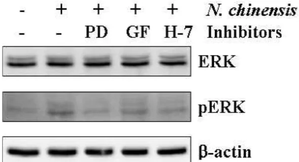
7. ERK 활성에 대한 PKC활성억제 효과
PKC는 세포분화의 MAPKs신호경로에서 upstream에 위치 해 있다. 甘松香 물추출물에 의해 유도된 U937세포의 분화에서 PKC/ERK 경로의 관련을 조사하기 위해, U937세포는 PD98059, GF 109293X, H-7를 1시간 전 처리한 후, 甘松香 물추출물을 처 리하였다. PKC inhibitor인 GF 109293X와 H-7은 甘松香 물추출 물에 의해 증가된 ERK 활성을 억제시켰다(Fig. 7).
Fig. 7. Involvement of PKC/ERK pathway in U937 cell differentiation induced byN. chineisis.The cells were treated with 20 μM PD98059 (PD), 4 μM GF 109203X (GF) or 10 μM H-7 for 1 h. The activation of ERK was determined by Western blot analysis with an anti-ERK and anti-p-ERK antibodies 3 day after treatment with N. chinensis.
고 찰
백혈병은 비교적 낮은 빈도로 발생되지만, 급성 또는 만성 백혈병 형태로 나타날 수 있는 혈액의 악성종양이며, 조혈세포의 분화장애로 인해 발병한다. 정상에서의 조혈 모세포는 self-renewal 기능이 있을뿐 아니라 다양한 혈액세포로 분화되며, 골수에서는 세포의 성장, 분화 및 사멸을 조절하여 정상적인 혈 액세포의 분포를 유지하게 한다. 그러나 백혈병이 발생하면 조혈 전구세포 등은 다양한 혈구세포로의 분화가 이루어지지 않고 immotalization 됨으로써 수많은 백혈병 세포가 만들어져 현재 백혈병치료는 과다 증식하는 백혈병세포를 세포사멸로 유도하거 나 분화를 유도하는 쪽으로 수행되고 있다12).
최근 연구에서 甘松香의 뿌리와 근경의 에탄올추출물로부터 새로운 diterpene구조의 10-isopropyl-2,2,6-tri- methyl-2,3,4,5-tetrahydro -naphtha[1,8-bc]oxocine-5,11-diol과 새 로운 monoteperne구조의, 6-hydroxy-7-(hydrox- ymethyl)-4-methylenehexahydro-cyclopenta[c]pyran-1(3H)-one 이 분리되었고 또한 이미 잘 알려진 sesquiterpenes, delta1(10)-a- ristolene-9beta-ol, debilon, nardosinone, and kanshone A도분리 되었다13). 하지만 새로이 분리된 terpenoid계열의 두화합물에 대 한 생물학적 활성은 보고되지 않았다. Sesquiterpenes는 endo-
peroxide에 대한 일부 효과로 인하여 antimalarial activity를 가 지는 것으로 보고되었다14). 특히 甘松香으로부터 분리된 nardo- sinone은 신경분화를 촉진시키는 화합물이다15). 또한 구16)등은 甘松香의 정유성분이 중추신경 억제효과가 있는 것으로 보고하 였으며, Yoon4)등은 甘松香 물추출물이 HL-60 세포의 분화를 유 도하는 것으로 보고하였고, Lee17)등은 甘松香 물추출물이 B16F10 세포의 멜라닌합성을 억제하는 효과가 있는 것으로 보고 하였다. 이 외에 항산화효과 및 항염효과와 수지상세포 성숙효과 가 있는 것으로 보고되었다18-20).
분화유도물질은 세포증식 억제효과를 가지고 있다. 항증식효과 는 말기분화에 밀접한 관련이 있다21-24). 말기분화는 분화된 세포 의 유형에 따라서 비가역적인 증식능의 상실이 자발적으로 일어 나거나 분화유도물질에 의하여 일어난다. 甘松香 물추출물은 U937세포에 대하여 세포증식 억제효과를 나타냈었고, 100 μ g/ml 농도의 甘松香 물추출물에서 과립성 백혈구표지자인 CD11b의 발현을 증가시켰다. 이 결과는 甘松香 물추출물이 U937세포의 증식억제와 분화유도효과를 가지는 것으로 보이고, 또한 甘松香 물추출물에 의한 U937세포의 분화가 과립백혈구 계 통으로 유도되었다는 것을 보여주고 있다.
세포의 분화유도는 PKC25,26)나 MAPKs28-31)와 같은 다양한 신호전달기전의 활성을 요구한다. MAPKs 신호전달기전은 백혈 병 세포주의 단핵백혈구와 과립백혈구분화에 수반되고 있다32,33). ERK경로는 백혈병세포를 단핵백혈구와 과립백혈구로 분화하는 데 요구된다. 하지만 p38경로는 단핵백혈구분화에서 음성적인 조절효과를 가지지만, 적혈구분화를 촉진시킨다32,33). 최근 연구 들은 all-trans retinoic acid (ATRA)에 의해 유도된 과립백혈구분 화는 ERK경로를 활성화 시키지만28,36), p38과 JNK경로와는 관련 이 없는 것으로 보고되었다. 대조적으로 1,25-dihydroxyvitamin D3에 의해 유도된 단핵밸혈구분화는 일시적으로 ERK 경로를 활 성화시켜서 분화초기에 관여하고29), 주로 JNK경로 활성이 밀접 한 관련이 있는 것으로 보고되었다30). 또한 JNK활성은 단핵백혈 구분화에서 p38 키나아제의 억제에 의해 강화된다31). 결국, ATRA에 의한 과립백혈구분화와 1,25-dihydroxyvitamin D3에 의한 단핵백혈구분화에서 ERK활성은 세포분화에 필수적이다.
甘松香 물추출물에 의해 유도된 U937세포의 분화에서 ERK 활성 은 증가하였지만, p38 활성과 JNK 활성에는 아무런 영향을 미치 지 못하였다. 또한 ERK 활성 억제는 甘松香 물추출물의 U937 세 포의 분화유도 효과를 감소시켰다. 이 결과는 ERK 활성 경로가 甘松香 물추출물에 의한 U937세포의 과립백혈구 분화를 중재하 고 있다는 것을 보여주고 있다.
PKC는 MAPKs신호경로에서 upstream에 위치해 있다. 세포 분화의 증진에서 PKC의 중요성은 현재 보편적으로 인정되고 있
다9,10). PKC는 최소 12개의 serine/threonine protein kinase
isoform들의 family로 이루어졌고 다양한 세포내의 반응을 수반
한다35,36). 특히 PKC β는 TPA에 의해 유도된 골수성백혈병세포
의 분화에 수반된다37-40). PKC β 발현의 저하와 PKC β의 기능적 결함은 TPA저항성 U937 변이세포에서도 발견된다41). 또한 세포 질에서 세포막으로 PKC β의 불완전한 전위는 TPA저항성 변이
U937세포에서 보여진다42,43). PKC β는 βⅠ과 βⅡ의 두가지 isoform으로 발현된다. 더 나아가, GF 109293X, chelerythrine, H-7과 같은 PKC inhibitor들은 세포분화의 증진을 막는다44-46). 甘松香 물추출물에 의해 유도된 U937세포의 분화에서 PKC 활성과 PKC βⅠ, βⅡ의 발현을 증가시켰다. 또한 PKC 활성억제 는 甘松香 물추출물에 의해 유도된 U937세포 분화를 감소시켰 다. 또한 PKC 활성억제는 甘松香 물추출물에 의해 증가된 ERK 활성을 억제시켰다. 이 결과들은 PKC가 ERK의 upstream에 존 재하면서 甘松香 물추출물이 U937세포에서 PKC활성과 발현을 증가시켜 ERK활성을 유도하여 HL-60세포의 과립성 백혈구 분 화를 유도한다는 것을 보여주고 있다.
결 론
본 연구에서는 甘松香 물추출물에 대한 U937세포의 분화유 도효과와 그에 대한 분화기전을 분자생물학적 방법을 통하여 유 의한 결과를 얻었으며, 다음과 같은 결론을 얻을 수 있었다.
甘松香 물추출물은 U937세포를 과립성 백혈구로 분화시켰 다. 甘松香 물추출물에 의해 유도된 U937세포 분화에서ERK 활 성이 관여하였다. 甘松香 물추출물에 의해 유도된 U937세포 분 화에서 PKC 활성이 관여하였다. 甘松香 물추출물에 의해 유도된 PKC 활성은 ERK 활성 증가를 유도하였다.
이상의 결과를 종합하면, 甘松香 물추출물은 U937세포의 분 화유도에 영향을 미치는 것을 확인하였으며, 甘松香 물추출물은 U937세포에서 PKC βⅠ/Ⅱ-ERK 신호전달경로를 통하여 과립백 혈구 분화를 유도하였고, 이 결과들은 백혈병치료에서 甘松香의 사용 가능성을 제시하고 있다.
감사의 글
이 논문은 2010년도 원광대학교의 교비 지원에 의해 수행됨.
참고문헌
1. Mayer, R.J., Davis, R.B., Schiffer, C.A., Berg, D.T., Powell, B.L., Schulman, P., Omura, G.A., Moore, J.O., McIntyre, O.R. and Frei, E. 3rd. Intensive post-remission chemotherapy in adult with acute myeloid leukemia, Cancer and Leukemia Group B. N. Engl. J. Med. 331:
896-903, 1994.
2. Hoffman, R., Benz, E.J. Jr., Shattil, S.J., Furie, B., Cohen, H.J., Silberstein, L.E. and McGlave, P. Hematology. Basic principles and practice. 3rd ed., New York, Churchill Livingstone, p 1577, 2000.
3. Pharmacopoeia of the People's Republic of China, Vol. I;
Chemical Industry Press: Beijing p 65, 2000.
4. Yoon, S.H., Ju, S.M., Kim, N.S., Park, S.C., Kim, S.H., Song, Y.S. and Jeon, B.H. Extracellular Signal-regulated Kinase
(ERK) is Required for Water Extract of Nardostachys chinersis-Induced Differentiation in HL-60 Cells Kor. J. Ori.
Physi. Patho. 20: 1415-1320, 2006.
5. Cross, T.G., Scheel-Toellner, D., Henriquez, N.V., Deacon, E., Salmon, M. and Lord, J.M. Serine/threonine protein kinases and apoptosis. Exp. Cell Res. 256: 34-41, 2005.
6. Pearson, G., Robinson, F., Beers Gibson, T., Xu, B.E., Karandikar, M., Berman, K. and Cobb, M.H.
Mitogen-activated protein (MAP) kinase pathways:
regulation and physiological functions. Endocr. Rev. 22:
153-183, 2001.
7. Cobb, M.H. MAP kinase pathways. Prog. Biophys. Mol.
Biol. 71: 479-500, 1999.
8. Xia, Z., Dickens, M., Raingeaud, J., Davis, R.J. and Greenberg, M.E.Opposing effects of ERK and JNK-p38 MAP kinases on apoptosis. Science 270: 1326-1331, 1995.
9. Caponigro, F., French, R.C. and Kaye, S.B. Protein kinase C:
a worthwhile target for anticancer drugs? Anticancer Drugs 8: 26-33, 1997.
10. Nishikawa, M. and Shirakawa, S. The expression and possible roles of protein kinase C in haematopoietic cells.
Leuk. Lymphoma 8: 201-211, 1992.
11. Komada, F., Nishikawa, M., Uemura, Y., Morita, K., Hidaka, H. and Shirakawa, S. Expression of three major protein kinase C isozymes in various types of human leukemic cells Cancer Res. 51: 4271-4278, 1991.
12. Tsiftsoglou, A.S., Pappas, I.S. and Vizirianakis, I.S.
Mechanisms involved in the induced differentiation of leukemia cells. Pharmacol. Ther. 100: 257-290, 2003.
13. Zhang, Y., Lu, Y., Zhang, L., Zheng, Q.T., Xu, L.Z. and Yang, S.L.from the roots and rhizomes of Nardostachys chinensis. J. Nat. Prod. 68: 1131-1133, 2005.
14. Tanitsu, M.A., Takaya, Y., Akasaka, M., Niwa, M. and Oshima, Y. Guaiane- and aristolane-type sesquiterpenoids of Nardostachys chinensis roots. Phytochemistry 59:
845-849, 2002.
15. Li, P., Yamakuni, T., Matsunaga, K., Kondo, S. and Ohizumi, Y. Nardosinone enhances nerve growth factor-induced neurite outgrowth in a mitogen-activated protein kinase- and protein kinase C-dependent manner in PC12D cells. J. Pharmacol. Sci. 93: 122-125, 2003.
16. 구병수, 김대근, 최정현, 이동웅 감송향 정유성분의 흡입 및 경구투여시의 중추신경 억제효과. 생명과학회지 16: 156-161, 2006.
17. Lee, S.J., Choi, Y.H. and Choi, B.T. Inhibitory Effects of Aqueous Extracts from Nardostachys chinensis on α -Melanocyte Stimulating Hormone-induced Melanogenesis in B16F10 Cells. Integrative Biosci. 10: 223-236, 2006.
18. 박철, 정민, 서은아, 권강범, 유도곤 감송향물추출물의 HO-1 발현 촉진을 통한 세포보호 작용 및 항염효과. 동의생리병리 학회지24: 624-629, 2010.
19. 백설, 최재혁, 고성훈, 이용재, 차동석, 박은영, 강양규 전훈 감송향의 in vitro 항산화 및 항염증 효과. 동의생리병리학회 지 23: 853-859, 2009.
20. 오광우, 정지해, 정현철, 조한백, 김송백, 최창민감송향이 수 지상세포 성숙에 미치는 영향. 대한한방부인과학회지 23:
14-25, 2010.
21. Furukawa, Y., Uenoyama, S., Ohta, M., Tsunoda, A., Griffin, J.D. and Saito, M.Transforming growth factor-beta inhibits phosphorylation of the retinoblastoma susceptibility gene product in human monocytic leukemia cell line JOSK-I. J. Biol. Chem. 267: 17121-17127, 1992.
22. Hui, E.K. and Yung, B.Y. Cell cycle phase-dependent effect of retinoic acid on the induction of granulocytic differentiation in HL-60 promyelocytic leukemia cells.
Evidence for sphinganine potentiation of retinoicacid-induced differentiation. FEBS Lett. 318:
193-199, 1993.
23. Cooper, S. Revisiting the relationship of the mammalian G1 phase to cell differentiation. J. Theor. Biol. 208: 399-402, 2001.
24. Dimberg, A. and Oberg, F. Retinoic acid-induced cell cycle arrest of human myeloid cell lines. Leuk. Lymphoma 44:
1641-1650, 2003.
25. Caponigro, F., French, R.C. and Kaye, S.B. Protein kinase C:
a worthwhile target for anticancer drugs? Anticancer Drugs 8: 26-33, 1997.
26. Nishikawa, M. and Shirakawa, S. The expression and possible roles of protein kinase C in haematopoietic cells.
Leuk. Lymphoma 8: 201-211, 1992.
27. Yen, A., Roberson, M.S., Varvayanis, S. and Lee, A.T.
Retinoic acid induced mitogen-activated protein (MAP)/extracellular signal-regulated kinase (ERK) kinase-dependent MAP kinase activation needed to elicit HL-60 cell differentiation and growth arrest. Cancer Res.
58: 3163-3172, 1998.
28. Yen, A., Roberson, M.S. and Varvayanis, S. Retinoic acid selectively activates the ERK2 but not JNK/SAPK or p38 MAP kinases when inducing myeloid differentiation. In Vitro Cell Dev. Biol. Anim. 35: 527-532, 1999.
29. Wang, X. and Studzinski, G.P. Activation of extracellular signal-regulated kinases (ERKs) defines the first phase of 1,25-dihydroxyvitamin D3-induced differentiation of HL60 cells. J. Cell Biochem. 80: 471-482, 2001.
30. Wang, X., Rao, J. and Studzinski, G.P. Inhibition of p38 MAP kinase activity up-regulates multiple MAP kinase
pathways and potentiates 1,25-dihydroxyvitamin D(3)-induced differentiationof human leukemia HL60 cells.
Exp. Cell Res. 258: 425-437, 2000.
31. Wang, X. and Studzinski, G.P. Inhibition of p38MAP kinase potentiates the JNK/SAPK pathway and AP-1 activity in monocytic but not in macrophage or granulocytic differentiation of HL60 cells. J. Cell Biochem. 82: 68-77, 2001.
32. Miranda, M.B., McGuire, T.F. and Johnson, D.E. Importance of MEK-1/-2 signaling in monocytic and granulocytic differentiation of myeloid cell lines. Leukemia 16: 683-692, 2002.
33. Yen, A., Roberson, M.S., Varvayanis, S. and Lee, A.T.
Retinoic acid induced mitogen-activated protein (MAP)/extracellular signal-regulated kinase (ERK) kinase-dependent MAP kinase activation needed to elicit HL-60 cell differentiation and growth arrest. Cancer Res.
58: 3163-3172, 1998.
34. Gate, L., Lunk, A. and Tew, K.D. Resistance to phorbol 12-myristate 13-acetate-induced cell growth arrest in an HL60 cell line chronically exposed to a glutathione S-transferase pi inhibitor. Biochem. Pharmacol. 65:
1611-1622, 2003.
35. Jaken, S. Protein kinase C isozymes and substrates. Curr.
Opin. Cell Biol. 8: 168-173, 1996.
36. Nishizuka, Y. The molecular heterogeneity of protein kinase C and its implications for cellular regulation.
Nature 334: 661-665, 1998.
37. MacFarlane, D.E. and Manzel, L. Activation of β-isozyme of protein kinase C (PKCβ) is necessary and sufficient for phorbol ester-induced differentiation of HL-60 promyelocytes. J. Biol. Chem. 269: 4327-4331, 1994.
38. Nishikawa, M., Komada, F., Uemura, Y., Hidaka, H. and Shirakawa, S. Decreased expression of type II protein kinase C in HL-60 variant cells resistant to induction of cell differentiation by phorbol diester. Cancer Res. 50:
621-626, 1990.
39. Tonetti, D.A., Henning-Chubb, C., Yamanishi, D.T. and Huberman, E. Protein kinase C-β isrequired for macrophage differentiation of human HL-60 leukemia cells.
J. Biol. Chem. 269: 23230-23235, 1994.
40. Tonetti, D.A., Horio, M., Collart, F.R. and Huberman, E.
Protein kinase C beta gene expression is associated with susceptibility of human promyelocytic leukemia cells to phorbol ester-induced differentiation. Cell Growth Differ.
3: 739-745, 1992.
41. Kiley, S.C., Adams, P.D. and Parker, P.J. Cloning and characterization of phorbol ester differentiation-resistant
U937 cell variants. Cell Growth Differ. 8: 221-230, 1997.
42. Hass, R., Hirano, M., Kharbanda, S., Rubin, E. and Kufe, D.
Resistance to phorbol ester-induced differentiation of a U-937 myeloid leukemia cell variant with a signaling defect upstream to Raf-1 kinase. Cell Growth Differ. 4:
657-663, 1993,
43. Yang, K.D., Kharbanda, S., Datta, R., Huberman, E., Kufe, D. and Stone, R. All-trans retinoic acid reverses phorbol ester resistance in a human myeloid leukemia cell line.
Blood 83: 490-496, 1994.
44. Kim, S.H., Oh, S.M. and Kim, T.S. Induction of human
leukemia HL-60 cell differentiation via a PKC/ERK pathway by helenalin, a pseudoguainolide sesquiterpene lactone. Eur. J. Pharmacol. 511: 89-97, 2005.
45. Kim, S.H., Kim, H.J. andKim, T.S. Differential involvement of protein kinase C in human promyelocytic leukemia cell differentiation enhanced by artemisinin. Eur. J. Pharmacol.
482: 67-76, 2003.
46. Kang. S.N., Lee, M.H., Kim, K.M., Cho, D. and Kim, T.S.
Induction of human promyelocytic leukemia HL-60 cell differentiation into monocytes by silibinin: involvement of protein kinase C.Biochem. Pharmacol. 61: 1487-1495, 2001.