DOI: 10.4196/kjpp.2009.13.6.417
417
ABBREVIATIONS: BMMs, bone marrow-derived macrophages; ERK, extracellular signal-regulated kinase; HF6-FC, hexane soluble fraction of Ficus carica; JNK, c-Jun-N-terminal kinase; NFATc1, nuclear factor of activated T-cells, cytoplasmic, calcineurin-dependent 1;
RANKL, receptor activator of NF-κB ligand; TRAF6, TNF receptor- associated factor 6.
Received September 15, 2009, Revised November 2, 2009, Accepted November 9, 2009
Corresponding to: Yunjo Soh, Department of Dental Pharmacology, School of Dentistry, Chonbuk National University, Duckjin-dong, Duckjin-gu, Jeonju 561-756, Korea. (Tel) 82-63-270-4038, (Fax) 82-63-270-4037, (E-mail) [email protected]
*The first two authors contributed equally to this work.
Hexane-Soluble Fraction of the Common Fig, Ficus carica, Inhibits Osteoclast Differentiation in Murine Bone Marrow-Derived Macro- phages and RAW 264.7 Cells
Young Ran Park1,*, Jae Soon Eun2,*, Hwa Jung Choi1, Manoj Nepal1, Dae Keun Kim2, Seung-Yong Seo2, Rihua Li2, Woo Sung Moon3, Nam-Pyo Cho4, Sung-Dae Cho4, Tae Sung Bae5, Byung Il Kim6, and Yunjo Soh1
1Department of Dental Pharmacology, School of Dentistry, and Institute of Oral Bioscience, Brain Korea 21 Project, Chonbuk National University, Jeon-Ju 561-756, 2College of Pharmacy, Woosuk University, Sam-Rye 565-701, 3Department of Pathology, School of Medicine,
4Department of Oral Pathology, School of Dentistry, 5Department of Dental Biomaterials, School of Dentistry, Chonbuk National University, Jeon-Ju 561-756, 6Department of Materials Science and Metallurgical Engineering, Sunchon National University, Sunchon 540-742, Korea
Osteoclasts, derived from multipotent myeloid progenitor cells, play homeostatic roles in skeletal modeling and remodeling, but may also destroy bone in pathological conditions such as osteoporosis and rheumatoid arthritis. Osteoclast development depends critically on a differentiation factor, the receptor activator of NF-κB ligand (RANKL). In this study, we found that the hexane soluble fraction of the common fig Ficus carica (HF6-FC) is a potent inhibitor of osteoclastogenesis in RANKL-stimulated RAW264.7 cells and in bone marrow-derived macrophages (BMMs). HF6-FC exerts its inhibitory effects by suppression of p38 and NF-κB but activation of ERK. In addition, HF6-FC significantly decreased the expression of NFATc1 and c-Fos, the master regulator of osteoclast differentiation. The data indi- cate that components of HF6-FC may have therapeutic effects on bone-destructive processes such as osteoporosis, rheumatoid arthritis, and periodontal bone resorption.
Key Words: Ficus carica, Osteoclast differentiation, RAW264.7 cells, Bone marrow-derived macrophages, Bone lytic diseases
INTRODUCTION
Ficus carica L. (Moraceae), the common Fig, is widely distributed in South Korea, and the leaves of this plant have been used as folk medicine for hemorrhoids, neuralgia, warts, diarrhea and carbuncles in Korea and China (Lee, 1996). The leaves of this plant contain terpenoid, saponin and flavonoid compounds (el-Kholy and Shaban, 1966; Ahmed et al., 1988), which may be related to their alleged antiviral effects on herpes simplex virus (HSV), and hypoglycemic activity in type-I diabetic patients (Serraclara et al., 1998;
Canal et al., 2000). But no study has yet reported the regu- latory effects of F. carica leaf on osteogenic differentiation.
A balance between osteoclast-regulated bone resorption and osteoblast-induced bone formation determines bone mass in adults. Perturbations in this balance contribute to diseases such as osteoporosis, osteomalacia, and osteopetrosis (Alliston et al., 2002; Oh et al., 2007). The multinucleated osteoclasts are generated from hematopoietic monocyte/
macrophage precursor cells under the control of two cyto-
kines, macrophage colony-stimulating factor (MCSF) and receptor activator of NF-κB ligand (RANKL). Osteoblasts produce these two cytokines in membrane-bound or secreted form, when activated by interleukin-1, prostaglandin E2, and vitamin D3 (Miyaura et al., 2003; Wei et al., 2005).
RANKL induces precursor cells to differentiate into os- teoclasts (Theill et al., 2002), whereas M-CSF provides an osteoclast survival signal (Yoshida et al., 1990). RANKL is expressed in osteoblasts and induces the signaling essential for precursor cells to differentiate into osteoclasts (Theill et al., 2002), whereas M-CSF, secreted by osteoblasts, pro- vides the survival signal to these cells (Yoshida et al., 1990).
Binding of RANKL to its receptor RANK activates TNF re- ceptor-associated factor 6 (TRAF6), which is linked to the nuclear factor κB (NF-κB) and mitogen-activated protein kinases (MAPKs) (Kobayashi et al., 2001; Lee et al., 2002).
Active extracellular signal-regulated kinase (ERK) can di- rectly phosphorylate c-Fos and active c-Jun-N-terminal kin- ase (JNK) phosphorylates c-Jun. Thus AP-1 transcription factor, a heterodimer composed of a Fos family (c-Fos, FosB, Fra-1, and Fra-2) and a Jun family (c-Jun, JunB, and JunD) protein, can be a target of ERK and JNK in response to
RANKL in osteoclast precursor cells. In addition, RANKL induces the key transcription factor for osteoclastogenesis, nuclear factor of activated T cells c1 (NFATc1) (Zhou et al., 2002; Yamashita et al., 2007).
In this study, we assessed the effects of the hexane solu- ble fraction of F. carica leaf (HF6-FC) on RANKL-induced osteoclastogenesis in murine monocytes/macrophage RAW 264.7 cells and bone marrow-derived macrophages (BMMs).
HF6-FC significantly suppressed osteoclast differentiation in both situations, and inhibited RANKL-induced activa- tion of NF-κB and c-Fos. Our results suggest that HF6-FC may be useful as a therapeutic agent for bone lytic diseases such as osteoporosis, osteomalacia, and osteopetrosis.
METHODS Reagents
The leaves of F. carica were collected in July 2008 in Youngam, Chonnam, Korea and air-dried. A voucher speci- men was deposited in the herbarium of the College of Pharmacy, Woosuk University (WSU-08-025). Murine mon- ocyte/marcrophage RAW 264.7 cells were purchased from the American Type Culture Collection (ATCC, Manassas, VA, USA). Cell culture media and supplements, including minimum essential medium-alpha (α-MEM), Dulbecco’s modified Eagle’s medium (DMEM), fetal bovine serum (FBS), penicillin/streptomycin, trypsin-EDTA and TRIzol were obtained from GIBCO (Invitrogen Inc, NY, USA).
RANKL was purchased from PeproTech (Rocky Hill, NJ, USA); macrophage-colony stimulating factor (M-CSF), from R&D Systems (Minneapolis, MN, USA); antibodies against Akt, ERK, p38, JNK, IκBα, phospho-ERK, phospho-p38, phospho-JNK and phospo-IκBα, from Cell Signaling Tech- nology (Beverly, MA, USA); and anti-β-actin antibody, from Sigma-Aldrich (St. Louis, MO, USA). All other chemicals were purchased from Sigma and/or the same distributors used in previous studies (Soh et al., 2000; Soh et al., 2003;
Choi et al., 2008), unless otherwise indicated.
Extraction, solvent fractionation and identification of fig leaf components
The shade-dried plant material (600 g) was extracted three times with methanol at room temperature and filtered. The extracts were combined and evaporated in va- cuo at 40oC. The resulting methanolic extract (60 g) was partitioned successively with n-hexane, methylene chloride, ethylacetate and n-butanol. The n-hexane-soluble extract, which had the most anti-osteoclastogenic activity, was frac- tionated on a silica gel column (n-hexane-ethyl acetate, 10:1-1:1) to give eight fractions (H1-H8). Of these eight, fraction H6, the most active one (HF6-FC), was analyzed by GS-MS to identify its constituents.
Cell culture and Isolation of bone marrow-derived macrophages
The murine monocyte/macrophage cell line RAW264.7 was cultured with DMEM containing 10% heat-inactivated FBS, penicillin (100 U/ml), and streptomycin (100 μg/ml).
All cells were grown in a humidified atmosphere containing 5% CO2 at 37oC. To induce osteoclast differentiation, RAW264.7 cells were suspended in α-MEM containing 10%
FBS, 2 mM L-glutamate, 100 U/ml penicillin, and 100 μg/ml streptomycin; seeded at 3×10³ cells/well in 96-well culture plates; and cultured with 50 ng/ml soluble RANKL for 6 days. The medium and HF6-FC were changed every two or three days. Six-week-old ICR (Institute of Cancer Research) mice were purchased from Damool Science (Daejeon, Korea).
Cells obtained from tibia and femur bone marrow were cul- tured in α-MEM with 10% fetal bovine serum (FBS) con- taining 30 ng/ml macrophage colony-stimulating factor (M-CSF). After culturing for three days, the nonadherent cells were removed by washing and adherent cells were used as bone marrow-derived macrophages (BMMs). BMM were cultured for three days in medium containing M-CSF (30 ng/ml) and RANKL (200 ng/ml).
MTT assay
Cells (5×10³ per well) were seeded in a 96-well plate with medium supplemented with 10% FBS and incubated for 24 h. Cells were treated with various concentrations of HF6-FC for 24 h, then washed three times with phos- phate-buffered saline (PBS) and treated with medium con- taining 100 μg/ml of 3-(4,5-dimethylthiazol-2-yl)-2,5-diphe- nyltetrazolium bromide (MTT) for 2 h at 37oC. Cells were then washed with PBS and solubilized in 200 μl of DMSO.
The intracellular purple formazan concentrations were de- termined from the absorbance at 540 nm.
Tartrate-resistant acid phosphatase (TRAP) staining TRAP staining was performed as described (Han et al., 2007). Briefly, cells were washed with PBS and fixed with 3.7% formaldehyde for 10 min. After washing with PBS, cells were made permeable with 0.1% (v/v) Triton X-100 for 1 min and washed with distilled water. Cells were in- cubated at 37oC in a humid and light-protected incubator for 40 min in the reaction mixture of the Leukocyte Acid Phosphatase Assay kit (Sigma, Cat No. 387), as directed by the manufacturer. Cells were washed three times with distilled water and TRAP-positive multinucleated cells con- taining three or more nuclei were counted under a light microscope.
RT-PCR analysis
Total RNA was isolated from cultured cells using TRIzol reagent (Invitrogen, Carlsbad, CA, USA), and cDNA was synthesized using SuperScript II reverse transcriptase (Invitrogen) according to the manufacturer’s instruction.
PCR was performed with mouse-specific primers, shown here with the corresponding gene sequences: c-Fos, 5’-atg- ggctctcctgtcaacac-3’ (forward) and 5’-tggagtttattttggcagcc-3’
(reverse); NFATc1, 5’-gggtcagtgtgaccgaagat-3’ (forward) and 5’-tccacccacttctgacttcc-3’ (reverse); GAPDH, 5’-accacagtccatg- ccatcac-3’ (forward) and 5’-tacagcaacagggtggtgga-3’ (reverse).
Thermal cycling parameters were 95°C for 5 min, followed by 25∼35 cycles for 30 sec at 95oC, 30 sec at 55∼60oC, and 30 sec at 72oC, and 10 min at 72oC for the final elongation. The number of cycles for each gene was de- termined to be in the range of linear amplification through an optimization experiment. PCR products were separated on 1.5% agarose gels, visualized by ethidium bromide stain- ing, and analyzed densitometrically using a Phosphoimager and Quantity One software (Version 4.3.1) (Bio-Rad, Hercules, CA). The optical densities for each gene were nor-
Fig. 1. GC/MS spectrum of HF6-FC.
malized to the corresponding values for glyceraldehyde-3- phosphate dehydrogenase (GAPDH).
Western blot analysis
Cells were harvested, washed three times with ice-cold phosphate buffered saline containing 1 mM sodium vana- date, and lysed in a buffer containing 20 mM Tris-HCl (pH 7.5), 137 mM NaCl, 10% glycerol, 1% Triton X-100, 1 mM Na3VO4, 1 mM phenylmethylsulfonylfluoride (PMSF), and 1×protease inhibitor cocktail. Total cell lystes were in- cubated for 20 min and centrifuged at 16,000× g for 15 min at 4oC, and supernatants were used as cell extracts. Cell extracts (30∼40 μg) were separated on an 8∼10% sodium dodecyl sulfate polyacrylamide gel (SDS-PAGE) and then transferred onto a polyvinylidene difluoride (PVDF) mem- brane (Bio-Rad). The membranes were blocked for 1 h at room temperature with 5% nonfat skim milk in Tris-buf- fered saline (TBS) containing 0.1% Tween-20 (TTBS), and then probed with specific antibodies in 5% nonfat skim milk in TTBS, for 16 h at 4oC. Horseradish peroxidase-conjugated anti-rabbit or mouse antibodies (Santa Cruz Biotechnology) were used as secondary antibodies (1:5,000∼1:10,000 dilution in 5% nonfat skim milk in TTBS) for 1 h incubation at room temperature. The antigen-antibody complexes were detected with an ECL Plus kit (Amersham Biosciences, Piscataway, NJ, USA).
Statistical analysis
All data are expressed as the mean±SD of three or more replicates, unless otherwise indicated. Group data were compared using one-way analysis of variance (ANOVA) fol- lowed by a Student’s t-test, and p values less than 0.05 were considered significant.
RESULTS
Characterization of the hexane soluble fraction of F.
carica
The n-hexane soluble fraction of F. carica showed a sig- nificant anti-osteoclastogenic activity in the murine mono- cyte/macrophage cell line RAW 264.7. Of the eight silica gel fractions (H1-H8), H6 (HF6-FC) exhibited the strongest anti-osteoclastogenic effect (data not shown). Analysis by GS-MS (Fig. 1) identified the following components of HF6- FC: octadecane, pentadecane, hexadecane, heptadecane, oc- tadecane, 2H-1-benzopyran-2-one, nonadecane, hexadecanoic acid methyl ester, octadecanoic acid methyl ester, tride- cane, tetradecane, eicosane, 9,12,-octadecadienoic acid meth- yl ester and 8-octadecenoic acid. HF6-FC was used for all cell experiments. DMSO was used as control vehicle to solu- bilize HF6-FC.
Effect of HF6-FC on osteoclastogenesis in murine mo- nocyte/macrophage RAW 264.7 cells
Using the murine monocyte/macrophage cell line RAW 264.7 as a model for osteoclastogenesis, we found that RANKL (50 ng/ml) induced TRAP-positive multinucleated osteoclasts. The HF6-FC, however, inhibited osteoclast dif- ferentiation in a concentration-dependent manner (Fig. 2A), reducing the numbers of TRAP-positive multinucleated cells by 34.3±4.2% and 88.6±1.5% at 1 and 10 μg/ml, re- spectively (Fig. 2B). To test the effects of HF6-FC on cell growth, cells were treated with various concentrations of HF6-FC for 24 h and growth and viability were tested in an MTT assay. In this study, the HF6-FC did not did not adversely affect the cell growth rate of RAW 264.7 cells (Fig. 2C). Hence, the anti-osteoclastogenic action of HF6-FC does not apparently stem from cell toxicity.
Fig. 2. Effect of HF6-FC on RANKL-induced osteoclastogenesis in RAW 264.7 cells. (A) RAW 264.7 cells were cultured in the presence of RANKL (50 ng/ml) for 6 days. HF6-FC was added to the culture medium at final concentrations of 0.1, 1, and 10 μg/ml, and cells were stained for TRAP activity on day 6. (B) TRAP-positive multi- nucleated osteoclasts were counted. Data represent the mean±SD of three independent experiments. *p<0.05 and **p<0.01, as com- pared to the control without HF6-FC, respectively. (C) RAW 264.7 cells were seeded into 96-well plates and incubated with HF6-FC for 24 h. Cell proliferation was evaluated with the MTT assay.
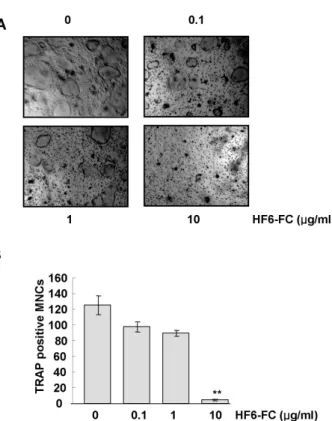
Fig. 3. Effect of HF6-FC on RANKL-induced osteoclastogenesis in bone marrow-derived macrophages (BMM). (A) BMMs were cultured in the presence of M-CSF (20 ng/ml) and RANKL (100 ng/ml) for 3 days. HF6-FC was added to the culture medium at final concentrations of 0.1, 1, and 10 μg/ml. (B) After three days, cells were fixed and stained for TRAP, and TRAP-positive multinuclear cells (MNC) were counted. Data represent the mean±SD of three independent experiments. **p<0.01 versus the control without HF6-FC.
Effect of HF6-FC on osteoclastogenesis in bone marrow- derived macrophages
Bone marrow-derived macrophages (BMMs) were induced to differentiate into osteoclasts in the presence of M-CSF and RANKL. In this system, HF6-FC reduced the formation of TRAP-positive multinucleated cell (MNC) in a concen- tration-dependent manner (Fig. 3A), with almost complete inhibition of osteoclastogenesis at 10 μg/ml and reductions in TRAP-positive multinucleated cells by 28.7±3.8% and 96.4±0.7% at 1 μg/ml and 10 μg/ml, respectively (Fig. 3B).
HF6-FC did not affect the growth of BMMs at the concen- trations used in this study (data not shown).
Effect of HF6-FC on MAPKs in RANKL-stimulated RAW 264.7 cells
The mitogen-activated protein kinases (MAPKs), which include the ERK, JNK, and p38 kinase families, participate in RANKL-induced osteoclast differentiation. To inves- tigate the role of MAPK signaling in the anti-osteoclasto- genic action of HF6-FC, we tested the effect of the extract on ERK, JNK, and p38 activities in RANKL-induced RAW264.7 cells. We measured these activities by immuno- blot analysis, using antibodies specifically directed against the phosphorylated forms of the enzymes, and for compar- ison, antibodies specific for the unphosphorylated forms. As shown in Fig. 4, RANKL induced both ERK and p38. HF6- FC further increased the ERK activity, but inhibited the RANKL-induced p38 kinase activation.
NF-κB expression in RANKL-stimulated RAW 264.7 cells
Activation of the transcription factor NF-κB is an essen- tial step in osteoclast differentiation (Wong, 1998; Kotake, 1999). NF-κB activation occurs through release from in- hibitory κB (IκB), which is phosphorylated, ubiquitinated,
Fig. 4. Effect of HF6-FC on MAPK activation by RANKL in RAW 264.7 cells. Cells were serum-starved for 16 h, pretreated with or without HF6-FC (10 μg/ml) for 30 min, and stimulated with RANKL (100 ng/ml) for times indicated. Whole cell lystes were used for western blotting with MAPK-specific antibodies. Blots were stripped and reprobed with other antibodies.
Fig. 5. Effect of HF6-FC on NF-κB activation by RANKL in RAW 264.7 cells. Cells were serum-starved for 16 h, pretreated with or without HF6-FC (10 μg/ml) for 30 min and stimulated with RANKL (100 ng/ml) for indicated times. Whole cell lysates were immunoblotted with antibodies specific for phospho-IκB-α and phospo- p65. Blots were stripped and reprobed with control antibodies. The histo- grams represent the level of the pho- spho-IκB-α (A) and phospo-p65 (B).
The asterisk (*) indicates a signifi- cant difference (p<0.05) compared with the control (#) at same time period.
and degraded in the proteasome. In RANKL-treated RAW 264.7 cells, the level of phosphorylated IκB-αprotein in- creased after a 15 min exposure. The presence of HF6-FC, however, significantly reduced that increase (Fig. 5A). At 30 min, the RANKL treatment also increased the phosphor-
ylation state (and hence the transactivation potential) of p65 (Vaira et al., 2008). Addition of HF6-FC significantly suppressed the level of phosphorylated p65 (Fig. 5B). These results suggest that HF6-FC suppresses the NF-κB in- duction by RANKL.
Fig. 6. Effects of HF6-FC on c-Fos protein and NFATc1 mRNA in RANKL-stimulated RAW 264.7 cells. Cells were serum-starved for 16 h, pretreated with or without HF6-FC (10 μg/ml) for 30 min and stimulated with RANKL (100 ng/ml) for times indicated. (A) Whole cell lysates were analyzed by immunoblotting with c-Fos-and β-actin-specific antibodies. (B) NFATc1 mRNA level was deter- mined by RT-PCR and compared with that of GAPDH. The asterisk (*) indicates a significant difference (p<0.05) compared with the control (#) at same time period.
c-Fos and NFATc1 expression in RANKL-stimulated RAW 264.7 cells
In osteoclast precursor cells, RANKL increases the level of c-Fos, a key transcription factor in osteoclast differ- entiation (Wong et al., 1998; Chang et al., 2007). As shown in Fig. 6A, RANKL induced c-Fos protein in RAW 264.7 cells at 12 h. HF6-FC significantly restricted this increase in c-Fos. We also examined the regulation of nuclear factor of activated T-cells, cytoplasmic, calcineurin-dependent 1 (NFATc1) in RANKL-induced RAW 264.7 cells, and found that HF6-FC significantly reduced RANKL-induced in- crease of NFATc1 mRNA (Fig. 6B). These results suggest that HF6-FC suppresses c-Fos as well as NFATc1 induction by RANKL in the RAW 264.7 cells.
DISCUSSION
Bone destruction and inflammation are closely linked in diseases such as periodontitis and rheumatoid arthritis (Jimi et al., 2004). Inflammatory cytokines and prosta- glandins up-regulate RANKL in osteoblasts, synovial fibro- blasts and activated T cells (Mino et al., 1998; Coon et al., 2007), and signaling interactions of RANKL and RANK stimulate osteoclast formation, resorption activity, and sur- vival (Han et al., 2007). Regulation of osteoclast develop- ment and the signaling pathway of RANK have been well characterized (Teitelbaum and Ross, 2003).
The signaling mechanism of RANKL has been extensively studied. Cells of the osteoblast lineage express a membrane- bound form of RANKL, a member of the TNF cytokine family. RANK, like other members of the TNF receptor su- perfamily, strongly activates the NF-κB pathway. Binding of RANKL to its receptor RANK in BMMs recruits TRAF family proteins such as TRAF6, which interacts with NF-κB and JNK pathways (Wong et al., 1998; Takayanagi et al., 2002; Yamashita et al., 2007). In mammals, the NF-κB family has five members: RelA/p65, RelB, c-Rel, NF-κB1/p50, and NF-κB2/p52 (Vaira et al., 2008). In the canonical NF-κB pathway, ligation of RANK activates the inhibitor of the IκB kinase (IKK) complex, which phosphorylates NF-κB- associated IκBα, and leads to its ubiquitination and proteo- somal degradation. These events release NF-κB dimers containing RelA and c-Rel into the cytosol, from which they enter the nucleus to enhance transcription of target genes (Luo et al., 2005). HF6-FC inhibited NF-κB transcriptional activity as well as phosphorylation of IκB and p65, which RANKL markedly induces.
In addition to the NF-κB pathways, RANKL activates ERK, JNK and p38 in osteoclasts and their precursor cells (Yoshida et al., 1990; Lee et al., 2002; Chang et al., 2007).
Downstream targets of ERK and JNK include the AP-1 transcription factor. While ERK can induce and activate c-Fos, JNK increases AP-1 transcriptional activity through c-Jun phosphorylation. In differentiated osteoclasts, ERK activity correlates with cell survival, but not with re- sorption function (Miyazaki et al., 2000). While HF6-FC in- hibited the RANKL-induced p38 kinase activation, it did not inhibit the ERK activation, but instead prolonged ERK activity, suggesting that HF6-FC influences osteoclast sur- vival through ERK, and osteoclastogenesis via p38 kinase.
Both RANKL and M-CSF activate Akt, a key regulator of osteoclast survival (Stern et al., 2007). RANKL activates Akt through the signaling sequence TRAF6-Src-PI 3-kinase (Wong et al., 1999).
The transcription factor NFATc1 also plays a critical role in RANKL-induced osteoclastogenesis (Takayanagi et al., 2002). Genes such as TRAP, cathepsin K, and MMP-9, which are specifically induced during terminal osteoclast differentiation, contain multiple NFAT and AP-1 binding sites (Reddy et al., 1995; Motyckova et al., 2001). Both AP-1 and NF-κB binding sites are present within the promoter region of the NFATc1 gene (Zhou et al., 2002). It is there- fore possible that these two transcription factors initiate induction of the gene, which is then followed by NFATc1- mediated autoamplification of gene induction. Accordingly, we investigated the up-regulation of specific genes such as MMP9, TRAP, RANK, and cathepsin K in RANKL-induced RAW 264.7 cells. However, HF6-FC did not affect the ex- pression of theses genes (data not shown).
In summary, HF6-FC inhibited the osteoclastic differ-
entiation of RANKL-stimulated macrophages and BMMs.
In RANKL-stimulated RAW 264.7 cells, HF6-FC inhibited the activation of c-Fos and p38 kinase, NFATc1 and NF-κB, possibly through the inhibition of IκB phosphorylation;
however, HF6-FC increased the ERK activity. Although further study is needed to confirm its effectiveness in vivo, HF6-FC may potentially provide a novel therapy for dis- orders associated with bone loss.
ACKNOWLEDGEMENTS
This work was supported by the Korea Research Foun- dation Grant funded by the Korean Government (MOEHRD) (The Regional Research Universities Program/Center for Healthcare Technology Development) and by the Korea Science and Engineering Foundation (KOSEF) grant fund- ed by the Korea government (MEST) (No. R01-2008-000- 20556-0).
REFERENCES
Ahmed W, Khan AQ, Malik A. Two Triterpenes from the Leaves of Ficus carica. Planta Med 54: 481, 1988.
Alliston T, Derynck R. Medicine: interfering with bone remodelling.
Nature 416: 686−687, 2002.
Canal JR, Torres MD, Romero A, Pérez C. A chloroform extract obtained from a decoction of Ficus carica leaves improves the cholesterolaemic status of rats with streptootocin-includede diabetes. Acta Physiol Hung 87: 71−76, 2000.
Chang EJ, Kim HJ, Ha J, Kim HJ, Ryu J, Park KH, Kim UH, Lee ZH, Kim HM, Fisher DE, Kim HH. Hyaluronan inhibits osteoclast differentiation via Toll-like receptor 4. J Cell Sci 120:
166−176, 2007.
Choi HJ, Song BJ, Gong YD, Gwak WJ, Soh Y. Rapid degradation of hypoxia-inducible factor-1alpha by KRH102053, a new activator of prolyl hydroxylase 2. Br J Pharmacol 154: 114−125, 2008 Coon D, Gulati A, Cowan C, He J. The role of cyclooxygenase-2 (COX-2) in inflammatory bone resorption. J Endod 33: 432−436, 2007.
El-Kholy IS, Shaban MA. Constituents of the leaves of Ficus carica, L. II. Isolation of a psi-taraxasteryl ester, rutin, and a new steroid sapogenin. J Chem Soc Perkin 13: 1140−1142, 1966.
Han KY, Yang D, Chang EJ, Lee Y, Huang H, Sung SH, Lee ZH, Kim YC, Kim HH. Inhibition of osteoclast differentiation and bone resorption by sauchinone. Biochem Pharmacol 74: 911−
923, 2007.
Jimi E, Aoki K, Saito H, D'Acquisto F, May MJ, Nakamura I, Sudo T, Kojima T, Okamoto F, Fukushima H, Okabe K, Ohya K, Ghosh S. Selective in hibition of NF-kappa B block osteoclas- togenesis and prevents inflammatory bone destructin in vivo.
Nat Med 6: 617−624, 2004.
Kobayashi N, Kadono Y, Naito A, Matsumoto K, Yamamoto T, Tanaka S, Inoue J. Segregation of TRAF6-mediated signaling pathways clarifies its role in osteoclastogenesis. EMBO 20: 1271−
1280, 2001.
Lee SE, Woo KM, Kim SY, Kim HM, Kwack K, Lee ZH, Kim HH.
The phosphatidylinositol 3-kinase, p38, and extracellular signal- regulated kinase pathways are involved in osteoclast differen- tiation. Bone 30: 71−77, 2002.
Lee WT. Coloured standard illustrations of Korean plants. Academy press, Seoul, p 624, 1996.
Luo JL, Kamata H, Karin M. IKK/NF-kappaB signaling: balancing life and death--a new approach to cancer therapy. J Clin Invest 115: 2625−2632, 2005.
Mino T, Sugiyama E, Taki H, Kuroda A, Yamashita N, Maruyama M, Kobayashi M. Interleukin-1alpha and tumor necrosis factor alpha synergistically stimulate prostaglandin E2-dependent
production of interleukin-11 in rheumatoid synovial fibroblasts.
Arthritis Rheum 41: 2004−2013, 1998.
Miyaura C, Inada M, Matsumoto C, Ohshiba T, Uozumi N, Shimizu T, Ito A. An essential role of cytosolic phospholipase A2alpha in prostaglandin E2-mediated bone resorption associated with inflammation. J Exp Med 197: 1303−1310, 2003.
Miyazaki T, Katagiri H, Kanegae Y, Takayanagi H, Sawada Y, Yamamoto A, Pando MP, Asano T, Verma IM, Oda H, Nakamura K, Tanaka S. Reciprocal role of ERK and NF-kappaB pathways in survival and activation of osteoclasts. J Cell Biol 148: 333−
342, 2000.
Motyckova G, Weilbaecher KN, Horstmann M, Rieman DJ, Fisher DZ, Fisher DE. Linking osteopetrosis and pycnodysostosis:
regulation of cathepsin K expression by the microphthalmia transcription factor family. Proc Natl Acad Sci U S A 98: 5798−
5803, 2001.
Mozar A, Haren N, Chasseraud M, Louvet L, Maziere C, Wattel A, Mentaverri R, Morliere P, Kamel S, Brazier M, Maziere JC, Massy ZA. High extracellular inorganic phosphate concentration inhibits RANK-RANKL signaling in osteoclast-like cells. J Cell Physiol 215: 47−54, 2008.
Oh SY, Aryal DK, Kim YG, Kim HG. Effects of R. Glutinosa and E. Senticosus on Postmenopausal Osteoporosis. Korean J Physiol Pharmacol 11: 121−127, 2007.
Reddy SV, Hundley JE, Windle JJ, Alcantara O, Linn R, Leach RJ, Boldt DH, Roodman GD. Characterization of the mouse tartrate-resistant acid phosphatase (TRAP) gene promoter. J Bone Miner Res 10: 601−606, 1995.
Rubnov S, Kashman Y, Rabinowitz R, Schlesinger M, Mechoulam R. Suppressors of cancer cell proliferation from Fig (Ficus carica) Resin: isolation and structure elucidation. J Nat Prod 64: 993−
996, 2001.
Serraclara A, Hawkins F, Pérez C, Domínguez E, Campillo JE, Torres MD. Hypoglycemic action of an oral fig-leaf decoction in type-I diabetic patients. Diabetes Res Clin Pract 39: 19−22, 1998.
Soh Y, Jeong KS, Lee IJ, Bae MA, Kim YC, Song BJ. Selective activation of the c-Jun N-terminal protein kinase pathway during 4-hydroxynonenal-induced apoptosis of PC12 cells. Mol Pharmacol 58: 534−541, 2000.
Soh Y, Shin MH, Lee JS, Jang JH, Kim OH, Kang H, Surh YJ.
Oxidative DNA damage and glioma cell death induced by tetrahydropapaveroline. Mutat Res 544: 129−142, 2003.
Stern PH. Antiresorptive agents and osteoclast apoptosis. J Cell Biochem 101: 1087−1096, 2007.
Teitelbaum SL, Ross FP. Genetic regulation of osteoclast develop- ment and function. Nat Rev Genet 4: 638−649, 2003.
Theill LE, Boyle WJ, Penninger JM. RANK-L and RANK: T cells, bone loss, and mammalian evolution. Annu Rev Immunol 20: 795−
823, 2002.
Vaira S, Alhawagri M, Anwisye I, Kitaura H, Faccio R, Novack DV. RelA/p65 promotes osteoclast differentiation by blocking a RANKL-induced apoptotic JNK pathway in mice. J Clin Invest 118: 2088−2097, 2008.
Wei S, Kitaura H, Zhou P, Ross FP, Teitelbaum SL. IL-1 mediates TNF-induced osteoclastogenesis. J Clin Invest 115: 282−290, 2005.
Wong BR, Besser D, Kim N, Arron JR, Vologodskaia M, Hanafusa H, Choi Y. TRANCE, a TNF family member, activates Akt/PKB through a signaling complex involving TRAF6 and c-Src. Mol Cell 4: 1041−1049, 1999.
Wong BR, Josien R, Lee SY, Vologodskaia M, Steinman RM, Choi Y. The TRAF family of signal transducers mediates NF-kappaB activation by the TRANCE receptor. J Biol Chem 273: 28355−
28359, 1998.
Yamashita T, Yao Z, Li F, Zhang Q, Badell IR, Schwarz EM, Takeshita S, Wagner EF, Noda M, Matsuo K, Xing L, Boyce BF.
NF-kappaB p50 and p52 regulate receptor activator of NF- kappaB ligand (RANKL) and tumor necrosis factor-induced osteoclast precursor differentiation by activating c-Fos and NFATc1. J Biol Chem 282: 18245−18253, 2007.
Yoshida H, Hayashi S, Kunisada T, Ogawa M, Nishikawa S,
Okamura H, Sudo T, Shultz LD, Nishikawa S. The murine mutation osteopetrosis is in the coding region of the macrophage colony stimulating factor gene. Nature 345: 442−444, 1990.
Zhou B, Cron RQ, Wu B, Genin A, Wang Z, Liu S, Robson P, Baldwin HS. Regulation of the murine Nfatc1 gene by NFATc2.
J Biol Chem 277: 10704−10711, 2002.