342
and Safety
Available online at http://www.foodhygiene.or.kr
http://dx.doi.org/10.13103/JFHS.2013.28.4. 342
감귤류 과피 추출물의 항염증 효과
이숙현·서석종·이경혜1*·양종범1·최성업1·박성수2
성균관대학교 생명과학과, 1동남보건대학교 식품생명과학과, 2제주대학교 식품영양학과
Anti-Inflammatory Effect of Peel Extracts from Citrus Fruits
Sook-Hyun Lee, Seok-Jong Suh, Kyoung-Hae Lee1
*, Jong-Beom Yang
1, Sung-Up Choi1, and Sung-Soo Park2Dept. of Biological Science, Sungkyunkwan University, Gyeonggi 440-746, Korea
1
Dept. of Food Science & Biotechnology, Dongnam Health College, Gyeonggi 440-714, Korea
2
Dept. of Food Science & Nutrition, Jeju National University, Jeju 690-708, Korea (Received July 31, 2013/Revised August 16, 2013/Accepted October 28, 2013)
ABSTRACT - The following study was presented to investigate the anti-inflammatory effect of peel extracts (PE) from three citrus fruits: Citrus unshiu, Citrus limonia Osbeck and Citrus hallabong. According to this study, cytotox- icity, NO-production and protein levels of iNOS (inducible nitric oxide synthase) in macrophage cell were analyzed, which had been incubated in murine macrophage cell line RAW 264.7 cell of PE from those three citrus fruits.
According to Citrus unshiu peel extracts (CUP), Citrus limonia Osbeck peel extracts (CHP) and Citrus hallabong peel extracts (CLP) treatment, the result showed that there was no cell growth inhibited below 2 mg/mL. Comparing the NO-production of the cell with LPS (100 ng/mL) and the treatment without LPS, significant increase of NO-produc- tion was detected. However NO-production also showed decrease trend, as the concentration increased. For each treat- ment, at the concentration of 1 mg/mL, NO-ihibitory activity showed significant result with following order: CUP >
CHP > CLP. According to the result from Western blot, the inhibitory activities of iNOS protein from CUP and CHP showed fairly similar performances. Also inhibitory activity of COX-2 showed the following order: CUP > CHP>
CLP. There was no doubt that all the treatments of CUP, CHP and CLP have anti-inflammatory effect and also that the inhibitory activity of the CUP treatment was the strongest among those three.
Key words : citrus fruit, peel extracts, anti-inflammatory effects
감귤은 운향과(Rutaceae), 감귤속(Citrus) 식용식물로 과피 에는 carotenoid, terpene류, bioflavonoid 등이 풍부하여 건 강기능성 소재로의 이용가치가 있다고 보고되고 있다
1-6). 이 러한 phytochemicals가 함유된 과일을 다량 섭취할 경우 항산화효과, 항암 및 항염증 작용 등이 있어 각종 만성질 환에 대한 예방효과가 있는 것으로 알려져 있다
7-10).
염증은 감염으로 인한 인체 조직손상을 막는 방어기전 으로 발전, 발열, 통증과 같은 증상을 수반한다. 이러한 염 증반응이 장기간 지속적으로 반복될 경우 신경퇴행성질환 과 같은 각종 만성질환과 암 등을 발병할 수 있는 요인이
된다
11-14). 대식세포(macrophage)는 인체내 면역반응에서 일
산화질소(nitric oxide; NO)와 프로스타글란딘(prostaglandin;
PG) 과 pro-inflammatory cytokine 등과 같은 염증매개물질
생성에 관여하고 이를 조절한다. 이러한 염증매개물질은 염증반응을 유도하며, 숙주의 면역반응이 적절하게 대응 하지 못할 경우 염증성 질환을 유발한다
15-17).
Nitric oxide synthase (NOS) 에는 neuronla NOS, en- dothelia NOS, inducible NOS (iNOS) 가 있는데, 이중 iNOS 에 의한 NO생성이 절대적으로 많으며, 과도하게 생성될 경우 존직손상, 유전자 변이 등을 야기한다
18,19). 그 외 cyclooxygenase (COX) 는 혈소판 형성, 신장기능 유지, 위 벽 보호 등의 정상적인 생체기능에 작용하는 COX-1과 발 열과 통증에 관여하며 염증반응에 발현하는 COX-2로 분
류된다
12,20,21). 이와 같이 염증반응에서 inducible nitric oxide
synthase (iNOS) 와 cyclooxygenase-2 (COX-2)를 합성하여 염증성 매개물질인 일산화질소(nitric oxide; NO)와 프로스 타글란딘(prostaglandin; PG)E
2을 생성한다
22,23). 따라서 염 증반응으로부터 생성되는 NO와 PGE
2와 같은 물질의 생 성 억제를 확인하여 항염증 효과를 확인할 수 있다
24,25).
최근에는 천연소재를 이용한 여러 가지 만성질환에 대
*Correspondence to: Kyoung-Hae Lee, Dept. of Food Science & Bio- technology, Dongnam Health College, Gyeonggi 440-714, Korea Tel: 82-31-249-6433, Fax: 82-31-249-6430
E-mail : [email protected]
한 예방·치료 효과 및 염증반응에 관여하는 효소들을 억 제하는 천연 항염증물질에 관한 연구들이 진행되고 있으 나, 아직까지 미흡한 실정이다
26). 또한 감귤류 관련 국내 외 연구로는 항산화 효과
1,27), 한라봉추출물에 대한 항균 유효성과 안전성
28), 레몬의 피부상재균에 대한 항산화 및 항균효과
29), 감귤과피의 활성산소종 소거활성
30), 건조밀감 - 녹차의 향기성분 분석
31)등이 보고된 바 있으나, 감귤류 를 이용한 항염증에 관한 연구는 전무한 실정이다.
본 연구에서는 감귤류 중 온주밀감(Citrus unshiu), 레몬 (Citrus limonia Osbeck), 한라봉(Citrus hallabong) 각각의 과피추출물을 이용하여 활성화된 대식세포주 Raw264.7에 대한 세포독성, 염증관련 매개물질인 lipopolysaccharide (LPS) 에 의하여 세포의 NO생성과 세포내 염증관련 단백 질 발현양상을 측정함으로서 항염증 작용에 미치는 영향 을 분석하였다.
재료 및 방법
재료
본 연구에서 사용된 감귤류는 온주밀감(Citrus unshiu), 레몬(Citrus limonia Osbeck), 한라봉(Citrus hallabong) 세 품종으로 2012년 제주도에서 재배 수확된 것을 제주감귤 농협을 통하여 구매하여 4
oC 의 저온저장고에 보관하여 사 용하였다.
세포 및 시약
대식기 murine macrophage cell line RAW 264.7세포는 한국세포주은행에서 구입하였고, DMEM high glucose medium, fetal bovine serum (FBS) 과 penicillin/streptomycin 은 WelGENE (Daegu, Korea)에서 구입하였다. Cell Pro- liferation kit II (2,3-bis(2-methoxy-4-nitro-5-sulfophenyl)-2- h-tetrazolium-5-carboxanilide; XTT) 는 Roche Molecular Bio- chemicals (Mannheim, Germany) 에서 구입하였고, lipopoly- saccharide (LPS), sodium nitrite, griess reagent, dimethyl sulfoxide (DMSO) 는 Sigma사(St. Louis, MO, USA)에서 구 입하였으며, Primary antibody (iNOS SC7271, COX-2 SC1745) 와 secondary antibody (HRP-conjugated anti-mouse, anti-goat immunoglobulin G) 는 Santa Cruz biotechnology, Inc. (Texas, USA) 에서 구입하였다.
시료 추출물 제조
본 실험에 사용된 온주밀감(Citrus unshiu), 레몬(Citrus limonia Osbeck), 한라봉(Citrus hallabong)은 제주도에서 수확된 것으로, 농약 및 불순물 제거를 위해 선별 세척 후 과피를 분리하여 사용하였다. 분리한 과피는 실온에서 3 시간 동안 건조한 후 고속믹서로 분쇄한 후, 각 분말 일 정량을 취하여 약 2배의 methanol을 가하여 추출한 후 진
공농축기(Centrivap benchtop centrifugal vacuum concen- trator, LABCONCO, MO, USA) 로 24 mmHg 조건하에서 감압농축하여 황색의 분말상태로 −20
oC 로 냉동저장하였 고, 사용 시에 DMSO로 녹여 분석시료로 사용하였다. 이 때 감귤류 세 품종 원료를 전처리하여 얻은 시료의 수율은 온주밀감과피추출물(CUP)은 0.78%, 레몬과피추출물(CLP) 은 0.63%, 한라봉과피추출물(CHP)은 0.76%이었다.
세포배양
한국세포주은행에서 분양받은 murine macrophage cell line RAW264.7 세포는 10% fetal bovine serum (FBS)과 1% penicillin-streptomycin 을 포함하는 DMEM (dulbeco's modified eagle medium) high glucose medium 배지를 사용 하여 37
oC, 5% CO
2조건에서 배양하였다.
세포독성 측정
감귤류 과피추출물에 대한 세포독성은 Roche사의 방법에 준하여 측정하였다. Murine macrophage cell line RAW264.7 세포를 96-well plate (SPL, Korea)에 세포수가 5 × 10
3cells/well 이 되게 분주하고 80% confluence로 CO
2incubator 에서 배양하였다(24hrs, 37
oC, 5% CO
2). 배양 후 serum- free media 로 세척하고, CUP, CHP, CLP를 0, 0.25, 0.50, 1, 2 mg/mL 농도별로 각각 처리하여 37
oC 에서 24시간 배 양하였다. Well 당 XTT 시약을 50 µl 처리하여 4시간 반 응 시킨 후 ELISA (enzyme linked immunosorbent assay) reader (Molecular Devices, USA) 로 490 nm에서 흡광도 변 화를 측정하여 세포의 생육억제율을 백분율로 나타내었다.
NO생성량 측정
NO 생성량 측정은 세포 배양액 내의 nitrite농도를 그리 스시약(Griess reagent)을 이용하였다. 실험에 사용된 대식 기 murine macrophage cell line RAW 264.7세포를 12-well plate 에 세포수가 1 × 10
5cells/well 이 되게 분주하고 80%
confluence 로 배양하였다. 그 후 CUP, CHP, CLP의 추출 물을 처리하고, LPS (100 ng/mL)로 24시간 동안 NO생성 을 유도한 뒤 배양 상등액과 Griess reagent를 1:1로 혼합 하여 ELISA reader를 사용하여 540 nm에서 NO생성량을 측정하였다. Sodium Nitrite (NaNO
2) 를 사용하여 standard curve 를 작성한 후 NO측정에 사용하였다. NO생성량을 백 분율로 나타내었다.
세포내 염증관련 단백질 발현 양상측정
세포내 염증관련 단백질발현 양상측정은 Western blot분
석을 적용하여 실시하였다. 감귤류 과피추출물의 세포염
증효과를 알아보기 위하여 inducuble nitric oxide synthetase
(iNOS) 와 cyclooxygenase (COX)-2 단백질의 양을 측정하
였다.
대식기 murine macrophage cell line RAW 264.7세포를 6-well plate 에 세포수가 4 × 10
5cells/well 가 되게 분주하고 80% confluence 로 배양한 후, 시료와 LPS (100 ng/ml)를 처리하여 24시간 배양하였다. 배양 후 well을 pH 7.4인 PBS 로 2회 세척한 후 세포를 채취하였다. Lysis buffer (50 mM Tris-HCl pH 7.5, 50 mM NaCl, 1% Triton X-100, 1M DTT) 를 사용하여 cell lysate를 준비하였고, 4
oC 13,000 rpm 에서 10분간 원심분리하여 얻은 상등액을 총단백질 추출 액으로 사용하였다. Bio-Radprotein assay (Bio-Rad Labo- ratories Hercules, CA, USA) 로 단백질을 정량하였으며, 단 백질 100 µg과 sample buffer (60 mM Tris-Cl; pH 6.8, 2%
SDS, 25% glycerol, 14.4 mM 2-mercaptaethanol, 0.1%
bromphenol blue) 를 혼합하여 100
oC 에서 10분간 끓인 후 냉각하였으며, 각 lane 별 150 V에서 전기영동하였다. PVDF 막으로 단백질을 부착하였고, 5% skim milk로 blocking하 였으며, 1차 항체로 iNOS SC7271과 COX-2 SC1745을 각 각 반응시킨 후, 2차 항체로 HRP-conjugated anti-mouse antibody 와 HRP-conjugated anti-goat antibody로 처리하였 다. 목적단백질을 동정하기 위하여 기질로 ECL (enhanced chemiluminescence, GE, Healthcare, Piscataway, NJ, USA) 을 사용한 후, membrane을 X-ray film에 노출시켜 LAS 4,000 image analyzer (Fujifilm Life Science, Tokyo, Japan) 를 사용하여 밴드를 현상하여 정량화하였다.
통계분석
모든 실험결과는 최소 3회 반복 측정하여 평균 ± 표준편 차로 나타내었고, 실험결과 자료의 통계분석은 one-way ANOVA 프로그램을 사용하여 Tukey HSD post-hoc test로 시료간의 유의성 검정은 p < 0.05 수준에서 실시하였다.
결과 및 고찰
감귤류 과피 추출물의 대식세포에 대한 독성
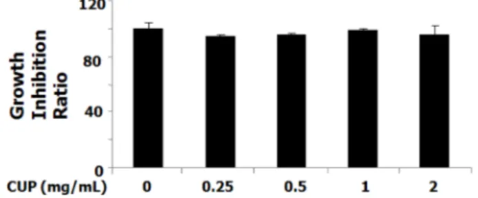
염증에 주요한 역할을 담당하는 대식세포를 이용하여 온 주밀감과피추출물(CUP), 레몬과피추출물(CLP), 한라봉과 피추출물(CHP)에 세포독성을 확인한 결과는 Fig. 1, Fig.
2, Fig. 3 과 같다. CUP, CHP, CLP 처리구 각각에 대한 세 포독성을 확인하기 위해 96 well plate에서 macrophage cell line RAW 264.7 세포를 배양하였고 대식기 세포에 각각의 추출물을 다양한 농도(0, 0.25, 0.5, 1 mg/mL)로 처리하였 다. 24시간 후 Cell Proliferation kit II (XTT)를 사용하여 생육억제 정도를 측정하였다.
그 결과 CUP, CHP, CLP 각 처리구 2 mg/mL 이하의 농 도에서는 세포생육도를 저해하지 않는 것으로 즉, 세포독 성이 낮음을 확인하였다. 대식세포는 독성물질에 대항하 여 면역조절에 중요한 역할을 담당하는 NO, PGE
2와 같은 2 차 염증매개믈질을 생산한다
32,33). 이러한 2차 매개물질의
생산이 과도할 경우 만성염증, 자가면역질환 등을 일으킨 다
34). 감귤류 3종 과피처리구의 추출물 최고농도 1 mg/mL 까지는 세포독성이 나타나지 않는 범위내에 있으므로 세 포사멸이 나타나지 않는 농도내에서 실험을 진행하였다.
감귤류 과피추출물의 NO억제능 검정
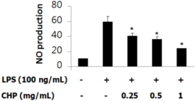
CUP, CHP, CLP 추출물의 항염증 효과를 확인하기 위 해, 그리스시약을 사용하여 NO생성 저해활성을 측정한 결 과는 Fig. 4, Fig. 5, Fig. 6과 같다. Murine macrophage Raw 264.7 세포에 CUP, CLP, CHP로 각각 0.25, 0.5, 1 mg/
mL CUP, CLP, CHP 추출물과 20 mg/mL LPS를 24시간 동안 처리하였고, CUP, CLP, CHP 추출물의 항염증효과를 확인한 결과, 생성된 NO측정치는 LPS를 100 ng/mL로 처 Fig. 1. Growth inhibition ratio of extracts from Citrus unshiu peel (CUP) on marcrophage cell (Raw 264.7).
Values are expressed as mean ± SD of three independent experi- ments.
Fig. 2. Growth inhibition ratio of extracts from Citrus limonia Osbeck peel (CLP) on marcrophage cell (Raw 264.7).
Values are expressed as mean ± SD of three independent experi- ments.
Fig. 3. Growth inhibition ratio of extracts from Citrus hallabong peel (CHP) on marcrophage cell (Raw 264.7).
Values are expressed as mean ± SD of three independent experi-
ments.
리한 세포의 NO생성량을 LPS를 처리하지 않은 대조구와 비교했을 때 상당량이 증가했음을 알 수 있었고, 추출물 을 농도별로 처리했을 때 농도가 증가할수록 NO생성량은 감소하였다.
세포내 염증반응에서 iNOS 및 COX-2에 의하여 유도된 염증인자인 NO생성은 신경독성, 신호전달 및 체내방어 등 의 생리기능을 갖고 있으며, 또한 염증과 암 발생에 관여 하는 등 병리적으로 중요한 작용을 한다
35,36). NO 는 아질 산염을 측정하는 griess assay를 이용해 간접적으로 측정 할 수 있으며, 염증반응 부위에서 NO의 과다발현은 많은 염증과 자가 면역 질환의 매개물질로 작용한다는 것은 잘 알려져 있다
37).
CUP 처리구는 LPS처리구에 비해 0.25 mg/mL 농도에서 는 약 80%, 0.5 mg/mL에서 약 60%, 1 mg/mL에서는 약 36% 감소하였음을 알 수 있었다(Fig. 4). CLP처리구 농도 에서 보면, LPS처리구에 비해 0.25 mg/mL 농도에서 약 90%, 0.5 mg/mL 에서는 약 84%, 1 mg/mL에서 약 69% 감
소하였음을 알 수 있었다(Fig. 5). CHP처리구를 나타낸 Fig. 6 의 결과를 보면, 각각은 LPS처리구에 비해 0.25 mg/
mL 농도의 경우 약 68%, 0.5 mg/mL에서는 약 61%, 1 mg/
mL 에서 약 40% 감소하였음을 알 수 있었다. Fig. 7은 CUP, CLP, CHP 각 처리구에서 각 1 mg/mL 농도에서의 저해활성을 비교한 값으로, CUP > CHP > CLP 순으로 NO 저해활성이 강하게 나타났다.
이러한 NO 등은 염증매개물질로서 다양한 염증관련 질 환을 유발한다고 보고된 바 있다
13,14). 이상의 결과로 감귤 류 과피의 NO저해작용은 레몬이나 한라봉보다 온주밀감 과피의 경우 NO저해활성이 크게 나타났다.
감귤류 과피추출물에 의한 대식세포의 iNOS, COX-2 단백 질의 저해활성 검정
CUP, CLP, CHP 추출물의 LPS로 유도된 iNOS, COX- Fig. 4. Effect of extracts from Citrus unshiu peel (CUP) on the
production of NaNO
2in marcrophage cell (Raw 264.7).
Values are expressed as mean ± SD of three independent experi- ments.
Data were analyzed by one-way ANOVA followed by the Tukey HSD post-hoc test.
Statistical significance was considered at P < 0.05.
Fig. 5. Effect of extracts from Citrus limonia Osbeck peel (CLP) on the production of NaNO
2in marcrophage cell (Raw 264.7).
Values are expressed as mean ± SD of three independent experi- ments.
Data were analyzed by one-way ANOVA followed by the Tukey HSD post-hoc test.
Statistical significance was considered at P < 0.05.
Fig. 7. Effect of extracts from various Citrus fruits peel on the production of NaNO
2in marcrophage cell (Raw 264.7) at 1 mg/
mL concentration.
Values are expressed as mean ± SD of three independent experi- ments.
Data were analyzed by one-way ANOVA followed by the Tukey HSD post-hoc test.
Statistical significance was considered at P < 0.05.
Fig. 6. Effect of extracts from Citrus hallabong peel (CHP) on the production of NaNO
2in marcrophage cell (Raw 264.7).
Values are expressed as mean ± SD of three independent experi- ments.
Data were analyzed by one-way ANOVA followed by the Tukey HSD post-hoc test.
Statistical significance was considered at P < 0.05.
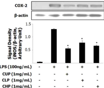
2 단백질의 저해활성을 확인하기 위해 western blotting assay 를 실시하였다. Murine macrophage Raw 264.7세포에 LPS (100 ng/mL) 를 처리하여, iNOS, COX-2 효소 발현을 유도하였고, 세포에 CUP, CLP, CHP 추출물을 1 mg/mL로 처리하였다. Fig. 8과 Fig. 9에서 알 수 있듯이 LPS (100 ng/
mL) 에 의해 iNOS, COX-2 단백질의 발현이 증가함을 확
인할 수 있으며, LPS를 처리하지 않은 대조구에서는 낮은 수준의 iNOS와 COX-2 단백질이 발현되고 있음을 확인할 수 있다. 1 mg/mL 농도의 CUP, CLP, CHP 추출물은 iNOS 와 COX-2 두 개의 단백질에 대해 동시에 저해활성을 가 지는 것으로 나타났으며, Western blot의 결과상으로는 CUP 와 CHP의 iNOS 단백질 저해활성이 비슷한 것으로 나타 났다. 따라서 NO생성 저해활성과 비교해 보았을 때도 CUP 와 CHP는 비슷한 정도의 NO생성 저해능을 가지는 것을 보아, CUP와 CHP는 LPS 로 유도된 NO를 비슷한 강도 로 저해하는 것으로 생각할 수 있다. COX-2 효소의 저해 활성은 CUP > CHP > CLP의 순으로 나타났으며, 이들 결 과를 종합해 볼 때 CUP, CHP, CLP는 모두 항염증 작용 을 가지고 있으며, 또한 CUP가 세 개의 추출물 중에서 가 장 저해활성이 강한 것으로 밝혀졌다. iNOS의 발현은 대 식세포에서 LSP에 의하여 염증매개물질이 과다 생성되는 주요한 기작이며, iNOS에 의하여 생성된 NO는 염증반응 을 촉진시키고, 염증을 심화시킨다
38).
Fig. 8 은 iNOS 단백질 발현량을 분석한 실험으로 murine macrophage Raw 264.7 세포에 1 mg/mL CUP, CLP, CHP 추출물과 100 ng/mL LPS를 24시간 동안 처리하였다. 동 일한 양의 단백질을 10% SDS-PAGE를 통해 분획하였고, PVDF membrane 에 electroblot을 해서 iNOS와 COX-2의 항 체를 사용하여 면역학적 방법으로 시그널을 확인하였고, 같 은 membrane을 reblot하여 β-actin의 시그널을 loading control 로 사용하였다. iNOS 단백질을 Raw 264.7세포에서 확인하였으며, iNOS와 β-actin의 값의 비를 그래프로 나타 내었다. 추출물 처리구는 LPS 값에 비해 CUP는 약 20%, CLP 는 약 46%, CHP는 약 17% 감소하였음을 알 수 있 다. Fig. 9는 COX-2 단백질을 Raw 264.7세포에서 확인하 였으며, COX-2와 β-actin의 값의 비를 그래프로 나타내었 다. 추출물 처리구는 LPS 값에 비해 CUP는 약 42%, CLP 는 약 59%, CHP는 약 50% 감소하였음을 알 수 있다.
iNOS 의 발현은 대식세포에서 LPS에 의하여 염증매개물질 이 과다 생성되는 주요한 기작이며, iNOS에 의하여 생성 된 NO는 염증반응을 촉진시키고, 염증을 심화시킨다
38,39). 염증반응에서 macrophage는 iNOS와 COX-2를 합성하여 NO 및 prostaglandin E2(PGE
2) 를 생성한다
22,23). 염증이 발 생하면 murine macrophage cell line RAW 264.7세포에 iNOS 의 발현양이 증가하여 다량의 NO가 생성된다. 과도하 게 생성된 NO에 의해 염증반응이 일어나고, COX-2에 의 해 PGE
2가 생성된다. 비스테로이드성 항염증제(nonsteroidal anti-inflammatory drugs; NSAIDs) 의 하나인 celecoxib의 경 우 10
−7µM 단위에서 Raw 264.7세포에 대한 COX-2 활성 정도
40)는 CUP 처리구 1 mg/mL에서와 유사한 결과를 보 였다. 따라서 염증반응으로부터 생성되는 NO와 COX-2 생성 억제를 확인하여 항염증 효과를 확인할 수 있었다.
Fig. 8. Inhibitory effect of Citrust peels extracts on the protein levels of iNOS in marcrophage cell (Raw 264.7).
Values are expressed as mean ± SD of three independent experi- ments.
Data were analyzed by one-way ANOVA followed by the Tukey HSD post-hoc test.
Statistical significance was considered at P < 0.05.
Fig. 9. Inhibitory effect of Citrust peels extracts on the protein levels of COX-2 in marcrophage cell (Raw 264.7).
Values are expressed as mean ± SD of three independent experi- ments.
Data were analyzed by one-way ANOVA followed by the Tukey HSD post-hoc test.
Statistical significance was considered at P < 0.05.
요 약
본 연구에서는 감귤류 중 온주밀감, 레몬, 한라봉의 과 피추출물(PE)에 대한 항염증 효과를 조사하였다. 감귤류 세 품종 과피추출물(PE)에 대한 murine macrophage cell line Raw 264.7 세포를 배양하여 세포독성, NO생성량, 세포내 염 증관련 단백질 발현 양상을 분석하였다. 그 결과 CUP, CHP, CLP 처리구의 경우 2 mg/mL 이하의 농도에서는 세포생 육을 저해하지 않았다. LPS (100 ng/mL)를 처리한 세포의 NO 생성량을 LPS를 처리하지 않은 대조구와 비교했을 때 상당량이 증가했음을 알 수 있었고, 추출물을 농도별로 처 리했을 때 농도가 증가할수록 NO생성량은 감소하였다. 각 처리구에서 각 1 mg/mL 농도에서는 CUP > CHP > CLP 순 으로 NO 저해활성이 강하게 나타났다. Western blot의 결 과상으로는 CUP와 CHP의 iNOS 단백질 저해활성이 비슷 한 강도로 나타남을 확인하였다. COX-2 효소의 저해활성 은 CUP > CHP > CLP의 순으로 나타났다. 따라서 CUP, CHP, CLP 처리구는 모두 항염증 작용을 가지고 있으며, 또한 CUP 처리구가 세 개의 추출물 중에서 가장 저해활 성이 강하였다.
감사의 글
본 연구는 2012년도 동남보건대학교 학술연구소 지원에 의하여 수행되었으며, 그 지원에 감사드립니다.
참고문헌