Korean J Vet Res(2017) 57(2) : 97~104 https://doi.org/10.14405/kjvr.2017.57.2.97
97
<Original Article>
Solanum lycopersicum (tomato) ethanol extract elicits anti-inflammatory effects via the nuclear factor kappa B pathway and rescues mice
from septic shock
Evelyn Saba
1, Mi-Ju Oh
1, Dongmi Kwak
1, Seong-Soo Roh
2, Hyuk-Woo Kwon
1, Sung-Dae Kim
1, Man Hee Rhee
1,*
1
Department of Veterinary Medicine, College of Veterinary Medicine, Kyungpook National University, Daegu 41566, Korea
2
College of Korean Medicine, Daegu Hanny University, Gyeongsan 38610, Korea (Received: March 21, 2017; Revised: May 16, 2017; Accepted: May 19, 2017)
Abstract: Solanum lycopersicum, commonly known as tomato, is widely used in raw, cooked, or liquid forms because it contains nutritional compounds that are beneficial for human health, including carotenoids, lycopene, ascorbic acid, vitamins, and minerals. The tomato is perhaps the most widely studied fruit, especially with respect to its cardioprotective effects. In this study, we aimed to identify the anti-inflammatory mechanisms by which the tomato elicits its anti- inflammatory properties. We treated murine macrophage RAW 264.7 cells with a tomato ethanol extract and performed various biochemical assays including nitric oxide inhibition, cell viability, RNA extraction, expression of pro- inflammatory mediators and cytokines, and immunoblotting, as well we assessed cell survival rates. Our results have shown for the first time that a tomato ethanol extract treatment can suppress nitric oxide production in a dose-dependent manner without cytotoxicity. Moreover, it inhibits the expression of pro-inflammatory mediators and cytokines and elicits its anti-inflammatory effects via the nuclear factor kappa-light-chain-enhancer of activated B cells (NF- κB) and mitogen-activated protein kinase (MAPK) pathways. In addition, administration of tomato syrup potently rescued mice from septic shock induced by lipopolysaccharide injection. Collectively, our results elucidate details regarding the anti- inflammatory mechanisms of tomato.
Keywords: anti-inflammation, cytokines, pro-inflammatory mediators, septic shock, tomato ethanol extract
Introduction
The growing incidence of cardiovascular, immunosuppres- sive and chronic inflammatory diseases due to the rapid pace of industrialization poses a serious threat for the well-being of mankind. The increased amounts of social, personal, and workplace stress cannot be ignored as factors predisposing humans to premature aging, mental illnesses and early deaths [19]. Rapid and ongoing global research towards a cure for various chronic diseases has altogether contributed posi- tively towards the health of mankind. However, the serious disadvantages of these chemical orientated medicines cannot be ignored. In order to avoid the side effects of these chemi- cal medications, scientists all over the world are struggling to improve health conditions by using natural products like fruits and vegetables. Solanum lycopersicum commonly known as tomato is widely consumed as fruit and vegetable all over the world. The anti-oxidant, anti-inflammatory, anti-diabetic, and anti-lipidemic activities of the tomato have been studied
extensively in the past and currently [1, 9]. The protective properties of tomato are mainly attributed to the carotenoid, lycopene, which is present in great quantities within the fruit.
There is much ongoing research on the tomato as a whole fruit, or isolated lycopene, which is the most abundant and active component of the tomato. Lycopene administration alleviated prostitis in a manner comparable to that of ciprof- loxacin [14]. Furthermore it reduced the chemical and histo- logical symptoms in iodoacetamide induced colitis in rats [28]. Anthocyanins from tomatoes have also been shown to exhibit inhibitory effects on inducible nitric oxide synthase (iNOS) and cyclooxygenase-2 (COX-2) [12].
Inflammation is the hallmark of almost every disease and if not controlled at proper time point, can lead to chronic dis- eases. The production of pro-inflammatory cytokines when the body encounters a foreign invader is a primary response of immune cells [21]. These pro-inflammatory cytokines and mediators further recruit more potent chemicals and cells, such as natural killer cells, that neutralize and destroy the for-
*Corresponding author
Tel: +82-53-950-5967, Fax: +82-53-950-5955
E-mail: [email protected]
eign particle. Macrophages are the cells that phagocytize for- eign particles and signal the recruitment of other cells via production of inflammatory mediators like iNOS, COX-2 and pro-inflammatory cytokines including interleukin (IL)- 1 β, 1L-6 and tumor necrosis factor (TNF)-α [29]. Produc- tion of pro-inflammatory mediators leads to the activation of pro-inflammatory cytokines [8]. All of these chemicals when released synchronously, fight against foreign materials and alleviate infection through the nuclear factor kappa-light- chain-enhancer of activated B cells (NF- κB) and mitogen- activated protein kinase (MAPK) pathways [30].
Previously we have shown the antiplatelet effects of the tomato extract [16], but here in this study, we geared to elu- cidate the detailed anti-inflammatory mechanisms of whole tomato ethanol extract in vitro using RAW 264.7 cells and in vivo using a septic shock ICR mouse model. It is a well- known and alarming fact that the cause of most post-opera- tive mortalities or bacterial infections is due to severe septic shock which is defined as the systemic distribution of bacte- rial toxins in body leading to death [22].
Materials and Methods
Materials
Dulbecco’s modified Eagle’s medium (DMEM) and fetal bovine serum (FBS) were purchased from WELGENE (Korea).
Total RNA extraction kit was purchased from Invitrogen (USA). Oligo dT, iNOS, COX-2, TNF- α, IL-6, and IL-1β primers were obtained from Bioneer (Korea). Lipopolysac- charide (LPS; Escherichia coli 055: B5) and 3-(4,5-dimethyl- thiazol-2-yl)-2,5-diphenyltetrazoliumbromide (MTT) were pur- chased from Sigma Aldrich (USA). Specific antibodies used against phospho- and/or total form of extracellular signal- regulated kinase (ERK), c-Jun N-terminal kinase (JNK), p38, nuclear factor of kappa light polypeptide gene enhancer in B cells inhibitor (I κB), IκB kinase (IKK) α/β, NF-κB p65 and β-actin as well as rabbit HRP linked secondary antibody were purchased from Cell Signaling Technology (USA). All other reagents and chemicals were obtained from Sigma Aldrich.
Animal studies
Male ICR mice (26–29 g) were purchased from Charles River, Orient Biotechnology, Gyeonggi-do, South Korea. The mice were housed in a specific pathogen free barrier facility at 21 ± 2
oC with a relative humidity of 60 ± 10% under a 12 h light and dark cycle. Feed and water were provided ad libitum. All animal care and experimental procedures were approved by Animal Care Committee (2015-0062) of Kyungpook National University, Daegu, South Korea. The mice were divided into 3 groups with each group (n = 10) for survival study. Group 1 was taken as control or vehicle treated group. Group 2 was LPS control group and Group 3 was treated with water soluble tomato concentrate (DSM Nutritional Product; DSM, Switzerland) [16], which we
named as tomato syrup for 3 days prior to LPS injection orally at a dose of 900 mg/kg once a day based on the equiv- alent human consumption of tomato per day which is 3 g/
day. The amount of lycopene present in the tomato syrup was around 3–5 mg/100 g fresh tomatoes [6, 32]. After the pre- treatment of mice with tomato syrup for three days, at the fourth day groups 2 and 3 were given LPS intraperitoneally at 30 mg/kg and then the survival rate was monitored for 96 h.
Tomato ethanol extract (TEE) preparation
Whole dried tomatoes were condensed using 70% ethanol in a 20-fold volume. After condensation, the ethanol extract was filtered through a 2 µm pore Whatman filter paper. The filtrate was then frozen at −70
oC for 2 days and then vacuum dried to obtain a powered form. The powdered form was then weighed and diluted in dimethyl sulfoxide (DMSO) and used accordingly for each experiment.
Cell culture
RAW 264.7 cells, murine macrophage cell line, originat- ing from American Type Culture Collection (ATCC-TIB-71) were cultured in complete DMEM supplemented with 5%
FBS, penicillin (100 IU/mL) and streptomycin sulfate (100 µg/mL) in humidified 5% CO
2incubator at 37
oC.
Nitric oxide assay
Nitric oxide (NO) measurements were performed on the basis of Griess reaction. In short, RAW 264.7 cells were seeded in 96-well plates and incubated with or without LPS (0.1 µg/mL) in the absence or presence of TEE at concentra- tions of 250–1,000 µg/mL for 18 h. Next day, the culture supernatants (100 µL) were mixed with Griess reagent (0.2%
naphthyl ethylene diamine dihydrochloride, and 2% sulpha- nilamide in 5% phosphoric acid) in double distilled water at equal volumes and incubated for 5 min at room temperature.
The absorbance in each well was then read at 540 nm in enzyme-linked immunosorbent assay reader (VersaMax ELISA Microplate Reader; Molecular Devices, USA).
Cell viability assay
Cytotoxic effects of TEE were examined using MTT rea- gent which was added to culture medium at a final concentra- tion of 0.1 mg/mL in 96-well plate. After 4 h of incubation at 37
oC in 5% CO
2, the violet coloured crystals were dissolved in DMSO (100 µL/ well) and absorbance values were read at 560 nm (VersaMax Microplate Reader; Molecular Devices).
RNA extraction and quantitative real-time polymerase chain reaction (qRT-PCR)
RAW 264.7 cells were pre-treated with or without TEE at
indicated concentrations for 30 min and then stimulated with
LPS (0.1 µg/mL) for 18 h in 6-well plates. Total RNA was
extracted using a TRIZOL reagent (Invitrogen, USA) follow-
ing the manufacturer’s instructions. Subsequent steps for
cDNA for qRT-PCR were according to previous study [31].
Quantitative PCR primer sequences are given in Table 1.
Western blot analysis
Cytosolic and nuclear proteins were extracted according to manufacturer’s instructions using NE-PER nuclear and cytosolic extraction reagents (No. 78833 and No. 78835; Thermo Sci- entific, USA) from RAW264.7 cells when they were treated or untreated with TEE (250–1,000 µg/mL) in the presence or absence of LPS (0.1 µg/mL) in 6-well plates. Protein quanti- fication was then performed using PRO-MEASURE assay kit (iNtRON Biotechnology, Korea). Proteins were then loaded onto 10% acrylamide gels, separated by SDS-PAGE and transferred onto PVDF membranes (Immobilon-P; Milli- pore, USA). Nonspecific binding on PVDF membranes was minimized with a blocking buffer containing 5% non-fat dry
milk and 0.1% Tween-20 in Tris-buffered saline. The mem- branes were then incubated with specific primary antibodies overnight at 4
oC followed by 1 h incubation with HRP-conju- gated anti-rabbit antibody (1:3,000 dilution, Cell Signaling Tech- nology). Bound antibodies were visualized using enhanced chemiluminescence (SUPEX ECL solution; Neuronex, Korea) and images were analyzed using ImageJ 2 software (National Center for Biotechnology Information, USA). β-actin was used as an internal control.
Statistical analysis
Data are presented as mean ± SEM. One-way analysis of variance followed by Dunnett’s -test was used for statistical analysis. SAS 9.3 (SAS Institute, USA) was applied for anal- ysis. P values less than 0.01 were considered statistically sig- nificant.
Fig. 1. Inhibition of nitric oxide (NO) by tomato ethanol extract (TEE). RAW 264.7 cells were preincubated with TEE for 30 min and then stimulated with lipopolysaccharide (LPS) for 18 h. Cell supernatant was then mixed with equal amounts of Griess reagent and NO production was measured (A). Effects of TEE on cell viability were measured by MTT assay (B). Values in bar graph are mean ± SEM of three independent experiments.
***p < 0.001 are considered significant compared to LPS group only.
Table 1. Primer sequences used for polymerase chain reaction
Gene Primer Oligonucleotide sequence (5'-3')
GAPDH F 5'CAATGAATACGGCTACAGCAAC3'
R 5'AGGGAGATGCTCAGTGTTGG3'
iNOS F 5'CCCTTCCGAAGTTTCTGGCAGCAGC3'
R 5'GGCTGTCAGAGCCTCGTGGCTTTGG3'
COX-2 F 5’-TCTCAGCACCCACCCGCTCA-3’
R 5’-GCCCCGTAGACCCTGCTCGA-3’
IL-1β F 5'CAGGGTGGGTGTGCCGTCTTTC3'
R 5'TGCTTCCAAACCTTTGACCTGGGC3'
TNF-α F 5'TTGACCTCAGCGCTGAGTTG3'
R 5'CCTGTAGCCCACGTCGTAGC3'
IL-6 F 5’-GTACTCCAGAAGACCAGAGG-3’
R 5’-TGCTGGTGACAACCACGGCC-3’
F, forward; R, reverse.
Fig. 2. TEE suppressed pro-inflammatory mediators. RAW 264.7 cells were preincubated with TEE for 30 min and then stimulated with LPS for 18 h. Total RNA was extracted and mRNA expression of inducible nitric oxide synthase (iNOS) and cyclooxygenase- 2 (COX-2) was determined both by real-time (A) and reverse transcriptase polymerase chain reaction (B). GAPDH was used as an internal control. Image is representative of three independent experiments. Values in bar graphs are mean ± SEM of three independent experiments.
***p < 0.001 and
**p < 0.05 are considered significant compared to LPS group only.
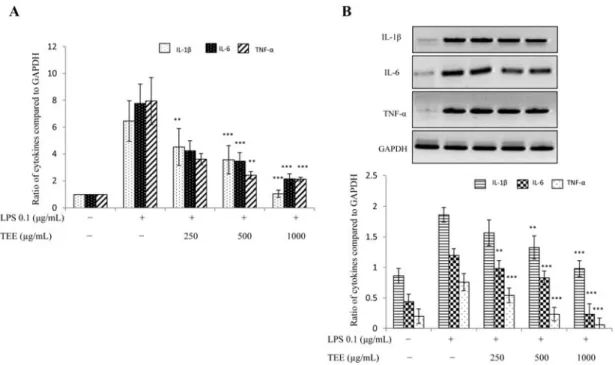
Fig. 3. Expression of pro-inflammatory cytokines was diminished by TEE. For mRNA expression, RAW 264.7 cells were pre-treated
with TEE for 30 min and then stimulated with LPS for 18 h. Total RNA was extracted and mRNA expression of interleukin (IL)-
1β, IL-6 and tumor necrosis factor (TNF)-α was determined by real-time PCR (A) and RT-PCR (B). GAPDH was used as an internal
control. Image is representative of three independent experiments. Values in bar graphs are mean ± SEM of three independent exper-
iments.
***p < 0.001 and
**p < 0.05 are considered significant compared to LPS group only.
Results
TEE inhibited LPS induced inflammation in RAW 264.7 cells
NO is produced when bacterial lipopolysaccharides bind to the Toll like receptors. Therefore, we sought to check the inhibition of NO by TEE. As shown in Fig. 1A, TEE signif-
icantly suppressed the NO production in a dose dependent manner without any cytotoxicity (Fig. 1B) .
Suppression of pro-inflammatory mediators by TEE In our result, we found that TEE suppressed the expres- sion of pro-inflammatory mediators that are iNOS and COX- 2 at transcriptional level as shown in Fig. 2A and B.
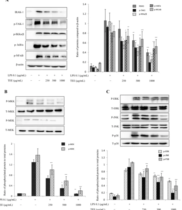
Fig. 4. TEE elicits anti-inflammatory effects via the nuclear factor kappa B (NF- κB) and MAPK pathways. For the protein expression, RAW 264.7 cells were treated with TEE and stimulated with LPS 30 min later. Nuclear and cytoplasmic proteins were extracted by NE-PER Nuclear and Cytoplasmic Extraction Kit (Thermo Scientific). β-actin was used as an internal control. Inhibition in the phos- phorylation of all downstream NF-κB (A). Suppression in the phosphorylation of MAPK factors downstream (B and C). Image is rep- resentative of three independent experiments. Values in bar graph are mean ± SEM of three independent experiments.
***p < 0.001,
**