RESEARCH ARTICLE
Received: November 2, 2015, Revised: November 11, 2015, Accepted: November 11, 2015 ISSN 1598-4478 (Print) / ISSN 2233-7679 (Online)
†
Correspondence to: Sang-Im Lee
Department of Dental Hygiene, School of Health Science, Dankook University, 119 Dandae-ro, Dongnam-gu, Cheonan 31116, Korea Tel: +82-41-550-1492, Fax: +82-41-559-4934, E-mail: [email protected]
Copyright © 2015 by the Korean Society of Dental Hygiene Science
NFATc Mediates Lipopolysaccharide and
Nicotine-Induced Expression of iNOS and COX-2 in Human Periodontal Ligament Cells
Sang-Im Lee † and Ji-Su Yu 1
Department of Dental Hygiene, School of Health Sciences, Dankook University, Cheonan 31116,
1 Department of Dental Hygiene, Gumi College, Gumi 39213, Korea
사람 치주인대세포에서 Lipopolysaccharide와 니코틴으로 유도된 iNOS와 COX-2 발현에 NFATc의 관여
이상임
†
ㆍ유지수1
단국대학교 보건과학대학 치위생학과,
1구미대학교 치위생과
Although nuclear factor of activated T cell (NFAT) plays a key role in inflammation, its anti-inflammatory effects and mechanism of action in periodontitis are still unknown. This study aimed to identify the effects of NFAT on the proinflammatory mediators activated by lipopolysaccharide (LPS) plus nicotine stimulation in human periodontal ligament cells (hPDLCs). The production of nitric oxide (NO) and prostaglandin E
2(PGE
2) was evaluated using Griess reagent and an enzyme immunoassay, respectively. The expression of inducible nitric oxide synthase (iNOS), cyclooxygenase-2 (COX-2) and NFAT proteins was evaluated by Western blot analysis. LPS plus nicotine synergistically induced the production of NO and PGE
2and increased the protein expression of iNOS, COX-2 and NFAT. Treatment with an NFAT inhibitor blocked the LPS plus nicotine-stimulated NO and PGE
2release as well as the expression of iNOS and COX-2. Our data suggest that the LPS plus nicotine-induced inflammatory effects on hPDLCs may act through a novel mechanism involving the action of NFAT. Thus, NFAT may provide a potential therapeutic target for the treatment of periodontal disease associated with smoking and dental plaque.
Key Words: Lipopolysaccharides, NFATc transcription factors, Nicotine, Periodontitis
Introduction
Periodontal disease initiation and progression occur as a consequence of the host response to pathogenic bacteria present in the dental biofilm. It is a chronic inflammatory disease that affects the periodontium resulting in the tooth supporting tissue destruction and even in the loss of the alveolar bone
1)
. Host immune response against this infection leads to the production of inflammatory mediators. Thelarge amounts of released proinflammatory mediators, in addition to nitric oxide (NO) and prostaglandins (PGs), has been shown to be associated with periodontal disease through the activity of inducible enzymes such as indu- cible nitric oxide synthase (iNOS) and cyclooxygenase-2 (COX-2)
2)
.The inducible isoform, iNOS, is involved in immune response, which catalyzes the oxidative deamination of L-arginine to produce NO and is thus responsible for
prolonged NO production
3)
. COX-2 is inducible in infla- mmatory conditions including periodontitis for the produ- ction of large amounts of proinflammatory PGs at the site of inflammation4)
. Based on these observations, understanding the regulation of proinflammatory mediators and their effects in periodontal tissues has been the objective of many studies5-7)
.Human periodontal ligament cells produce cytokines and chemokines in response to inflammation promoters, including lipopolysaccharide (LPS)
8)
. Also, it is reasonable to suggest that periodontal ligament cells play a key role as promoters of periodontal inflammation through these mechanisms9)
. Since a recent study suggested that periodontal ligament cells may have important implications for the development of new therapeutic strategies to treat perio- dontitis10)
.Smoking is a major environmental risk factor in the development and progression of periodontal disease
11)
, including gingivitis and periodontitis. It is associated with increased pocket depths, a loss of periodontal attachment and alveolar bone and a higher rate of tooth loss12)
.Nicotine, the major toxic component of tobacco smoke, and its metabolites make the early signs of periodontal disease by the inflammatory response. Previous reports demonstrated that nicotine detected on the root surfaces of teeth, in the saliva
13)
and in the gingival crevicular fluid of smokers14)
. Although, nicotine is a cytotoxic agent to gingival fibroblasts and periodontal ligament cells, inhibiting their viability, attachment, and proliferation11)
, little is known about the effect of both nicotine and LPS on inflammatory responses in periodontal ligament cells.The nuclear factor of activated T cell (NFAT) is com- posed of a family of transcription factors that includes 5 members, 4 of which are regulated by Ca
2+
signaling; NFAT1 (NFATc2 or NFATp), NFAT2 (NFATc or NFATc1), NFAT3 (NFATc4), NFAT4 (NFATx or NFATc3), and calcineurin-independent NFAT5 (TonE-BP or NFATL1).The NFAT family of transcription factors plays a fun- damental role in the transcriptional regulation of the immune response and is best known that regulate cytokine production after T-cell activation
15)
. Furthermore, NFATs are not only restricted to the cells of the immune system, because their expression can be found in other tissues,including the heart
16)
, muscle17)
, brain18)
, and endothelium19,20)
. Although, a key function of NFAT in immune cells is to regulate the expression of potent immunomodulatory cytokines, the role of NFAT signaling pathway in periodontal ligament cells remains unclear. Thus, the purpose of the present study was to investigate whether NFATc1 could regulate on LPS and nicotine-induced iNOS and COX-2 expression in hPDLCs.Materials and Methods
1. Cell culture
Immortalized hPDLCs
21)
transfected with human telo- merase catalytic component (hTERT), were kindly pro- vided by Professor Takashi Takata (Hiroshima University, Japan). The cells were cultured in -modified Eagle medium (-MEM) supplemented with 10% fetal bovine serum (FBS), 100 U/mL penicillin, and 100 g/mL streptomycin in a humidified atmosphere of 5% CO2
at 37o
C. -MEM, FBS, and penicillin/streptomycin were purchased from Gibco BRL Co. (Grand Island, NY, USA).2. Cell viability assay
The cytotoxicity of LPS (from Porphyromonas gingivalis) plus nicotine was determined by a 3-(4,5-dimethylthiazol- 2-yl)-2,5-diphenyl tetrazolium bromide (MTT; Sigma- Aldrich Chemical Co., St. Louis, MO, USA) assay. Cells seeded on 96-well microplates at 1×10
4
cells per well were incubated with LPS plus nicotine for the indicated time period. Medium was removed and then incubated with 100 L MTT assay solution for 4 hours. Absorbance was measured in an microplate reader (Bio-Rad, Hercules, CA, USA) at 595 nm. The percentage of cell viability was calculated as the ratio of the absorbance of treated media that of the control media ×100.3. Quantification of nitric oxide
Thawed 50 L aliquots of culture supernatant were mixed with 50 L Griess reagent, comprising: 5% phos- phoric acid (Fisher Scientific, FairLawn, NJ, USA), 1%
sulfanilamide and 0.1% N-naphthylethylenediamine (Sigma Aldrich). Samples were incubated at room temperature for approximately 10 minutes and then read on an enzyme-
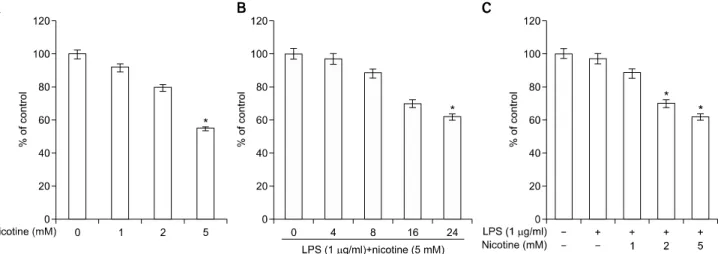
Fig. 1. Effects of lipopolysaccharide (LPS) plus nicotine on cytotoxicity. Cell viability was determined by MTT. Data were obtained from three independent experiments. Values are mean±standard deviation of three experiments. *Statistically significant difference com- pared with control (p<0.05).
linked immunosorbent assay microplate plate reader (Bio- Rad) at 570 nm.
4. Determination of PGE 2 levels
The culture medium of control and treated cells was collected, centrifuged, and stored at −70
o
C until tested.The level of PGE
2
released into culture medium was quantified using a specific enzyme immunoassay according to the manufacturer’s instructions (Amersham, Arlington Heights, IL, USA).5. Western blotting assay
The treated cells were washed with PBS and cytosolic and nuclear protein extracts were prepared using 1× Cell Lysis buffer (Santa Cruz Biotechnology, Santa Cruz, CA, USA) supplemented with a protease inhibitor cocktail.
Protein concentrations were determined using the Bradford assay (Bio-Rad) as per the manufacturer's protocol.
Proteins (30 g) were mixed with an equal volume of 2×
sodium dodecyl sulphate (SDS) sample buffer, boiled for 5 minutes, and then resolved by SDS-polyacrylamide gel electrophoresis (10% acrylamide) and transferred to poly- vinylidene difluoride membrane, immobilon-P (Millipore Co., Milford, MA, USA). Protein bands were detected using an enhanced chemiluminescence system (Amersham Biosciences, Buckinghamshire, UK, USA) according to the manufacturer’s instructions and exposed to X-ray film.
6. Statistical analysis
Differences among groups were analyzed using one- way analysis of variance with the IBM SPSS Statistics ver.
20.0 (IBM Co., Armonk, NY, USA) computer program.
Statistical significance was determined at p<0.05.
Results
1. Effects of LPS plus nicotine on cytotoxicity
We first assessed the effects of LPS plus nicotine on periodontal ligament cell viability via MTT assay. Peri- odontal ligament cells exposed to different concentrations of LPS plus nicotine for various lengths of time showed a concentration- and time-dependent reduction in cell viability compared with control cells (Fig. 1).2. Effect of LPS plus nicotine on the iNOS/COX-2 expression and of NO/PGE 2 production
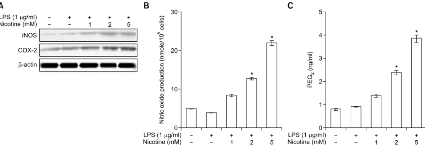
Next, we examined the time course of LPS plus nicotine- induced changes in iNOS/COX-2 levels and NO/PGE
2
production in hPDLCs. Co-treatment with LPS (1 g/mL) plus nicotine (5 mM) resulted in a time- dependent increase of iNOS and COX-2 expression, with maximal induction after 18 or 24 hours of incubation (Fig. 2A). This combina- tion of LPS plus nicotine also increased iNOS-derived NO (Fig. 2B) and COX-2-derived PGE
2
(Fig. 2C), with maximal induction after 18 or 24 hours of incubation.Fig. 3. Effects of lipopolysaccharide (LPS) plus nicotine on the NFATc1 activation and the phosphorylation of MAP kinase (MAPK). NFATc1 protein levels (A) and the MAPK phosphor- ylation were analyzed by western blotting (B). The data presented are representative of three independent experiments.
Fig. 2. Effects of lipopolysaccharide (LPS) plus nicotine on the iNOS/COX-2 expression and of NO/PGE
2production. Cells were in- cubated for 24 hours with the indicated concentrations of LPS plus nicotine. The levels of expression were determined by western blotting, ELISA and Griess assay. Data were obtained from three independent experiments. Values are the mean±standard deviation of three experiments. *Statistically significant difference compared with control (p<0.05).
3. Effects of LPS plus nicotine on the NFATc1 activation and the phosphorylation of MAP kinase (MAPK)
NFATc1 protein activation induced by LPS plus nicotine, because the induction of NFATc1 by various stress stimuli have been implicated in inflammation. As shown in Fig. 3A, the NFATc1 expression induced by nicotine plus LPS was comparable to that in the control cultures in hPDLCs. To elucidate the molecular basis of the responses to NFAT, we examined the effects of LPS plus nicotine on the MAPK signaling pathways in hPDLCs.
As shown in Figure 3B, LPS plus nicotine treatment induced the phosphorylation of p38 MAPK but not ERK or JNK.
4. Effect of NFATc1 the inhibitor, cyclosporine A (CsA), on LPS plus nicotine induced iNOS/COX-2 expression and of NO/PGE 2 production
Moreover, cyclosporine A (CsA) pretreatment revealed a significant inhibitory effect on LPS plus nicotine- induced iNOS proteins and NO production in hPDLCs (Fig. 4A). In addition, CsA decreased the LPS plus nicotine-induced synthesis of COX-2 proteins and PGE
2
production in hPDLCs (Fig. 4B).
Fig. 4. Effects of NFATc1 the inhibitor, cyclosporine A (CsA), on lipopolysaccharide (LPS) plus nicotine induced iNOS/COX-2 expression and of NO/PGE
2production. Cells were pretreated for 4 hours with CsA, and then incubated for 24 hours with the indicated concen- trations of LPS plus nicotine. The levels of expression were determined by western blotting, ELISA and Griess assay. Data were obtained from three independent experiments. Values are the mean±standard deviation of three experiments. *Statistically significant difference compared with control (p<0.05). **Statistically significant difference compared with LPS plus nicotine (p<0.05).
Discussion
As the major constituent of the particulate phase of tobacco smoke, nicotine is a major contributor to peri- odontitis
12)
. Although previous studies reported that nicotine causes injury to periodontal ligament cells via multiple mechanisms, including c-Fos, COX-2, heme oxygenase-1 and oxidative stress22-24)
, the molecular mechanisms of these effects have not been fully elucidated. Elevated P.gingivalis levels are present in periodontal lesions and are
significantly reduced by successful therapy25)
. LPS is a well-known endotoxin which elicits a variety of inflam- matory responses. Furthermore, previous studies have reported that LPS from a periodontal pathogenic bacterium stimulated interleukin (IL) 6 and IL-8 production from hPDLCs26)
, and these inflammatory cytokines play a role in the destruction and disintegration of periodontium27)
. Previous studies have shown that LPS and nicotine is a cytotoxic agent to human fibroblasts derived from perio- dontium28,29)
. In this study, we present evidence of LPS plus nicotine-induced cytotoxicity on hPDLCs by inhibitingcell growth and proliferation in a dose-dependent manner.
Our results also showed that nicotine enhanced LPS plus nicotine-induced NO, iNOS, COX-2 and PGE
2
synthesis in hPDLCs.NFAT is highly phosphorylated and remain in the cytoplasm in unstimulated cells
30)
. When various physio- logical processes results in an increase in intracellular calcium level, dephosphorylation of NFAT translocate to the nucleus, and induces expression of NFAT target genes31,32)
. The NFAT family of transcription factors regulates a various biological processes such as cell survival, proliferation, migration, invasion33)
, angiogenesis34)
, and neural development and function35)
. NFAT activation was first identified from the extracts of activated T lympho- cytes. However, NFAT proteins also play key roles of diverse cellular functions within the cardiovascular system, skeletal muscle, bone, and nervous system35)
. In addition, recent studies have demonstrated that NFATc1 not only a master transcriptional factor for induced in osteoclast precursors36)
but also regulates bone homeostasis. NFATc1 over-expression in osteoblasts stimulates transcriptionalactivity of osterix, which are major osteoblastogenic trans- cription factors
37)
. Furthermore, our previous studies demonstrated that the first report of the expression of NFATc1 mRNA and protein being induced during osteo- blastic differentiation in hPDLCs38)
. However, the induction of NFAT, COX-2 and iNOS by LPS plus nicotine has not been verified in hPDLCs. Thus, we hypothesized that the induction of NFATc1 by LPS plus nicotine in hPDLCs may be responsible for the COX-2- and iNOS-inducing effects of treatment with LPS plus nicotine. Our first finding was that treatment of LPS plus nicotine syner- gistically induced NFATc1 activation in hPDLCs.Moreover, MAP kinases play a critical role in the regulation of cell growth and differentiation and in the control of cellular responses to cytokines and other stresses.
In the present study, LPS plus nicotine induced MAPK phosphorylation (Fig. 3B). In the present study, p38 MAPK was slightly activated in LPS plus nicotine- stimulated cells, while the level of phosphorylated ERK and JNK was basal level, indicating a constitutive activa- tion of these pathways in hPDLCs. This suggests that, even though LPS plus nicotine-activated p38 MAPK play differential roles in iNOS and COX-2 induction.
In addition, the NFATc1 inhibitor CsA blocked the LPS plus nicotine-induced increases in iNOS/ COX-2 expression and NO/PGE
2
production. CsA, widely used pharmacolo- gical inhibitor, are known to block the calcineurin/NFATc1 signaling pathway39)
. Our results are in agreement with Mena et al.40)
, who reported that COX-2 upregulation was inhibited by the NFAT antagonist CsA in endothelial cells.These results suggest that LPS plus nicotine induced iNOS and COX-2 as well as NO and PGE
2
production via the NFATc1-dependent pathway in hPDLCs. Based on these findings, we propose that NFATc1 as an inflammatory mediator represents a novel preventive or therapeutic target in periodontitis.Summary
Host immune response and immune system are the main causes of individual susceptibility to periodontal diseases.
Bacterial infection and smoking is an important envi- ronmental risk factor involved in the causation and
progression of periodontal tissue destruction. Therefore, the aim of this study was to identify the effects of NFAT signaling pathway on LPS plus nicotine stimulated iNOS and COX-2 expression, a therapeutic target in periodontal disease, in hPDLCs. We also examined NO and PGE
2
synthesis in LPS plus nicotine-stimulated hPDLCs. This study is the first to demonstrate that the inhibition of NFATc1 in LPS plus nicotine-stimulated hPDLCs results in the suppression of iNOS and COX-2 expression, as well as the reduction of NO and PGE
2
. Therefore, targeted NFATc1 regulation during inflammation, by modulating NO and PGE2
levels in PDL cells, may represent a novel pharmacological approach in the prevention or treatment of periodontal diseases caused by dental plaque and smoking.요 약
숙주 면역 반응과 면역 체계는 치주 질환에 대한 개인의 감수성의 주요 원인이다. 세균 감염과 흡연은 치주 조직의 파괴의 원인과 진행에 관여하는 중요한 환경 위험 요인이 다. 따라서, 본 연구는 사람 치주인대세포에서 LPS와 니코 틴이 전염증성 사이토카인인 iNOS/COX-2의 발현과 NO/
PGE
2
생산에 미치는 영향을 알아보고 NFATc1가 어떤 기전으로 항염작용을 하는지 밝히고자 하였다. LPS와 니코틴 을 처리한 사람 치주인대세포에서 iNOS/COX-2의 발현과 함께 NO/PGE
2
생산은 증가되었다. NFATc1 inhibitor인 CsA는 LPS와 니코틴에 의해 유도되는 iNOS/COX-2의 발현과 함께 NO/PGE
2
생산을 감소시켰다. 이러한 연구 결과로 볼 때, NFAT signaling pathway가 LPS와 니코틴에 의한 iNOS/COX-2의 발현을 조절하여 NO/PGE
2
매개 염증에 대해 방어할 수 있다고 생각된다.Acknowledgements
This research was supported by the National Research Foundation of Korea (NRF) grant funded by the Korea government (MSIP) (No. 2014R1A1A2058805).