Kor. J. Pharmacogn.
46(1) : 12∼ 16 (2015)
12
몰약으로부터 분리된 1β,6α-dihydroxyeudesm-4(15)-ene의 LPS로 유도된 BV2 미세아교세포에서의 항염증효과
김동철1,2#·윤치수1,2#·고원민1,2·이동성3·김대성4·조형권4·서정원1,2· 김성연1,2·오현철1,2*·김윤철1,2*
1원광대학교 약학대학, 2원광대학교약품연구소, 3인하대학교 의과대학 의과학연구소, 4(유)한풍제약
Anti-inflammatory Effects of 1β‚6α-Dihydroxyeudesm-4(15)-ene Isolated from Myrrh on LPS-induced Neuroinflammation in BV2 cells
Dong-Cheol Kim1,2#, Chi-Su Yoon1,2#, Wonmin Ko1,2, Dong-Sung Lee3, Dae-Sung Kim4, Hyoung-Kwon Cho4, Jungwon Seo1,2, Sung Yeon Kim1,2, Hyuncheol Oh1,2, and Youn-Chul Kim1,2
*
1 College of Pharmacy, Wonkwang University, Iksan 570-749, Korea
2Institute of Pharmaceutical Research and Development, College of Pharmacy, Wonkwang University, Iksan 570-749, Korea
3Inha Research Institute for Medical Sciences, Inha University School of Medicine, Incheon 400-712, Korea
4Hanpoong Pharm & Foods Co., Ltd., Jeonju, 561-841, Korea
Abstract − Myrrh is a resinous substance obtained from Commiphora trees, which has long been used as an antiseptic agent.
A sesquiterpene, 1β, 6α-dihydroxyeudesm-4(15)-ene (DE), was isolated from the hot water extract of Myrrh. In the present study, we found that DE attenuates the lipopolysaccharide (LPS)-induced inflammation in BV2 microglial cells. DE sig- nificantly inhibited LPS-induced production of pro-inflammatory mediators such as nitric oxide (NO) and prostaglandin E2 (PGE2) in BV2 microglia in a concentration-dependent manner without cytotoxic effect. Furthermore, DE dose-dependently suppressed the protein expression of inducible nitric oxide synthase (iNOS) and cyclooxygenase-2 (COX-2). These results sug- gest that DE may be a good candidate to regulate LPS-induced inflammatory response.
Key words – Myrrh, 1β, 6α-Dihydroxyeudesm-4(15)-ene, Anti-neuroinflammation, BV2 cells
몰약(Myrrh)은 몰약수(沒藥樹) Commiphora myrrha Engler 또는 합지수(哈地樹) Commiphora molmol Engler(감 람나무과 Burseraceae)에서 얻은 고무수지로서, 엷은 황색~
어두운 갈색의 고르지 않은 덩어리이며, 방향성의 냄새가 있고 맛은 쓰며 약간 떫고 맵다.1) 몰약은 고대 이집트의 미 라제작에 방부제로 사용된 이래 지금까지 오랫동안 향료와 의약용으로 사용되고 있다. 몰약의 주요성분으로는 α-, β-, γ-commiphoric acid 등의 수지산과 elemol, furanodiene를 비 롯한 정유를 함유하는 것으로 보고되었다.2-4) 몰약의 추출물 또는 성분에 대한 약리작용은 항암,5) 항산화,6) 항균 및 파 두유 유발 염증억제 효과7) 등이 알려져 있으나, 뇌세포에서 의 항염증 효과에 대해서는 아직까지 보고된 바 없다.
최근 고령화 사회에 빠르게 진입함과 더불어 퇴행성 뇌질 환의 유병률이 급격하게 증가하고 있으며 이에 따른 사회 적, 경제적 문제점들이 발생함으로써 이러한 퇴행성 뇌질환 의 예방 또는 치료기술의 개발이 시급하게 요구되고 있다.
염증은 신체의 중요한 방어시스템으로 작용하지만, 조절되 지 않는 염증반응은 다양한 질병을 초래한다. 대식세포는 이러한 염증반응에서 중요한 역할을 한다고 알려져 있다.
뇌에 존재하는 대식세포인 미세아교세포는 유해요인이 발 생하는 경우 활성화되어 체내 이상반응을 제거하여 항상성 을 유지하는 중요한 역할을 한다. 그러나, lipopolysaccharide (LPS), interferon-gamma(IFN-γ), tumor necrosis factor- alpha(TNF-α)와 같은 자극으로8) 미세아교세포가 과도하게 활성화되면 nitric oxide(NO), prostaglandin E2(PGE2)와 같 은 전염증매개체(proinflammatory mediators), 사이토카인 (cytokine), 활성산소종(reactive oxygen species, ROS) 등이
#These authors contributed equally to this work.
*교신저자(E-mail):[email protected], [email protected] (Tel):+82-63-850-6823, +82-63-850-6823
분비되어 뇌조직을 파괴하고 퇴행성 뇌질환인 알츠하이머, 파킨슨씨병 및 헌팅턴증후군이 발생하는 것으로 알려져 있
다.9-13) 이들 전염증매개체 중 NO와 PGE2 는 염증유발인자
인 LPS, IFN-γ, TNF-α 등에 의해 발현되는 inducible nitric oxide synthase(iNOS)와 cyclooxygenase-2(COX-2)에 의해 생성되기 때문에 이들 단백질 조절이 염증의 조절에 중요 한 기전으로 인식되고 있다.14) 본 연구실에서는 퇴행성 뇌 질환의 예방 또는 치료효능을 가지는 천연물을 발견하기 위 하여 노력하고 있으며, 본 논문에서는 LPS로 유발한 BV2 세포의 염증반응을 억제하는 효과를 나타내는 몰약으로부 터 분리한 1β, 6α-dihydroxyeudesm-4(15)-ene에 대한 연구 를 진행하였다.
재료 및 방법
실험재료 − 본 실험에 사용된 몰약은 2014년 3월 전북 진 안군 원광허브에서 구입하였으며, (유)한풍제약 조형권 박 사가 동정하였다. 확증표본(HP-2014-12)은 (유)한풍제약 표 본실에 보관되어 있다. 분쇄한 몰약 500 g을 20배의 증류수 에 넣고 2시간 동안 환류추출하였다. 추출액을 여과하고 여 과 후 60oC이하에서 감압농축하여 물 추출물 360 g을 얻었 다(수득률 72.0%).
시약 및 기기 − DMEM 배지, trypsin-ethylenediamine- tetraacetic acid(EDTA), fetal bovine serum(FBS)는 Invitrogen (Carlsbad, CA, USA)에서 구입하였다. 3'-(4,5-dimethylthiazol- 2-yl)-2,5-diphenyltetrazolium bromide(MTT)는 Sigma사에서 구입하였다. 96-well tissue culture plates와 기타 tissue culture dishes는 Nunc사 제품을 이용하였다. 흡광도는 BioRad사의 Microplate Reader를 이용하여 측정하였다.
NMR 스펙트럼은 JEOL JNM ECP-400 spectrometer(400 MHz for 1H and 100 MHz for 13C)기기로 CDCl3용매를 이 용하여 측정하였다.
몰약 추출물로부터 DE의 분리 및 구조 동정 − 몰약 물 추출물 60 g을 MeOH로 현탁시킨 후 MeOH에 녹는 부분 을 EtOAc, BuOH을 이용하여 순차적으로 분획한 후 각 분 획들을 감압농축하여, MeOH 불용부(2.65 g), EtOAc 분획 물(23.3 g), BuOH 분획물(18.7 g) 및 H2O 분획물(22.4 g)을 얻었다. EtOAc 분획물 3.5 g을 Sephadex-LH20 column chromatography를 이용하여 n-hexane:acetone(3:1)의 용매조 건으로 용출하고 TLC 양상이 비슷한 것들을 합하여 3개의 소분획물(Fr. 1-3)을 얻었다. Fr. 2(1.9 g)는 MeOH:H2O(1:1) 의 용매를 이용하여 역상(RP-C18) column chromatography를 실시하여 9개의 소분획물(Fr. 21-29)을 얻었다. Fr. 25(520 mg) 는 n-hexane:acetone(4:1)의 용출용매를 사용하여 silica gel column chromatography를 실시하여 1β,6α-dihydroxyeudesm- 4(15)-ene (22 mg)을 얻었다.
1β,6α-Dihydroxyeudesm-4(15)-ene: 1H NMR (400 MHz, CDCl3) δ: 5.00 (1H, br s, 15-Ha) , 4.72 (1H, br s, 15-Hb), 3.70 (1H t, J=9.9, 9.8 Hz, 6-H), 3.40 ( 1H, dd, J=11.7, 4.7 Hz, 1-H), 2.31 (1H, ddd, J=13.2, 4.8, 2.2 Hz, 3-Ha), 2.22 (1H, ddd, J=13.9, 7.0, 2.6 Hz, 11-H), 2.05 (1H, td, J=26.9, 7.6, 5.5, 5.1 Hz, 3-Hb), 1.91(1H, m, 8- Ha), 1.73 (1H, d, J=9.9 Hz, 5-H), 1.53-1.54 (2H, m, 2, 8- H), 1.28 (1H, m, 7-H), 1.16-1.23 (2H, m, 9-H2), 0.95 (3H, d, J=7.0 Hz, 13-CH3), 0.85 (3H, d, J=7.0 Hz, 12- CH3), 0.68 (3H, s, 14-CH3); 13C NMR (100 MHz, CDCl3) δ: 146.3 (4-C), 107.9 (15-CH2), 79.0 (1-CH), 67.0 (6-CH), 55.9 (5-CH), 49.3 (7-CH), 41.8 (10-C), 36.3 (9-CH2), 35.2 (3-CH2), 31.9 (2-CH2), 26 (11-CH), 21.2 (13-CH3), 18.2 (8-CH2), 16.2 (12-CH3), 11.7 (14-CH3).
세포배양 − BV2 미세아교세포(5×10⁵cells/well)를 10%
heat-inactivated FBS, penicillin G(100 IU/ml), streptomycin (100µg/ml), L-glutamine(2 mM)을 함유한 DMEM배지에 분 주하고 5% CO2 배양기내에서 37oC의 온도로 배양하였다.
세포독성 − 본 실험에서 BV2 미세아교세포에 대한 세포 독성 및 실험 시 처리농도를 결정하고, DE의 세포독성을 측 정하기 위하여 MTT assay를 사용하였다. 간단히 기술하면 먼저 96 well plate에 세포를 1×10⁴cells/well로 동일하게 분 주하고 배양하였다. 24시간 동안 배양한 후 기존의 배지를 제거하고 새로운 배지를 넣어준 다음 DMSO에 녹인 시료 를 다양한 농도(20, 40, 80, 160 µM)로 DMEM배지에 희석 하여 첨가하였다. DMSO의 처리농도는 배지 대비 0.1% 이 하가 되도록 하였다. 이를 다시 24시간 배양한 다음에 배지 를 제거하고 MTT 시약(5 mg/ml)을 넣고, 4시간 동안 방치 한 후 상등액을 제거하였다. 각 well에 DMSO 150 µl를 첨 가하여 생성된 formazan을 orbital shaker를 이용하여 녹이 고, 30분 후 595 nm에서 흡광도를 측정하였다. 실험은 3회 반복 실시하여 평균값을 구하였으며, 대조군의 흡광도값을 기준으로 세포생존율을 비교하였다.
Western Blot Analysis − BV2 세포를 60 mm dish에 3×10⁵cells/well 밀도로 24시간 배양한 후 시료를 농도별로 처리하였다. BV2 세포에 RIPA buffer를 첨가한 다음, 4oC, 14,000×g에서 25분 동안 원심분리하고 상등액을 튜브에 옮 겼다. 단백질정량은 BSA 단백질 실험키트를 이용하였고, 각 각의 시료를 7.5% SDS-polyacrylamide gel에서 영동하고 nitrocellulose membrane(NC membrane)으로 전사하였다. 전 사된 NC membrane을 5% 무지방유가 포함된 신선한 blocking buffer(0.1% Tween 20 in Tris-buffered saline)에 서 blocking한 후 iNOS, COX-2 antibody를 1:1000으로 희 석하여 넣고 1시간30분 동안 반응시켰다. 다시 2차 antiboby (Anti-mouse IgG)를 1:1000으로 희석하여 넣고 1시간 동안 반응시킨 다음, ECL용액을 1:1 비율로 잘 섞어서 NC
membrane위에 가하여 발광시킨 다음 암실에서 X선 필름에 감광한 후 현상하였다. 같은 방법으로 actin antibody를 이 용하여 actin을 측정하였다.
Nitrite Assay − 배양된 세포를 5×10⁵ cells/well 수준으로 96 well plate에 100 µl씩 접종한 다음 24시간 동안 배양하 였다. 24시간 동안 배양을 한 다음 medium을 제거하고 DMEM으로 희석된 각 농도별 시료를 넣은 다음 이어서 LPS(1µg/ml)를 처리하였다. 24시간 동안 배양을 마친 다음 세포에서 배양액으로 분비된 NO의 양을 Griess시약[0.1%
(w/v) N-(1-naphathyl)-ethylenediamine and 1%(w/v) sulfa- nilamide in 5%(v/v) phosphoric acid]을 사용하여 반응시켰 다. 반응이 완료된 후 ELISA micro plate reader(Bio Rad Laboratories Inc., CA, USA)를 사용하여 540 nm에서 측정 하였다.
EIA에 의한 PGE2 측정 − PGE2의 측정은 항체(lyophilized prostaglandin E2 conjugate to horseradish peroxidase)를 사 용하여 prostaglandin E2 enzyme immunoassay system (EIA, Amersham Pharmacia Biotech, Inc., Piscataway, NJ, USA)을 이용하였다.
통계처리 − 본 실험의 통계처리는 GraphPad Prism, version 3.03(GraphPad Software Inc., San Diego, CA, USA)을 사용하였다. 각 실험군간의 결과는 평균치와 표준 오차로 나타내었으며, 각 실험군간의 결과는 ANOVA test 를 사용하여 분석하고 유의적인 차이가 있는 항목에 대해 서만 검정하였다. 실험군간의 차이는 95% 수준(p<0.05)에 서 유의성 있는 것으로 하였다.
결과 및 고찰
염증반응은 다양한 내인적 또는 병원균 등의 외인적 위험 요소에 대한 중요한 생체방어 역할을 수행한다. 미세아교세 포는 뇌와 척수에 존재하는 면역 감시적 역할을 하는 대식
세포로서 정상적인 상태에서는 뇌의 항상성을 유지시키는 숙주 방어기능을 수행한다고 알려져 있다.15) 그러나, 물리 적 손상, 감염 및 염증 등의 유해반응에 의하여 쉽게 활성 화되어 NO, PGE2와 같은 전염증매개체를 분비하고 이들의 조절이상 또는 과도한 활성화는 알츠하이머질환, 파킨슨씨 병 등의 심각한 신경질환을 일으키는 것으로 보고되어 있 다.16) 또한, iNOS와 COX-2는 각각 NO와 PGE2의 생성에 관여하는 것으로 알려져 있다.17,18)따라서, 미세아교세포에 서 전염증매개체의 생성을 감소시키는 물질의 발견은 퇴행 성 뇌질환 치료제 개발에 있어서 중요한 목표 중의 하나로 인식되고 있다.
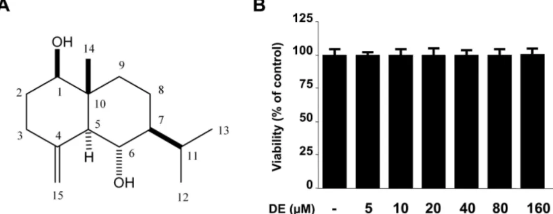
본 연구에서는 선행연구를 통해 몰약 물 추출물이 LPS로 자극한 BV2 세포에서 NO생성을 억제하는 효과가 있음을 확인하였고, 이 결과를 바탕으로 물 추출물을 분획하여 얻 은 EtOAc, BuOH, 물 분획물에 대하여 LPS로 자극한 BV2 세포에서 NO 억제효과를 검토한 결과 EtOAc 분획물이 유 의한 효과를 나타냈다. 이 결과를 바탕으로 EtOAc 분획물 의 일부인 3.5 g을 Sephadex LH-20, RP-18 및 silica gel column chromatography를 실시하여 하나의 화합물을 분리 하였고, 이 물질의 1H NMR과 13C NMR 스펙트럼 data를 문헌19)과 비교하여 eudesmane type sesquiterpene계 물질인 1β,6α-dihydroxyeudesm-4(15)-ene (DE)으로 동정하였다 (Fig. 1A).
몰약으로부터 분리한 DE가 미세아교세포에서 항염증 효 과를 나타내는지 확인하기 위하여 LPS로 유도된 BV2 미세 아교세포에서의 염증관련인자들을 측정하였다. 먼저 MTT assay를 이용하여 DE의 BV2 미세아교세포에서의 독성을 확인한 결과, 5-160 µM의 농도 범위에서 세포독성을 나타 내지 않았다(Fig. 1B). 이 결과를 바탕으로 160 µM을 최고 농도로 설정하여 실험을 진행하였다. 즉, LPS를 BV2 세포 에서의 염증유도물질로 사용하여 전염증매개체인 NO와 PGE2의 생성량을 측정하기 위하여 nitrite assay와 EIA실험
Fig. 1. Chemical structure of DE (A) and effects of DE on cell viability (B). BV2 microglia were incubated for 24 hr with various concentrations of DE (5-160µM). Cell viability was determined as described under Materials and methods. Data represent the mean values of three experiments±SD.
을 실시하였다. BV2 미세아교세포에 DE를 20, 40, 80, 160µM의 농도로 각각 처리한 후, 3시간 동안 CO2 incubator 에서 배양하고 LPS(1 µg/ml)를 처리하였을 때, DE는 농도 의존적으로 NO와 PGE2의 생성을 감소시켰다(Fig. 2).
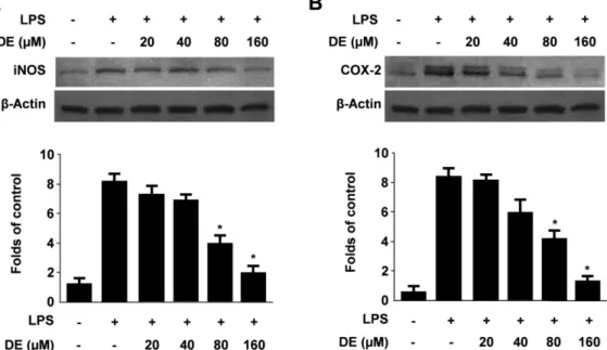
iNOS는 L-arginine으로부터 NO를 생성시키는 효소로 알 려져 있으며, 과도하게 생성된 NO는 염증부위에서 해당 조 직의 염증성 파괴를 유발하는 것으로 알려져 있다.20) 또한, COX에 의해 합성되는 prostaglandin류 역시 염증반응의 매
개역할을 수행하는 중요한 물질이며, COX-2는 염증과정 동 안 전염증매개체인 PGE2를 다량으로 생성시키는 것으로 보 고되어 있다.21) 저자들은 DE의 NO와 PGE2 생성 억제효과 가 이들 단백질의 조절에 의한 것인지를 확인하고자 하였 다. BV2 미세아교세포에 DE를 20, 40, 80, 160 µM의 농도 로 3시간 동안 전처리한 다음, LPS(1 µg/ml)를 처리하고 24 시간 후에 iNOS와 COX-2의 단백질발현을 western blot analysis를 통하여 관찰하였다. DE는 NO와 PGE2생성 억
Fig. 2. Effects of DE on nitrite (A), PGE2 (B) production in BV2 microglia stimulated with LPS. Cells were pre-treated for 3 hr with indicated concentrations of DE, and stimulated 24 hr with LPS (1µg/ml). The concentration of nitrite, PGE2 were determined as described under Materials and methods. Data represent the mean values of three experiments±SD. *p<0.05 compared to the group treated with LPS.
Fig. 3. Effects of DE on protein iNOS (A) and COX-2 (B) expression in BV2 microglia stimulated with LPS. Cells were pre- treated for 3 hr with indicated concentrations of DE, and 24 hr with LPS (1 ìg/ml). Western blot analyses were performed as described in Materials and methods, and representative blots of three independent experiments are shown. *p<0.05 compared to the group treated with LPS.
제와 유사한 양상으로 농도의존적인 iNOS와 COX-2의 단 백질발현 감소를 나타냈다(Fig. 3). 이상의 결과로부터 몰약 의 sesquiterpene계 성분인 DE는 iNOS와 COX-2 단백질 발 현의 감소를 통하여 NO 및 PGE2의 생성을 감소시킴으로 써 LPS로 유발한 미세아교세포의 염증을 억제하는 것으로 추정된다.
결 론
몰약 물 추출물의 EtOAc 분획물에 대하여 각종 컬럼 크 로마토그래피를 실시하여 sesquiterpene계 물질인 1β,6α- dihydroxyeudesm-4(15)-ene(DE)을 분리, 동정하였다. DE는 LPS로 자극한 BV2 미세아교세포에서 전염증매개체인 NO 와 PGE2의 생성을 각각 농도의존적으로 감소시켰으며, 이 는 iNOS와 COX-2 단백질의 발현을 감소시키는 것에 의하 여 기인되는 것으로 추정된다. 따라서, 몰약의 성분인 DE 는 퇴행성 뇌질환의 원인 중의 하나인 과도한 염증반응을 억제할 수 있는 후보물질로 연구가치가 있다고 생각된다.
사 사
본 연구는 산업통상자원부와 한국산업기술진흥원의 지역 특화산업육성사업 (R0002267)으로 수행된 연구결과입니다.
인용문헌
1.생약학교재편찬위원회 (2014) 생약학, 674. 동명사, 서울 2. Brieskorn, C. H. and Noble, P. (1982) Constituents of the
essential oil of myrrh. Planta Med. 44: 87-90.
3. Craveiro, A., Corsano, S., Proietti, G. and Strappaghetti, G.
(1983) Constituents of essential oil of Commiphora guidotti.
Planta Med. 48: 97-98.
4. Marongiu, B., Piras, A., Porcedda, S. and Scorciapino, A.
(2005) Chemical composition of the essential oil and super- critical CO2 extract of Commiphora myrrha (Nees) Engl. and of Acorus calamus L. J. Agric. Food Chem. 53: 7939-7943.
5. Chen, Y., Zhou, C., Ge, Z., Liu, Y., Liu, Y., Feng, W., Li, S., Chen, G. and Wei, T. (2013) Composition and potential anti- cancer activities of essential oils obtained from myrrh and frankincense. Oncol. Lett. 6: 1140-1146.
6. Siddiqui, M. Z., Thomas, M. and Prasad, N. (2013) Phys- icochemical characterization and antioxidant activity of essential oils of Guggul (Commiphora wightii) collected from Madhya Pradesh. Indian J. Pharm. Sci. 75: 368-372.
7. Fraternale, D., Sosa, S., Ricci, D., Genovese, S., Messina, F., Tomasini, S., Montanari, F. and Marcotullio, M. C. (2011) Anti-inflammatory, antioxidant and antifungal furanosesquit- erpenoids isolated from Commiphora erythraea (Ehrenb.) Engl. resin. Fitoterapia 82: 654-661.
8. Fujihara, M., Muroi, M., Tanamoto, K., Suzuki, T., Azuma, H. and Ikeda, H. (2003) Molecular mechanisms of mac- rophage activation and deactivation by lipopolysaccharide:
Roles of the receptor complex. Pharmacol. Ther. 100: 171- 194.
9. Doyle, S. L. and O’Neill, L. A. (2006) Toll-like receptors:
From the discovery of NFkappaB to new insights into tran- scriptional regulations in innate immunity. Biochem. Phar- macol. 72: 1102-1113.
10. Brown, G. C. and Bal-Price, A. (2003) Inflammatory neu- rodegeneration mediated by nitric oxide, glutamate, and mito- chondria. Mol. Neurobiol. 27: 325-355.
11. Good, P. F., Werner, P., Hsu, A., Olanow, C. W. and Perl, D.
P. (1996) Evidence of neuronal oxidative damage in Alzhe- imer's disease. Am. J. Pathol. 149: 21-28.
12. Pratico, D. and Trojanowski, J. Q. (2000) Inflammatory hypotheses: novel mechanisms of Alzheimer's neurodegen- eration and new therapeutic targets. Neurobiol. Aging. 21:
441-445.
13. Hald, A. and Lotharius, J. (2005) Oxidative stress and inflam- mation in Parkinson's disease: is there a causal link. Exp.
Neurol. 193: 279-290.
14. Liao, C. H., Sang, S., Liang, Y. C., Ho, C. T. and Lin, J. K.
(2004) Suppression of inducible nitric oxide synthase and cyclooxygenase-2 in downregulating nuclear factor-kappa B pathway by Garcinol. Mol. Carcinog. 41: 140-149.
15. Gonzalelz-Scarano, F. and Baltuch, G. (1999) Microglia as mediators in inflammatory and degenerative disease. Annu.
Rev. Neurosci. 22: 219-240.
16. Xu, K., Xu, Y. H., Chen, J. F. and Schwarzschild, M. A.
(2010) Neuroprotection by caffeine: time course and role of its metabolites in the MPTP model of Parkinson’s disease.
Neuroscience. 167: 475-481.
17. Nathan, C. F. and Hibbs, Jr. JB. (1991) Role of nitric oxide synthesis in macrophage antimicrobial activity. Curr. Opin.
Immunol. 3: 65-70.
18. Chen, Y. C., Shen, S. C., Lee, W. R., Hou, W. C., Yang, L. L.
and Lee, T. J. (2001) Inhibition of nitric oxide synthase inhib- itors and lipopolysaccharide induced inducible NOS and cyclooxygenase-2 gene expressions by rutin, quercetin, and quercetin pentaacetate in RAW264.7 macrophages. J. Cell.
Biochem. 82: 537-548.
19. Taichi, O., Keiji, I., Sachiyo, N., Masaru, S. and Sachi, S.
(1987) Studies on the sesquiterpenes from Ambrosia elatior LINNE. Chem. Pharm. Bull. 35: 2272-2279.
20. Hou, Y. C., Janczuk, A. and Wang, P. G. (1999) Current trends in the development of nitric oxide donors. Curr.
Pharm. Des. 5: 417-441.
21. Hammond, R. A., Hannon, R., Frean, S. P., Armstrong, S. J., Flower, R. J. and Bryant, C. E. (1999) Endotoxin induction of nitric oxide synthase and cyclooxygenase-2 in equine alve- olar macrophages. Am. J. Vet. Res. 60: 426-431.
(2015. 2. 24 접수; 2015. 3. 16 심사; 2015. 3. 17 게재확정)