The Anti-Inflammatory Effects of Persicaria thunbergii Extracts on Lipopolysaccharide-Stimulated RAW264.7 Cells
Sang-Bo Kim
1, Yeong Ae Seong
1, Hee Jae Jang
2and Gun-Do Kim
1*
1
Department of Microbiology, College of Natural Sciences, Pukyong National University, Busan 608-737, Korea
2
Department of Chemistry, College of Natural Sciences, Changwon National University, Changwon 641-773, Korea
Received November 23, 2011 /Revised December 8, 2011 /Accepted December 8, 2011In this study, we investigated the anti-inflammation effect of Persicaria thunbergii (P. thunbergii) on RAW 264.7 murine macrophage cells. The anti-inflammatory activity of P. thunbergii was determined by measuring expression of the LPS-induced inflammatory proteins, inducible nitric oxide synthase (iNOS), cyclooxygenase-2 (COX-2) and nuclear factor-κB (NF-κB), and the production of nitric oxide (NO) and prostaglandin E
2(PGE
2). Methanol extract of P. thunbergii decreased the expression of iNOS, COX-2 and NF-κB, and increased the expression of HO-1 in LPS-stimulated RAW264.7 cells. Methanol extract was fractioned by n-butanol, hexane and ethyl acetate (EtOAc) and each fraction was tested for inhibitory effects on inflammation. Among the sequential solvent fractions, the EtOAc soluble frac- tion was investigated by the expression of prostaglandin E
2(PGE
2), and showed decreasing form to the dose-dependent manner. EtOAc extract showed the most effective inhibitory activity of the ex- pression of iNOS, COX-2 and NF-κB, and the production of NO. The study showed that P. thunbergii has anti-inflammatory activity through the decrease of NO and inhibition of iNOS, COX-2, PGE
2and NF-κB expression, and by the increase of HO-1 enzyme. This study needs for more investigation to find out the most effective single compound with anti-inflammatory activity.
Key words : Persicaria thunbergii, anti-inflammation, nitric oxide, cyclooxygenase-2 (COX-2), nuclear factor-κB (NF-κB)
*Corresponding author
*Tel:+82-51-629-5618, Fax:+82-51-629-5619
*E-mail : [email protected]
Introduction
Inflammation is a major defense mechanism against pathogens and is stimulated by a range of microbial products. In macrophages and dendritic cells, toll-like re- ceptors (TLRs) are expressed at high levels for the attach- ment of various microbial products [1]. This binding triggers a wide spectrum of responses from phagocytosis to various cytokine productions, which enhances the inflammatory and adaptive immune responses [12,23].
LPS is a product and a major constituent of gram negative bacteria. It initiates a number of major cellular responses that play critical roles in the pathogenesis of inflammatory re- sponses and is also employed to induce the activation of RAW264.7 cells [26]. In addition, this eventually triggers nu- clear factor-κB (NF-κB), inducible nitric oxide synthase (iNOS) and cyclooxygenase-2 (COX-2) enzymes. However, the production of excessive inflammatory cytokines and pro- teins needs to be regulated, since it can lead to harmful in- flammatory responses such as rheumatoid arthritis, septic
shock and other chronic inflammatory diseases [5].
During the inflammatory processes, Nitric oxide (NO) and Prostaglandin E
2(PGE
2) are produced by iNOS and COX-2 enzymes, respectively [24]. NO is a short-lived free radical and is a signaling molecule that mediates many phys- iological and pathophysiological processes, including neuro- transmission and inflammation [6,18]. NO, as generated in activated macrophages by iNOS, is an important event in host defenses and it modulates the synthesis of prosta- glandins, tormboxans and other inflammatory molecules [17]. Despite the beneficial roles of NO in host defense mech- anism against tumor cells, viral replication and other factors, and over expression of NO can be harmful to the host, lead- ing to rheumatoid arthritis [22], experimental allergic ence- phalomyelitis [4] and allograft rejection [27]. Thus, selective inhibition of iNOS can be beneficial to control the pro- duction of NO.
COX is an enzyme that catalyzes the conversion of arach-
idonic acid to prostaglandin H
2, the precursor of a variety
of biologically active mediators such as PGE
2, prostacyclin
and thromboxane A2 [10]. Two isoforms of COX are de-
scribed as COX-1 and COX-2. COX-1 is ubiquitously ex-
pressed and it produces prostanoids that are involved in
normal cellular functions. COX-2 expression, on the other hand can be induced in several cell types by cytokines, mi- togens, bacterial endotoxins and other growth factors. Also, it plays a critical role in the damage produced by in- flammation [2].
Prostaglandins and glucocorticoids are potent mediators of inflammation. Non-steroidal anti-inflammatory drugs (NSAIDs) exert certain effects by the inhibition of prosta- glandin production. The pharmacological target of NSAIDs is cyclooxygenase, which catalyze the first committed step in arachidonic acid metabolism. NSAIDs act on the active site of cyclooxygenase and inhibit expression of both COX-1 and COX-2 with little specificity. This eventually leads to serious side effects such as gastric lesions and renal toxicity.
COX-2 selective inhibitors are also identified to show potent anti-inflammatory activity, in vivo, with minimal gastric side effects [14]. It was then investigated whether P. thunbergii extracts can suppress the expression of COX-2.
Inactive NF-κB is constitutively present as a homo- or a hetero-dimer, and binds to inhibitory IκB proteins. Pro-in- flammatory cytokines or bacterial infections can induce phosphorylation, ubiquitination and proteasome mediated degradation of the IκB proteins. This is followed by the translocation of NF-κB to the nucleus, binding to the rele- vant DNA sites on the promoter region of the genes and induction of gene transcription [23]. Lipopolysaccharide (LPS) is known to stimulate the degradation of one of the isoforms of IκB, IκBα and to promote the activation of NF-κB DNA binding activity [25]. The effects of P. thun- bergii extracts on the nuclear translocation of NF-κB in LPS-stimulated RAW264.7 cells were examined through immunofluorescence.
As an annual plant, P. thunbergii is widely distributed in Korea and it has been used as a folk medicine to treat rheu- matism, hemorrhage and measles in both Korea and China.
In addition, the antioxidative and antitumor effects of P.
thunbergii were reported recently [15,19]. Thus, we did experi- ment to investigate the effects of P. thunbergii on the pro-in- flammatory response in LPS-stimulated RAW264.7 cells.
Materials and Methods Plant materials
The aerial parts of P. thunbergii were collected in August 2010 at Yangsan, Gyeongnam in Korea. Collected plants were preserved at -80℃ freezer and used for this research.
Extraction and Isolation
Plant materials were extracted with MeOH for 4 weeks at room temperature and filtered. The filterate was evapo- rated in vacuo. The resultant of methanol extract was fol- lowed by successive partitioning with n-butanol, n-hexane and ethyl acetate. After the adsorption of silica gel, each ex- tract was fractioned in MPLC (Combifresh-RF, ISCO) using Hexane/EtOAc solvent and evaporated in vacuo.
Cell culture and treatments
RAW264.7 cells from the mouse macrophage cell line were obtained from the American Tissue Culture Collection (Manassas, VA, USA). The RAW264.7 cells were grown in Dulbecco’s Modified Eagle’s Medium (DMEM), supple- mented with high glucose, 10% heat inactivated fetal bovine serum, penicillin and streptomycin (Cellgro, Manassas, VA, USA). The cells were incubated in a humidified atmosphere of 5% CO
2at 37℃. The P. thunbergii extract was then added to the culture media with the final concentration as indicated.
Cell viability assay (WST-1)
For the cell viability study, RAW264.7 cells, approx- imately 1×10
5cells/ml in number were resuspended in me- dium and were plated in a 96-well plate. The cells were treat- ed with 50~150 μg/ml of P. thunbergii extract with LPS for 24 hr. After the treatment, 10 μl of WST-1
®(Daeil Lab serv- ice, Jong-No, Korea) solution was added into each well and the cells were incubated at 37℃ for 3 hr. The absorbance was read at 460 nm.
Nitric oxide (NO) assay
The cells (5×10
4cells/ml) were pre-incubated with P.
thunbergii extract for 2 hr and were incubated with indicated concentrations of LPS for 24 hr. The nitrite accumulation in the supernatant was assessed by Griess regent (Sigma- Aldrich, St. Louis, MO, USA). Each 100 μl of the culture su- pernatant was mixed with an equal volume of Griess reagent and was incubated at room temperature for 10 min. The ab- sorbance was then measured at 540 nm in a microplate ab- sorbance reader and a series of known concentrations of so- dium nitrite was used as standard.
Western blot analysis
RAW264.7 cells were cultured as described above, were
pre-incubated with P. thunbergii extract (100 μg/ml) for 2
hr and were incubated with LPS (1 μg/ml). The RAW264.7 cells were harvested and were lysed after 24 hr with ice-cold cell lysis buffer (Intron Biotechnology Inc., Gyeonggi, Korea). After incubation on ice for 30 min, the insoluble ma- terials were removed by centrifugation at 14,000 rpm and 4℃ for 20 min. The protein content of the cell lysates was determined by using a Protein Quantification Kit (CBB sol- ution) (Biosesang Inc., Gyeonggi, Korea) with bovine serum albumin (BSA) (Thermo Scientific, Rockford, Il, USA) was used as standard. An aliquot from each sample (50 μg of protein) was boiled with the sample buffer for 5 min, and then, was resolved in 12% SDS-polyacrylamide gel electro- phoresis (SDS-PAGE). The protein was electrotransferred to a nitrocellulose membrane (Pall, Pensacola, Fl, USA) and was blocked overnight in PBST buffer (135 mM sodium chloride, 2.7 mM potassium chloride, 4.3 mM sodium phos- phate, 1.4 mM potassium dihydrogen phosphate and 0.5%
Tween-20) containing 5% skim milk powder at 4℃. The western blots were then probed overnight with primary anti- bodies (anti-HO-1, anti-NF-κB p65, anti-iNOS, anti-COX-2 and anti-β-actin) (Cell Signaling Technology Inc. Beverly, MA, USA), were washed three times with PBST and then were incubated for 1 hr with anti-rabbit IgG or anti-mouse IgG conjugated with HRP. The blots were washed in PBST and were visualized using an enhanced chemiluminescent (ECL) detection solution (Pierce, IL, USA).
Immunofluorescence
The cells were cultured (37℃, 5% CO
2) in coverglass bot- tom dishes (SPL Lifesciences, Gyeonggi, Korea) for 24 hr.
For the experiment, cells were fixed with 4% formaldehyde (Junsei Chemical Ltd., Tokyo, Japan) for 15 min at room tem- perature and were blocked for 1 hr in 5% normal serum of the host against primary antibodies and 0.3% Triton X-100. The fixed and blocked cells were then incubated with 0.1 μg/ml of primary antibodies (anti-NF-κB p65) for 3 hr and then, with 0.1 μg/ml of anti-rabbit IgG Fab2 fragment Alexa Fluor 488 conjugate (Cell Signaling Technology Inc.) for 1 hr. Stained cells on the slides were mounted with Prolong Gold Antifade Reagent (Invitrogen, Eugene, OR, USA) and were observed under Nikon FCLIPS 50i micro- scope, equipped with a charged-coupled device (CDD) camera. Images were captured and were processed with High-Content Analysis Software (Cambridge Healthtech Institute, Needham, MA, USA).
PGE
2measurement
For the quantitative determination of Prostaglandin E
2(PGE
2) in cell culture supernatants, the cells were pre-in- cubated with P. thunbergii extract for 2 hr, and were in- cubated with indicated concentrations of LPS for 24 hr. The PGE
2Parameter Assay Kit (R&D systems, Minneapolis, MN, USA) was used to measure PGE
2concentrations in cell su- pernatants based on the manufacturer’s protocol. The ab- sorbance was then measured at 450 nm within 30 min in a microplate absorbance reader.
Results
Effects of LPS and P. thunbergii extracts on cell proliferation
For the cell viability study, the LPS and the methanol ex- tract were treated to RAW264.7 cells. The LPS (1 μg/ml) was employed to induce RAW264.7 cell activation and to pro- duce nitric oxide without cell cytotoxicity. After 24 hr, the LPS and the methanol extract did not influence cell survival (Fig. 1A, 1B). Therefore, 100 μg/ml concentration of meth- anol extract was selected for the anti-inflammatory experiment. The methanol extract of P. thunbergii was frac- tioned more by n-butanol, hexane and ethyl acetate. After 24 hr of incubation, the viabilities of the cells were de- termined by WST-1 assay. The various concentrations of P.
thunbergii fractions did almost not influence cell survival (Fig. 1C). The ethyl acetate extract was fractioned and their effect for cell viability then measured. Among of them, the 2, 4 and 6 fractions have little cytotoxicity. However, other fractions have over the 80% cellular viability (Fig. 1D).
Inhibitory activity of the extracts on LPS-induced NO generation
As shown in Fig. 2A, the LPS-induced NO generation was
suppressed by the methanol extract of P. thunbergii. Most
of all, the concentration of 100 μg/ml has a high inhibitory
activity. The more fractioned methanol extracts of P. thunber-
gii by n-butanol, hexane and ethyl acetate were measured
regarding the nitric oxide generation. Among them, the ethyl
acetate extract suppressed NO generation more than the oth-
ers in a dose-dependent manner (Fig. 2B). Each of the frac-
tioned ethyl acetate extracts of P. thunbergii was examined
for the NO inhibitory activity at 100 μg/ml and was labeled
(fraction number). All of the fractions more inhibited NO
generation than those treated with the LPS only (Fig. 2C).
A)
B)
C )
D )
Fig. 1. Cytotoxicity of LPS and the extracts of
P. thunbergii
in RAW264.7 cells. Cell viability was measured by WST-1 assay. (A) Cells were treated with various concentrations of LPS for 24 hr. (B) The filtered and evaporated meth- anol extract was treated with indicated concentrations for 24 hr. (C) The cells were pre-treated with the various concentrations ofP. thunbergii
extracts (n
-butanol, hex- ane and ethyl acetate) for 2 hr and then treated with LPS (1 μg/ml) for 24 hr. (D) The fractions of ethyl acetate extract were pre-treated with 100 μg/ml for 2 hr and then treated with LPS (1 μg/ml) for 24 hr.The 5 and 7 fractions were adopted for anti-inflammation study without cell cytotoxicity.
A)
B)
C )
Fig. 2. Inhibitory activity of the extract on NO generation in LPS-induced RAW264.7 cells. RAW264.7 cells were seed- ed in 24 well plates at a density of 5×10⁵cells/ml for 24 hr. (A) The cells were pre-treated with indicated con- centrations of methanol extract for 2 hr and then treated with LPS (1 μg/ml) for 24 hr. (B) RAW264.7 cells were pre-treated with
n
-butanol, hexane and ethyl acetate ex- tract respectively for 2 hr and then treated with LPS (1 μg/ml) for 24 hr. (C) NO generation by the fractioned ethyl acetated extract (fraction number 1~8, 100 μg/ml) was measured. The amounts of NO were determined by Griess assay and a standard curve created using NaNO2in culture medium.
Down-regulation of pro-inflammatory protein and PGE
2by the extracts
For investigating anti-inflammatory effects of P. thunber-
gii, western blot analysis was conducted. When the cells
were treated with methanol extract, pro-inflammatory pro-
tein such as iNOS, COX-2 and NF-κB were decreased gradu-
ally in a dose-dependent manner. The HO-1 is known for
its beneficial protective effect in inflammation [20]. It is ex- pected that an increased HO-1 protein can inhibit in- flammation (Fig. 3). The ethyl acetate extract of P. thunbergii was measured as to iNOS and COX-2 expression levels.
According to the result, iNOS was highly suppressed at the concentration of 50, 100 μg/ml and the ethyl acetate extract inhibited COX-2 expression levels gradually (Fig. 4A). In ad- dition, generation of Prostaglandin E
2was decreased. These results suggest that the ethyl acetate extract suppressed Prostaglandin E
2by inhibiting COX-2 expression levels (Fig.
4B). The 5 and 7 fractions of the more fractioned ethyl acetate extracts inhibited the expression levels of iNOS, COX-2 and NF-κB at the concentration of 100 μg/ml (Fig. 5).
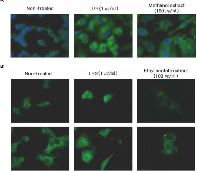
Effects of P. thunbergii extracts on LPS-induced nuclear translocation of NF-κB
NF-κB is one of the principal factors for the expression of COX-2 and iNOS as mediated by the LPS or pro-in- flammatory cytokines [11]. The present study identified that NF-κB is responsible for the maintenance of iNOS ex- pression [9]. To identify NF-κB translocation from the cyto- sol to the nucleus, immunofluorescence was performed.
Translocation of NF-κB showed that treating RAW264.7 cells with the LPS (1 μg/ml) enhanced NF-κB activation.
However, when the methanol and the ethyl acetate extracts (100 μg/ml) were added to the RAW264.7 cells, the NF-κB activity was suppressed markedly than that treated with the
Fig. 3. Down-regulation of pro-inflammatory proteins by meth- anol extract of
P. thunbergii
in LPS-stimulated RAW264.7 cells. RAW264.7 cells were pre-treated with methanol extract ofP. thunbergii
(10, 50, 100 μg/ml) for 2 hr and then treated with LPS (1 μg/ml) for 24 hr. The cells were lysed and the expression levels of proteins were then measured by western blot analysis. Beta actin was used as an internal control.A)
B)
Fig. 4. Effects of ethyl acetate extracts of
P. thunbergii
on the expression levels of COX-2 and iNOS and PGE2 in LPS-stimulated RAW264.7 cells. (A) RAW264.7 cells were pre-treated with ethyl acetate extract ofP. thunber- gii
(10, 50, 100 μg/ml) for 2 hr and then treated with LPS (1 μg/ml) for 24 hr. (B) The quantitative determi- nation of PGE2 in cell supernatants was measured by using the prostaglandin E2parameter assay kit. The cells were pre-treated with ethyl acetate extract ofP. thunber- gii
(10, 50, 100, 150, 200 μg/ml) for 2 hr and then treated with LPS (1 μg/ml) for 24 hr.Fig. 5. Effects of fractioned ethyl acetate extract (fraction no 5, 7) of
P. thunbergii
on the levels of pro-inflammatory pro- teins in LPS-stimulated RAW264.7 cells. The cells were pre-treated with fractioned ethyl acetate extract (no 5, 7) ofP. thunbergii
(100 μg/ml) for 2 hr and then treated with LPS (1 μg/ml) for 24 hr. The cells were lysed and the expression levels of proteins were then measured by western blot analysis. Beta actin was used as an internal control.A)
B)
Fig. 6. Translocation of NF-κB in LPS-induced RAW264.7 cells. (A) The cells were cultured with methanol extract of
P. thunbergii
(100 μg/ml) for 2 hr and then treated with LPS (1 μg/ml) for 24 hr. (B) The cells were pre-treated with ethyl acetate extract ofP. thunbergii
(100 μg/ml) for 2 hr and then treated with LPS (1 μg/ml) for 24 hr. The cells were treated as indicated and then fixed. The fixed cells were pre-treated with 0.1 μg/ml of primary antibodies (anti-NF-κB p65) for 3 hr and then treated with 0.1 μg/ml of anti-rabbit IgG Fab2 fragment Alexa Fluor 488 conjugate for 1 hr. Stained cells on the slides were mounted and then observed under microscope, equipped with a charged-coupled device camera. Blue color indicates DAPI stained nuclear and green show the expression and location of NF-κB.LPS (1 μg/ml) only (Fig. 6A, 6B).
Discussion
Macrophages are produced by the differentiation of mon- ocytes in tissues. Their role is to phagocytose (engulf and digest) cellular debris and pathogens. They also stimulate lymphocytes and other immnue cells to respond to patho- gens [13]. In theory, LPS induces the activation of macrophages. Over activated macrophages generates pro-in-
flammatory responses that release nitric oxide, PGE
2, in- flammatory cytokines and ROS, which can all lead to harm- ful inflammatory responses.
Nitric oxide has beneficial roles in host defense system against tumor cells, viral replication and other factors.
However, over production of NO causes various in- flammatory diseases. Relative to the inflammatory response, iNOS and COX-2 are the most important target proteins that produce NO and PGE
2. iNOS produces NO in the cytoplasm.
COX-2, on the other hand, activates the synthesis of prosta-
glandins, prostacyclin and thromboxanes. Generally, COX-2 is barely detectable under normal physiological conditions, but the lipopolysaccharide induces over expression of the COX-2 protein [16]. Hence, it plays a vital role on the in- duction of inflammatory responses.
In this study, the effects of P. thunbergii on anti-in- flammatory responses were investigated. The inflammatory responses was induced by LPS (1 μg/ml) in RAW264.7 cells.
For the study, the dried P. thunbergii powder was extracted by methanol and was partitioned with n-butanol, hexane and ethyl acetate. In order to select non-cytotoxic concen- trations for the cells, cell viability was measured by WST-1 assay. Various concentrations of P. thunbergii methanol ex- tracts and LPS did not influence the survival of RAW264.7 cells (Fig. 1A, 1B). Under the same conditions, the effects of NO production on LPS-stimulated RAW264.7 cells were determined. As shown in Fig. 2A, NO production was de- creased by P. thunbergii methanol extracts in a dose-depend- ent manner. Following the NO analysis, western blot analy- sis was employed to analyze pro-inflammatory protein ex- pression levels. The expression of iNOS, COX-2 and NF-κB were decreased, but the beneficial protective HO-1 protein was increased (Fig. 3).
For further fractionation, methanol extract of P. thunbergii was partitioned by sequential use of solvents (n-butanol, hexane and ethyl acetate). Among them, the ethyl acetate extract had the highest activation against NO production and had decreased by half on the 100 μg/ml concentration without cell cytotoxicity (Fig. 1C, 2B). Following the NO analysis, expression levels of iNOS and COX-2 as pro-in- flammatory marker proteins were determined by western blotting. As shown in Fig. 4A, cells treated with the ethyl acetate extract were more efficient than cells treated with the methanol extract. iNOS was completely decreased at 50~100 μg/ml concentration and COX-2 expression was sup- pressed in a dose-dependent manner. Considering the re- sults of NO analysis and western blotting, the decreased iNOS expression levels influenced NO production. The de- creased COX-2 expression levels down regulated PGE
2of up to 40% at 150 μg/ml concentration (Fig. 4B).
The NF-κB family of proteins comprises several tran- scription factors that regulate inducible gene expression in various immunological and antioxidant protective re- sponses, including the up-regulation of major pro-in- flammatory cytokines, adhesion molecules and antioxidant stress proteins [8,21]. Under basal conditions, NF-κB is lo-
cated in the cytoplasm in conjugation with the inhibitory NF-κB (I-κB). In response to a wide range of signals, the regulatory NF-κB subunits, p50 and p65, dissociate NF-κB from I-κB and subsequently translocate to the nucleus [3,7].
In LPS-treated RAW264.7 cells, translocation of NF-κB in the nucleus was observed by immunofluorescence. However, the methanol extract of P. thunbergii suppressed the trans- location of NF-κB into the nucleus (Fig. 6A). To identify the translocation of NF-κB on the ethyl acetate extract treated cells, the cells were treated with LPS (1 μg/ml) and then the translocation of NF-κB in the nucleus were observed.
When the ethyl acetate extract (100 μg/ml) was added to the LPS-stimulated RAW264.7 cells, NF-κB translocation was dramatically suppressed. (Fig. 6B). The ethyl acetate extract was partitioned and arranged in an orderly manner. The number 5 and 7 fractions decreased NO production without cytotoxicity at 100 μg/ml concentration (Fig. 1D, 2C). The effects of 5 and 7 fractions were further examined by western
Fig. 7. Effects of
P. thunbergii
on inflammation signaling. LPS induces the activation of RAW264.7 macrophage cells.NF-κB protein is one of the transcription factors for the expression of COX-2 and iNOS by the LPS or pro-in- flammatory cytokines. Following NF-κB activation, the expression levels of iNOS and COX-2 were increased.
The activation of sequential upstream signaling resulted in increased in PGE2and NO productions.
P. thunbergii
suppressed NO production, decreased the expression level of pro-inflammatory proteins and inhibited trans- location of NF-κB on LPS stimulated RAW264.7 cells.blotting and results showed the down-regulated expression of pro-inflammatory proteins (Fig. 5). In the inflammation signaling, LPS activates the upstream signal proteins such as NF-κB, iNOS and COX-2, leading to the over expression of NO and PGE
2.
In this study, P. thunbergii showed that it could be a poten- tial anti-inflammatory agent by suppressing NO production, decreasing the expression level of pro-inflammatory proteins and inhibiting NF-κB translocation on LPS stimulated RAW264.7 cells (Fig. 7). However, the more fractioning of P. thunbergii extracts and additional researches about its an- ti-inflammatory effects are needed.
References
1. Akira, S. and H. Hemmi. 2003. Recognition of patho- gen-associated molecular patterns by TLR family.
Immunol.
Lett.
85, 85-95.2. Araki, E., C. Forster, J. M. Dubinsky, M. E. Ross, and C.
Iadecola. 2001. Cyclooxygenase-2 inhibitor NS-398 protects neuronal nultures from lipopolysaccharide-induced neurotoxicity.
Stroke
32, 2370-2375.3. Chen, L. F. and W. C. Greene. 2004. Shaping the nuclear action of NF-kappaB.
Nat. Rev. Mol. Cell Biol.
5, 392-401.4. Cross, A. H., T. P. Misko, R. F. Lin, W. F. Hickey, J. L.
Trotter, and R. G. Tilton. 1994. Aminoguanidine, an in- hibitor of inducible nitric oxide synthase, ameliorates ex- perimental autoimmune encephalomyelitis in SJL mice.
J
.Clin. Invest.
93, 2684-2690.5. D’Aiuto, F., M. Parkar, G. Andreou, J. Suvan, P. M. Brett, D. Ready, and M. S. Tonetti. 2004. Periodontitis and sys- temic inflammation: control of the local infection is asso- ciated with a reduction in serum inflammatory markers.
J.
Dent. Res.
83, 156-160.6. Garthwaite, J. 1995. Neural nitric oxide signaling.
Trends Neurosci.
18, 51-52.7. Ghosh, S. and M. S. Hayden. 2008. New regulators of NF-kappaB in inflammation.
Nat. Rev. Immunol.
8, 837-848.8. Gilmore, T. D. 2006. Introduction to NF-kappaB: players, pathways, perspectives.
Oncogene
25, 6680-6684.9. Gookin, J. L., S. Chiang, J. Allen, M. U. Armstrong, S. H.
Stauffer, C. Finnegan, and M. P. Murtaugh. 2006. NF-κB mediated expression of iNOS promotes epithelial defense against infection by
Cyptosporidium parvum
in neonatal piglets.J. Physiol.
290, 164-174.10. Hawkey, C. J. 1999. COX-2 inhibitors.
Lancet.
353, 307-314.11. Jung, W. K., S. J. Heo, Y. J. Jeon, C. M. Lee, Y. M. Park, H. G. Byun, Y. H. Choi, S. G. Park, and I. W. Choi. 2009.
Inhibitory effects and molecular mechanism of dieckol iso- lated from marine brown alga on COX-2 and iNOS in micro- glial cells.
J. Agric. Food Chem.
57, 4439-4446.12. Kaisho, T. and S. Akira. 2006. Toll-like receptor function and signaling.
J. Allergy Clin. Immunol.
117, 979-987.13. Khazen, W., J. P. M’bika, C. Tomkiewicz, C. Benelli, C.
Chany, A. Achour, and C. Forest. 2005. Expression of macro- phage-selective markers in human and rodent adipocytes.
FEBS. Lett.
579, 5631-5634.14. Kurumbail, R. G., A. M. Stevens, J. K. Gierse, J. J. Mcdonald, R. A. Stegeman, J. Y. Pak, D. Gildehaus, J. M. Miyashiro, T. D. Penning, K. Seibert, P. C. Lsakson, and W. C. Stallings.
1996. Structural basis for selective inhibition of cyclo- oxygenase-2 by anti-inflammatory agents.
Nature
384, 644-648.15. Lee, K. T., C. H. Ku, J. S. Eun, T. Y. Shin, J. P. Lim, D.
O. Eom, O. P. Zee, and D. K. Kim. 2001. Antioxidative com- ponents from the aerial parts of
Persicaria thunbergii
.Arch.
Pharm. Res.
6, 611-616.16. Lee, S. H., J. S. Han, S. J. Heo, J. Y. Hwang, and Y. J. Jeon.
2010. Protective effects of dieckol isolated from
Ecklonia cava
against high glucose-induced oxidative stress in human um- bilical vein endothelial cells.Toxicol. In Vitro
24, 375-381.17. Moncada, S., R. M. J. Plamer, and E. A. Higgs. 1991. Nitric Oxide: Physiology, pathophysiology, and pharmacology.
Pharmacol.
43, 109-142.18. Nathan, C. and Q. W. Xie. 1994. Nitric oxide sythases: Roles, tolls, and controls.
Cell
78, 915-918.19. Oh, H. M., B. M. Kwon, N. I. Baek, S. H. Kim, I. S. Chung, M. H. Park, H. W. Park, J. H. Lee, H. W. Park, E. J. Kim, and D. K. Kim. 2005. Inhibitory activity of isorhamnetin from
Persicaria thunbergii
on farnesyl protein transferase.Arch. Pharm. Res
. 28, 169-171.20. Paine, A., B. Eiz-Vesper, R. Blasczyk, and S. Immenschuh.
2010. Signaling to heme oxygenase-1 and its anti-in- flammatory therapeutic potential.
Biochem. Pharmacol.
80, 1895-1903.21. Perkins, N. D., and T. D. Gilmore. 2006. Good cop, bad cop:
the different faces of NF-kappaB.
Cell Death Differ.
13, 759-772.22. St Clair, E. W., W. E. Wilkinson, T. Lang, L. Sanders, M.
A. Misukonis, G. S. Gilkeson, D. S. Pisetaky, D. I. Granger, and J. B. Weinberg. 1996. Increased expression of blood mononuclear cell nitric oxide synthase type 2 in rheumatoid arthritis patients.
J. Exp. Med.
184, 1173-1178.23. Tak, P. P. and G. S. Firestein. 2001. NF-kappaB: a key role in inflammatory diseases.
J. Clin. Invest.
107, 7-11.24. Vane, J. R., J. A. Mitchell, I. Appleton, A. Tomlinson, D.
Bishop-Bailey, J. Croxtall, and D. A. Willoughby. 1994.
Inducible isoforms of cyclooxygenase and nitric-oxide syn- thase in inflammation.
Proc. Natl. Acad. Sci. USA
91, 2046-2050.25. Watters, J. J., J. A. Sommer, Z. A. Pfeiffer, U. Prabhu, A.
N. Guerra, and P. J. Bertics. 2002. A differential role for the mitogen-activated protein kinases in lipopolysaccharide sig- naling: the MEK/ERK pathway is not essential for nitric ox- ide and interleukin 1β production.
J. Biol. Chem.
277, 9077-9087.26. Woo, M. S., S. H. Jung, J. W. Hyun, and H. S. Kim. 2004.
Differential regulation of inducible nitric oxide synthase and cytokine gene expression by forskolin and dibutyryl-cAMP
초록:Lipopolysaccharide로 처리 된 RAW264.7 세포에서 고마리 추출물의 항염증 효과 김상보
1․성영애
1․장희재
2․김군도
1*
(
1부경대학교 미생물학과,
2창원대학교 화학과)
본 연구는 고마리 추출물이 가지는 항염증 활성을 알아보기 위하여 쥐의 대식세포(RAW264.7 cell)에 Lipopolysaccharide (LPS)를 처리하여 염증반응을 유도하고 이때 발생되는 Nitric oxide (NO)의 생성 억제를 확 인하였다. 또한 염증에서 중요하게 알려져 있는 Inducible nitric oxide synthase (iNOS), Cyclooxygenase-2 (COX-2), Nuclear factor-kappa B (NF-κB) 단백질들의 발현을 비교하였고, 추가적으로 NF-κB 단백질의 핵 내부 로의 이전 및 활성을 확인하였다. 메탄올 추출물은 NO 생성 및 iNOS, COX-2, NF-κB 단백질의 발현을 억제하고, 세포를 보호하는 효과를 가지는 Heme oxygenase-1 (HO-1) 단백질의 발현을 증가시켰다. 위 결과를 바탕으로 하여 n-butanol, hexane, ethyl acetate 용매를 이용한 추가적인 분획을 실시하였다. 이들 분획 중 고마리의 ethyl acetate 추출물은 Prostaglandin E
2(PGE
2), NO 생성을 억제 하였으며, iNOS, COX-2 단백질들의 발현을 감소, NF-κB의 핵 내부로의 이동을 억제하는 효과가 높다는 것을 확인하였다. 이러한 연구결과는 고마리 식물이 좋은 항염증 활성을 가지고 있음을 나타내며, 지속적인 분획으로 고마리 식물이 가지는 항염증 활성 물질을 선별하여 그 작용기작을 규명하는 연구가 필요하다.
in lipopolysaccharide-stimulated murine BV2 microglial cells.
Neurosci. Lett.
356, 187-190.27. Worrall, N. K., W. D. Lazenby, T. P. Misko, T. S. Lin, C.
P. Rodi, P. T. Manning, R. G. Tilton, J. R. Williamson, and
T. B. Ferguson. 1995. Modulation of
in vivo
alloreactivity by inhibition of inducible nitric oxide synthase.J. Exp. Med.
181, 63-70.