Anti-inflammatory Effect of HaGF peptide of Harmonia axyridis
Dong-Hee Kim
2, Hyeon-Jeong Kim
1,3, Jin-Young Lee
4, Jae-Sam Hwang
5, In-Woo Kim
5, Seul-Gi Lee
1, Hyeon-Guk Jeong
1and Bong-Jeun An
1*
1
Department of Cosmeceutical Science, Daegu Haany University, Gyeongsan 712-715, Korea
2
Korea Promotion Institute for Traditional Medicine Industry, Gyeongsan 712-260, Korea
3
Herbnoori, Daegu 702-062, Korea
4
Department of Herbal Cosmetic Science, Hoseo University, Asan 336-795, Korea
5
Department of Agricultural Biology, National Academy of Agricultural Science, Suwon 441-100, Korea Received November 12, 2012 /Revised April 3, 2013 /Accepted April 22, 2013
Harmonia axyridis is known to display diverse biological activities, such as growth promotion. However, few studies have investigated the effect of H. axyridis on inflammation of the skin. In this study, we explored the anti-inflammatory effect of the Harmoniasin gene fragment (HaGF) peptide from H. axyr- idis on macrophage cells. During the entire experimental period, 5, 25, 50, and 100 µg/ml of HaGF showed no cytotoxicity. However, at these concentrations, HaGF inhibited the activity of iNOS and COX-2 by 51% and 49%, respectively. In addition, the HaGF extract reduced the release of in- flammatory cytokines, including TNF-a and IL-6. Therefore, HaGF has been LPS-induced macrophage Raw 264.7 cells could be expected from the inhibitory effects of the inflammatory.
Key words : Anti-inflammatory, COX-2, Harmonia axyridis, iNOS, lipopolysaccharide
*Corresponding author
*Tel:+82-53-819-1429, Fax:+82-53-819-1429
*E-mail : anbj@dhu.ac.kr
This is an Open-Access article distributed under the terms of the Creative Commons Attribution Non-Commercial License (http://creativecommons.org/licenses/by-nc/3.0) which permits unrestricted non-commercial use, distribution, and reproduction in any medium, provided the original work is properly cited.
Journal of Life Science 2013 Vol. 23. No. 4. 495~500 DOI : http://dx.doi.org/10.5352/JLS.2013.23.4.495
서 론
예로부터 곤충은 동양에서 약용·식용으로 많이 이용되었는 데, 고서의 기록에 의하면 동의보감은 약 95종의 약용 곤충이, 본초강목습유(本草綱目拾遺)에는 106종의 약용 곤충이 기록 되어있다[22]. 이러한 곤충자원은 최근 생명공학 기술의 발달 과 함께 다양화 되고 있는데, 그 예로 각종 유용물질을 생산하 는 누에(실크, 누에가루, 동충하초), 꿀벌(꿀, 로얄제리. 프로폴 리스), 곤충의 외골격(chitin의 추출) 등이 좋은 예라 할 수 있 다. 또한 곤충의 추출물은 전통적인 생약으로 이용되었는데, 최근 이러한 곤충 추출물에 대한 효능 및 유효성분 분석에 대한 연구가 진행되고 있다. 이 외에도 곤충 자원은 유용생물 자원으로 현재 세계적으로 여러 분야에서 매우 광범위하게 개발 활용되고 있다[6, 7, 8, 12, 16].
염증은 생체 혹은 조직에 물리적 자극이나 화학적 물질, 세 균 감염 등의 기질적 변화를 가져오는 침습이 가해질 때 그 손상부위를 수복 재생하려는 복구기전으로서 자극이 가해지 면 국소적으로 염증성 성분이 증가되면서 염증이 유발된다 [14]. 임상적으로는 발적, 발열, 종창, 동통, 기능장애 등의 증
상이 나타난다[11]. 대식세포는 선천면역뿐만 아니라 획득면 역 등 다양한 숙주반응에 관여하여 항상성 유지에 관여하는 것으로 알려져 있으며, 염증반응 시에는 nitric oxide (NO)와 cytokine을 생산하여 감염초기에 생체방어에 중요한 역할을 한다[3]. 포유동물 세포의 nitric oxide synthase (NOS)의 경 우, 유사형태가 3가지 존재하는데 neuronal NOS (nNOS), en- dothelial NOS (eNOS), 그리고 inducible NOS (iNOS)이다.
그 중에서 특히 iNOS가 염증반응에 관여한다. nNOS와 eNOS 는 항상 발현되어 있으며, iNOS의 경우 interferon-γ, lip- opolysaccharide (LPS), 그리고 여러 가지 염증 cytokine의 자 극이 있을 때 발현된다. 또한 NO는 급성, 만성 염증반응을 조절하기도 한다. COX에 대해서는 1990년대 초반에 주로 연 구되었는데, 이 또한 유사형태가 두 가지 존재한다. COX-1은 거의 모든 조직에 발현되어 있고, prostagrandin을 생산하여 신장의 혈액흐름을 조절하거나 위장의 세포를 보호하는 등의 생리적인 기능을 조절한다. 반대로 COX-2의 경우는 미생물에 의한 감염이나 손상 혹은 여러 요인의 스트레스에 반응한 대 식세포 (macrophage)에서 발현된다. 즉 iNOS와 COX-2의 발 현유무는 면역 세포의 염증 반응에 관여한다. 또한 염증 사이 토카인(proinflammatory cytokines)인 tumor necrosis factor- α (TNF-α), interleukin-1β (IL-1β), interleukin-6 (IL-6), mono- cyte chemoattractant protein-1 (MCP-1) 등이 염증 반응에 관 여하는 인자로 알려져 있다[9, 24].
따라서 본 연구에서는 무당벌레 유충으로부터 분리된 펩타
이드 유전자 Harmoniasin의 일부 영역 HaGF을 이용하여 LPS
로 활성 된 Raw 264.7 cell로 염증에 관련된 다양한 기전과
단백질 발현을 측정하였다.
재료 및 방법
재료
본 실험에서 사용한 펩타이드 HaGF는 AnyGen (Gwang-ju, Korea)사에서 98% 이상의 순도로 합성하였고, 0.01% acetic acid에 녹여 사용하였다
시약 및 기기
세포 배양액인 dulbeco's modifide eagle medium (DMEM), fetal bovine serum (FBS), streptomycin penicillin 등의 세포배양용 시약들은 invitrogen (Carlsbad, CA, USA)사 에서 구입하였다. 실험에 사용된 시약 중 sodium dodesyl sul- fate (SDS), acrylamide, bisacrylamide는 Bio-rad사 (Hercules, CA, USA)에서 구입하였고, NP-40, protease inhibitor, RIPA buffer, griess reagent 등은 Sigma Chemical Co. (St. Louis, MO, USA)에서 구입하였다. 실험에 사용된 1차 항체인 iNOS 는 BD bioscience (Sanjose, CA, USA)에서 구입하였으며, COX-2는 cayman (Ann arbor, MI, USA)에서 -β-actin과 2차 항체인 anti-rabbit Ig-G horseradish peroxidase (HRP)-conjugated antibody는 Santa cruz사 (CA, USA)에서 구입하였다. PGE
2측정을 위한 ELISA kit는 R&D systems Inc (Minneapolis, MN, USA)에서 구입하여 실험에 사용하였다.
세포 배양
마우스의 대식세포주인 Raw 264.7 cell은 한국세포주은행 (KCLB)에서 분양 받아 10% FBS과 1% penicillin-streptomycin 을 포함하는 DMEM 배지를 사용하여 37℃, 5% CO
2조건에서 배양하였다.
MTT assay에 의한 세포 생존율 측정
세포 생존율을 측정하기 위하여 MTT 실험 방법을 이용하 였다. Raw 264.7 cell 5x10
4cells/ml의 농도로 96- well plate에 분주하고, 37℃, 5% CO
2incubator에서 24시간 배양 후 HaGF 를 5, 10, 25, 50, 100, 500 μg/ml 농도로 24시간 동안 처리하였 다. Well당 20 μl의 MTT용액을 첨가하여 37℃, 5% CO
2in- cubator에서 4시간 동안 반응시킨 후 MTT solution을 제거하 고 각 well에 DMSO 200 μl을 첨가하여 생성된 formazan 결정 을 용해시켰다. 용해 시킨 후 microplate reader를 이용하여 540 nm에서 측정하여 세포생존율을 백분율로 표시하였다.
Nitric Oxide 생성량 측정
NO의 농도는 배양액 내의 nitrite 농도를 griess reagent를 이용하여 측정하였다. Raw 264.7 cell은 DMEM 배지를 이용 하여 5x10
5cells/ml로 6 well plate 조절해 분주 하였다. 5%
CO
2incubator에서 24시간 배양한 후 세포에 1 μg/ml의 LPS 를 처리하고 1시간 뒤에 5, 10, 25, 50, 100 μg/ml 농도로 HaGF 을 처리하여 24시간 배양하였다. 배양액의 상층액을 얻은 후 griess 시약과 반응 시킨 후 ELISA reader로 540 nm에서 흡광 도를 측정하여 NO 생성율을 백분율로 표시하였다
Western blot을 이용한 inducible NO synthase (iNOS), cyclooxygenase-2 (COX-2) 활성 측정
iNOS protein 활성 측정을 확인하기 위하여 대식세포주인 Raw 264.7 cell은 DMEM 배지를 이용하여 5x10
5cells/ml로 조절한 후 100 mm cell culture dish에 접종하고, 5% CO
2in- cubator에서 24시간 전 배양하였다. 세포에 1 μg/ml의 LPS를 처리하고 1시간 뒤에 5, 25, 50, 100 μg/ml의 HaGF를 처리하 여 24시간 배양 한 후 배지를 제거하고 phosphate buffered saline (PBS)로 2번 세척해 준 후, lysis buffer를 이용하여 단백 질을 추출한 후 원심 분리하여 상등액을 취하였다. 상등액을 bradford assay로 단백질 농도를 정량 한 후 10% SDS-PAGE 를 시행하고, 전개된 단백질을 nitrocellulose membrane으로 transfer시켰다. 이 membrane을 5% skim milk로 1시간 동안 blocking하고, 1차 항체를 희석(1:1,000)하여 4˚C에서 over night 한 다음, TBST로 10분 간격으로 3회 세척하고, 각각의 2차 항체를 1:1,000으로 희석하여 실온에서 2시간 동안 상온에 서 반응시켰다. 다시 TBST로 10분간 3회 세척 후 ECL용액으 로 반응시켜 LAS 4000 chemiluinescence detection system (Fuji, Tokyo, Japan)을 이용하여 현상 및 정량을 하였다.
TNF-α, IL-6, IL-1β 생성량 측정
세포배양액 내의 TNF-α, IL-6, IL-1β 생성량은 ELISA kit를 이용하여 측정하였다. Raw 264.7 cell은 DMEM 배지를 이용 하여 5x10
5cells/ml로 조절한 후 6 well plate에 접종하고, 5%
CO
2incubator에서 24시간 배양하였다. 세포에 1 μg/ml의 LPS를 처리 한 뒤 1시간 후에 5, 25, 50, 100 μg/ml 농도로 HaGF를 처리하여 24시간 배양하였다. 배양배지를 취하여 TNF-α, IL-1β, IL-6를 측정하였다.
통계처리
모든 실험은 3회 반복하여 측정하였고, 그 결과는 평균값
± 표준편차로 나타냈으며 통계적 분석은 SPSS 10.0 프로그램 을 이용하여 각 처리군 간의 유의성(p<0.05, 0.01) 검증을 위해 분산분석(analysis of variance, ANOVA) 후 tukey test로 다중 비교를 실시하였다.
결과 및 고찰
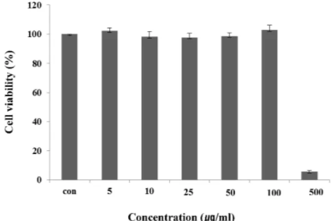
Raw 264.7 cell에 대한 독성
마우스 대식세포인 Raw 264.7 cell에 대한 HaGF의 세포독
성을 확인하기 위하여 MTT assay를 수행하였다. HaGF를 농 도 별로 24시간 동안 처리한 결과 100 μg/ml의 농도까지는 독성이 나타나지 않았지만, 500 μg/ml의 농도에서는 세포의 생존율을 95% 감소시켰다(Fig. 1). HaGF는 100 μg/ml 이하의 농도에서는 세포독성이 낮아 세포의 생존율에 영향을 주지 않는다는 사실을 확인할 수 있었다.
Nitric oxide (NO) 생성억제 효과
NO는 박테리아를 죽이거나 종양을 제거시키는 역할을 하 지만, 병리적 원인에 의해 과도하게 생성, 분비되면 염증을 유발시키게 되며, 조직 손상, 유전자 변이 및 신경손상 등을 일으키는 것으로 알려져 있다[13, 20]. 또한, NO는 NO 합성효 소에 의해 l-arginine으로부터 생성되는 무기 유리체로 면역반 응, 세포독성, 신경 전달계 및 혈관이완 등 여러 가지 생물학적 인 과정에 관여하는 것으로 알려져 있으며, 농도에 따라 세포 기능유지에 중요한 작용을 하기도 하고 세포독성을 일으키기 도 한다[14, 17]. NO는 그 자체로 염증유발과 조직손상을 일으 킬 뿐만 아니라, 활성산소인 superoxide와 결합하게 되면 보다 반응성이 강한 peroxynitrite의 형태로 전환되어 생체조직의 괴사를 야기할 수 있다[19]. 이에 본 실험에서는 NO 생성에 대한 HaGF의 효과를 알아보았다. 생성된 NO양을 griess 시약 을 이용하여 세포배양액 중에 존재하는 NO
2-의 형태로 측정하 여 Fig. 2에 나타내었다. 그 결과 LPS 처리 후 NO 생성량은 정상세포에 비하여 약 4배 이상 증가되었다. HaGF를 100 μg/
ml의 농도로 처리한 군에서는 LPS로 증가된 NO 생성량이 LPS 처리군에 비하여 50% 이상 감소함을 확인할 수 있었다.
TNF- α, IL-1β, IL-6 생성 억제 효과
TNF-α를 포함한 매개물질인 cytokine IL-6와 IL-1β는 NF-κ
Fig. 1. Cell viability of HaGF on Raw 264.7 cell. Raw 264.7 cells were treated with 5, 10, 25, 50, 100, 500 μg/ml of HaGF dissolved in media for 1 hr prior to the addition of LPS (1 μg/ml), and the cells were further incubated for 24 hr. Data represent the mean±S.D. with eight separate experiments.
B를 통해 활성화 되고 또한 NF-κB를 활성화 시켜서 cytokine cascade를 증폭하고 염증상태를 확장한다[2]. 특히, TNF- α는 염증반응에 있어서 중요한 역할을 하며 macrophage와 mast cell 등에서 분비되며, LPS반응의 주요 매개체로서 내재면역 에 있어서도 중요한 역할을 하며 만성 염증 반응과도 관련되 어 있다[10]. IL-1β는 T-cell의 활성화, B-cell의 성숙, NK cell의 activity를 활성화 하며, IL-6는 림프구를 활성화시켜 항체생산 을 증가시키는 것으로, IL-6의 level은 염증 반응에서 항상 증 가하는 것으로 보고되고 있다[1]. 본 실험에서 LPS는 TNF-α, IL-1β, IL-6의 생성을 증가 시켰지만, HaGF를 농도 별로 처리 한 군에서는 억제되는 것을 확인할 수 있었다(Fig. 3).
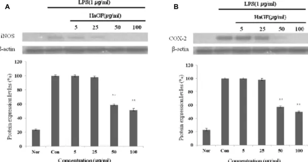
iNOS, COX-2의 단백질 발현 저해 효과
iNOS는 평소에는 세포 내에 존재하지 않으나 일단 유도되 면 장시간 동안 다량의 NO를 생성하며, 생성된 NO는 혈관 투과성, 부종 등의 염증 반응을 촉진 시킬 뿐만 아니라 염증매 개체의 생합성을 촉진하여 염증을 심화시키는 것으로 알려져 있다[5, 21]. 염증반응과 관련된 조직 손상에서 NO와 iNOS의 발현이 증가되어 있음이 보고된 바 있다[15, 18, 23] COX-2는 cyclooxygenase와 peroxidase 활성을 모두 가지고 있는 효소 이다. Cyclooxygenase 기능으로서 arachidonic acid를 pros- tagland로 변환하고, peroxidase 기능으로서는 endoperoxide 를 prostaglandin로 변환시키며, prostaglanin는 prosta- glandins, thromboxane 및 prostacyclins의 전구체로 사용된 다. COX-1은 모든 세포에 존재하면서, 정상세포의 항상성을 유지하며, COX-2는 급성염증반응에서 prostaglandin의 합성 에 관여하고, LPS 및 cytokine에 의해 발현이 유도된다[4, 9]
Fig. 2. Inhibitory effects of HaGF on the production of nitric
oxide Raw 264.7 cells. Raw 264.7 cells were cultured
with LPS (1 μg/ml) in the presence or absence of HaGF
for 24 hr to determine the level of NO. (Nor: LPS not
induced group, Con: LPS induced group) The data rep-
resent the mean±SD of three separate experiments
(significant as compared to control * p <0.05).
A B C
Fig. 3. Effect of HaGF on the production of cytokines stimulated by LPS. Production of TNF-α (a), IL-6 (b), IL-1β (c) were measured in the medium of Raw 264.7 cells cultured with LPS (1 μg/ml) in the presence or absence of HaGF for 24 hr. The amount of TNF-α was measured by immunoassay as described in materials and methods. (Nor : LPS not induced group, Con: LPS induced group) Data represent the mean±S.D. with three separate experiments. One-way ANOVA was used for comparisons of multiple group means followed by t-test (significant as compared to control * p <0.05, ** p <0.01).
A B
Fig. 4. Inhibitory effects of HaGF on the protein levels of iNOS and COX-2 in Raw 264.7 cells. Raw 264.7 cells (5x10
5cells/ml) were pre-incubated for 24 hr, and the cells were stimulated with lipopolysaccharide (1 μg/ml) in the presence of complex extracts sample (5, 25, 50, 100 μg/ml) for 24 hr. (Nor: LPS not induced group, Con: LPS induced group) Data represent the mean±S.D. with three separate experiments. One-way ANOVA was used for comparisons of multiple group means fol- lowed by t-test (significant as compared to control * p <0.05, ** p <0.01).
따라서, 본 연구에서는 HaGF에 의한 NO 생성 저해 기전을 확인하기 위해 western blot을 실시하여 iNOS와 COX-2 단 백질 발현을 측정 하였다. 그 결과 LPS에 의해 증가된 iNOS 와 COX-2의 단백질 발현양이 농도 의존적으로 유의성 있게 감소되었다. β-Actin의 band density 비율에 따라 iNOS 단 백질 생성을 49%와 COX-2의 단백질 발현을 51% 저해함을 확인 하였다(Fig. 4). 이를 통해 LPS 자극에 의해 발현된 iNOS와 COX-2는 많은 양의 NO를 생성하게 되며 HaGF는 이를 저해함으로서 결과적으로 NO 생성을 억제함을 확인할 수 있었다.
감사의 글
본 논문은 농촌진흥청 차세대 바이오그린21사업(과제번호:
PJ008158)의 지원에 의해 이루어진 것임.
References
1. Delgado, A. V., McManus, A. T. and Chambers, J. P. 2003.
Production of tumor necrosis factor-alpha, interleukin 1-be-
ta, interleukin 2, and interleukin 6 by rat leukocyte sub-
populations after exposure to substance. P Neuro 37,
355-361.
2. Hanada, T. and Yoshimura, A. 2002. Regulation of cytokine signaling and inflammation. Cytokine Growth Factor Rev 13, 412-421.
3. Higuchi, M., Hisgahi, N., Taki, H. and Osawa, T. 1990.
Cytolytic mechanisms of activated macrophages. Tumor ne- crosis factor and L-arginine-dependent mechanisms act syn- ergistically as the major cytolytic mechanisms of activated macrophages. J Immunol 144, 1425-1431.
4. Hume, D. A., Wells, C. A. and Ravasi, T. 2007.
Transcriptional regulatory networks in macrophages.
Novartis Found Symp 281, 2-18.
5. Kim, R. G., Shin, K. M., Chun, S. K., Ji, S. Y., Seo, S. H., Park, H. J., Choi, J. W. and Lee, K. T. 2002. In vitro anti-in- flammatory activity of the essential oil from ligularia fischeri var. spiciformis in murine macrophage RAW 264.7 cells.
Yakhak Hoeji 46, 343-347.
6. Kou, J., Ni, Y., Li, N., Wang, J., Liu, L. and Jiang, Z. H.
2005. Analgesic and anti-inflammatory activities of total ex- tract and individual fractions of Chinese medicinal ants Polyachis lamellidens. Biol Pharm Bull 28, 176-180.
7. Kurioka, A. and Uamazaki, M. 2002. Purification and identi- fication of flavonoids from the yellow green cocoon shell (Sasamayu) of the silkworm, Bombys mori. Biosci Biotechnol Biochem 66, 1396-1399.
8. Li, N. G., Osakovskii, V. L. and Ivanova, S. S. 2003. Chemical composition and cryoprotective activity of ethanol extract from winter caterpillars Aporia crataegi L . Izv Akad Nauk Ser Biol 5, 547-552.
9. Lin, W. J. and Yeh, W. C. 2005. Implication of Toll-like re- ceptor and tumor necrosis factor alpha signaling in septic shock. Shock 24, 206-209.
10. Lee, A. K., Sung, S. H., Kim, Y. C. and Kim, S. G. 2003.
Inhibition of lipopolysaccharide -inducible nitric oxide syn- thase, TNF-α and COX-2 expression by sauchinone effects on I-κBα phosphorylation, C/EBP and AP-1 activation.
British J Pharmacol 139, 11-20.
11. Lee, T. H., Kwak, H. B., Kim, H. H., Lee, Z. H., Chung, D. K., Baek, N. i. and Kim, J. 2007. Methanol extracts of Stewartia Korean inhibition cyclooxygenase-2 (COX-2) and inducible nitric oxide synthase (iNOS) gene expression by blocking NF-Kappa B transactivation in LPS-activated Raw 264.7 cells. Mol Cells 23, 398-404.
12. Ma, Y., Wang, X., Zhao, Y., Kawabata, T. and Okada, S.
1997. Inhibitory effects of Chinese and extract (CAE) on
nephrotoxycity induced by ferric-nitrilotriacetate (Fe-NTA) in Wistar rats . Res. Commun Mol Pathol Pharmacol 96, 169-178.
13. Marin, J. and Rodriquez-Martinez, M. A. 1997. Role of vas- cular nitric oxide in physiological and pathological conditions. Pharmacol Ther 75, 111-134.
14. Moncada, S., Palmer, R. M. and Higgs, E. A. 1991. Nitric oxide: physiology, pathophysiology and pharmacology.
Pharmacol Rev 43, 109-142.
15. Mori, M. 2007. Regulation of nitric oxide synthesis and apop- tosis by arginase and arginine recycling. J Nutr 137, 1616-1620.
16. Nakatani, T., Konishi, T., Miyahara, K. and Noda, N. 2004.
Three novel cantharidin-related compounds from the Chinese blister beetle, Mylabris Phalerata Pall. Chem Pharm Bull 52, 807-809.
17. Nathan, C. and Xie, Q. W. 1994. Nitric oxide synthases: roles, tolls and controls. Cell 78, 915-918.
18. Palmer, R. M., Ashton, D. S. and Moncada, S. 1988. Vascular endothelial cells synthesize nitric oxide from L-arginine.
Nature 333, 664-666.
19. Radi, R., Beckman, J. S., Bush, K. M. and Freema, B. A. 1991.
Peroxynitrite oxidation of sulfhydryls the cytotoxic potential of superoxide and nitric oxide. J Biol Chem 266, 4244-4250.
20. Rodeberg, D. A., Chaet, M. S., Bass, R. C., Arkoviz, M. S.
and Garcia, V. F. 1995. Nitric oxide: An overview. American J Surgery 170, 292-303.
21. Tezuka, Y., Irikawa, S., Kaneko, T., Banskota, A. H., Nagaoka, T., Xiong, Q., Hase, K. and Kadota, S. 2001.
Screening of Chinese herbal drug extracts for inhibitory ac- tivity on nitric oxide production and identification of an ac- tive compound of zanthoxylum bμgeanum . J Ethnopharmacol 77, 209-217.
22. Wang, C. and Leger, R. J. 2005. Developmental and tran- scriptional responses to host and nonhost cuticles by the specific locust pathogen Metarhizium anisoplae var.
acridum. Eukaryot Cell 4, 937-947.
23. Weisz, A., Cicatiello, L. and Esumi, H. 1996. Regulation of the mouse inducible-type nitric oxide synthase gene pro- moter by interferon-γ, bacterial lipopolysaccharide and NG-monomethyl-L-arginine. Biochem J 316, 209-215.
24. Yun, H. J., Heo, S. K., Yi, H. S., Kim, C. H., Kim, B. W.
and Park, S. D. 2008. Anti-inflammatory effect of in-
jinho-tang in RAW264.7 Cells. Korean J Herbol 23, 169-178.
초록:무당벌레에서 유래된 HaGF peptide를 이용한 염증 억제 효과 김동희
2․김현정
1,3․이진영
4․황재삼
5․김인우
5․이슬기
1․정현국
1․안봉전
1*
(
1대구한의대학교 화장품약리학과,
2한국한방산업진흥원,
3허브누리,
4호서대학교 한방화장품과학과,
5