67
Biomarkers for Lung Cancer
Over the last decade, intense interest has been focused on discovery of biomarkers and their clinical uses. Lung cancer biomarker discovery has particular eminence in this field due to its anticipated critical role in risk stratification, early detection, treatment selection, prognostication, and monitoring for recurrence of cancer. Significant progress has been made in our understanding of the steps involved in lung carcinogenesis and in development of novel technologies for biomarker discovery. The most active areas of research have been in promoter hypermethylation, proteomics, and genomics. Many investigators have adopted panels of serum biomarkers in an attempt to increase sensitivity. Markers for identification of lung cancer patients who may benefit from targeted therapy have been developed more rapidly. Development of targeted lung cancer therapy has engendered interest in markers for identification of optimal candidates for these therapies. Despite extensive study to date, few have turned out to be useful in the clinic. Even those used in the clinic do not show enough sensitivity, specificity, and reproducibility for general use. All biomarkers identified so far must be validated in larger clinical cohorts. (J Lung Cancer 2009;8(2):67 77)
Key Words: Biological markers, Lung neoplasms
Sei Hoon Yang, M.D., Ph.D.
Department of Internal Medicine, Wonkwang University College of Me- dicine, Iksan, Korea
Received: November 13, 2009 Accepted: November 20, 2009 Address for correspondence Sei Hoon Yang, M.D., Ph.D.
Department of Internal Medicine, Wonkwang University Hospital, 344-2, Shinyong-dong, Iksan 570-711, Korea Tel: 82-63-859-2582 Fax: 82-63-855-2025
E-mail: [email protected]
This work was supported by Won- kwang Clinical Medical Research Center Grant in 2006 (WCMRC-2006- 01).This paper was reported to Lung Cancer Symposium 2009 in the Korean Academy for Tuberculosis and Respiratory Diseases.
서 론
폐암은 흔한 질환 중 하나이며, 전세계적으로 생명을 가 장 위협하는 종양이다. 1년에 120만 명이 발병하며, 암으로 인한 사망의 25%에 해당된다. 한국의 경우도 폐암과 관련 된 발생률, 사망률이 매년 증가되는 추세이다(1). 이는 대부 분 말기단계에서 진단되기 때문이다. 폐암에서 컴퓨터 전 산화 단층촬영, 기관지내시경, 객담검사 등을 시행하고 있 으나, 폐암의 조기진단에는 효과적이지 못하다. 전체 비소 세포폐암의 5년 생존율이 15%인데 비해 IA 병기처럼 조기 에 진단된 경우 5년 생존율이 80%로 높은 생존을 보이므로 (2), 폐암에 특이적인 새로운 바이오 마커(biomarker)의 발굴 이 필연적이며, 이는 암 진단과 표적치료(targeted therapy)에 중요한 역할을 할 수 있다. 본문에서는 폐암의 생물학, 분자 생물학적 과정을 이해하고, 폐암의 바이오마커에 대한 최 근 연구들에 대하여 정리해 보고자 한다.
암 바이오마커
바이오마커란 정상과 비정상을 구별할 수 있는 생물학적 항상성(biological homeostasis)의 정량을 위한 모든 수단을 총칭한다(3). 즉, 모니터링 할 수 있는 조직, 세포, 생화학, 분자학적인 변화로, DNA, RNA, protein, antibody를 말한다.
2001년에 NIH Biomarker Definition Working Group에서 바이 오마커는 정상적인 생물학적 과정, 병리학적 과정, 치료적 중재에 대한 약물학적 반응을 객관적으로 측정하고 평가하 는데 사용되는 특성으로, 대상의 행동, 생리학적 변화로부 터 하나의 분자에 이르기까지 다양하게 개체의 생물학적인 상태를 추적할 수 있는 표지자로 정의하였고, 암 바이오마 커(cancer biomarkers)는 인체에서 암의 존재를 의미하는 물 질 또는 일련의 과정을 말한다(4). 즉, 암 그 자체 또는 암에 대한 인체의 특별한 반응에 의해 분비되는 물질이다. Table 1은 암 환자로부터 얻어진 다양한 체액에서 검출된 암 바이 오마커이다.
Table 1. Potential Biomarkers from Specimens of Human Various Cancers
MGMT (methylguanine methyl-transferase), hnRNP (heterogeneous nuclear ribonucleoprotein), LOH (loss of heterozygosity), FHIT (fragile histidine triad), TMS-1 (target of methylation inducing silencing), RASSF1A (ras association domain family 1A gene), DAPK (death-associated protein kinase), APC (adenomatous polyposis coli), hTERT (human telomerase catalytic subunit), CEA (carcinoembryonic antigen) NSE (neuron-specific enolase)
Sputum:
Mutations of K-ras and p53; epigenetic changes; methylayion of p16 and MGMT; overexpression of hnRNP A2/B1 and other members of hnRNP family
Circulating genes in plasma/serum:
Mutations of K-ras, p53 and β-tubulin genes; LOH of FHIT; promoter hypermethylation of TMS-1, RASSF1A, DAPK, APC genes;
detection of hTERT mRNA; presence of elevated cell-free circulating DNA and RNA levels in cancer as compared with healthy controls and patients with benign diseases; detecting abnormal proteins/peptides, for example CEA and NSE
Autoantibodies in serum:
Detection of antibodies against p53: glycosylated annexins I and/or II; anti-p40; antineural and antinuclear antibodies; MUCI; livin and survivin; c-Myc and L-myc
Breath analysis:
Detection of volatile organic compounds (VOCs), mainly alkanes and aromatic compounds
폐암에서 암 바이오마커의 유용성
암 바이오마커는 암 생물학의 많은 분야에서 좋은 길잡 이가 될 수 있다. 암의 조기 진단뿐만 아니라 암 치료에 중 요한 정보를 제공할 수 있기 때문이다. 현재 종양병기 (cancer staging)는 1940년에 Pierre Denoix가 발표한 TNM 병 기로 정해지지만, 암 바아오마커는 TNM 병기로 구분할 수 없는 생물학 분야에서 많은 정보를 제공할 수 있다.
암 바이오마커의 유용성에 대하여 다음과 같이 요약할 수 있다.
(1) Risk assessment: 대상이 종양발생의 위험이 있는지 평 가하기 위하여 환자가 얼마나 자주 검사를 해야 하는지, 예 방조치가 필요한지 판단하는 과정
(2) Screening: 정상 또는 고위험군 인구집단에서 종양을 최대한 발견하는 단계
(3) Differential diagnosis: 병리학적, 영상의학적 검사와 같 은 임상지표와 함께 질환의 상태를 정확히 평가하기 위한 진단과정
(4) Prognosis: 치료방법과 별개로 종양의 재발 또는 진행 속도 등을 예측하기 위한 지표
(5) Prediction: 특정한 치료방법에 대한 반응을 예측하기 위한 지표
(6) Monitor course: 치료 후 환자에서 재발되는 종양을 추 적하거나 치료효과를 추적할 수 있는 지표
폐암에서 혈청 또는 혈장 바이오마커는 최소의 침습적 방법과 적은 비용으로 고위험군에서 폐암을 식별할 수 있
어, CT 스크리닝보다 효과적이라고 할 수 있다. 이런 바이 오마커로 폐암으로 갈 확률이 적은 양성 비석회화 결절과 조기폐암 또는 진행이 느린 작은 암을 구별할 수 있다.
폐암의 경우 조직학적 특징에 따라 소세포폐암과 비소세 폐포암으로 분류하나, 더 다양한 기준으로 분류할 수 있다 (예, EGFR mutation-induced lung cancers). 특정 바이오마커 에 의한 분류는 보다 정확한 암 진단에 유용하고, 분자생물 학적인 근거에 의한 환자 치료에 몇 가지 이점을 기대할 수 있다. 첫째, 암 바이오마커는 암화과정을 예측하여, 암 전구단계를 가진 개인을 찾아내고, 폐암에 대한 예방화학 요법을 시행할 수 있으며(3), 유발인자 예측, 초기에 암 진 단을 통해 암을 치료할 수 있는 기회를 제공할 뿐만 아니라 사망률 또한 낮출 수 있다. 둘째, 암 바이오 마커는 암 치료 의 길잡이가 될 수 있다. 임상의사가 최적의 치료 도구를 선택할 수 있게 도와 줄 수 있고, 치료에 대한 반응을 여부 와 전이 가능성을 결정하는데 도움을 준다.
일부 바이오마커는 치료 반응에 따라 발현 정도가 변하 여, 중요한 임상적 종점(clinical endpoint)이 될 수 있어 약물 표적치료에도 이용될 수 있다. 조직에서 바이오마커는 영 상적 진단 바이오마커(imaging diagnostic biomarkers) 뿐만 아니라 가능성 높은 약물 표적(potential drug targets)으로 사 용될 수 있다. 예를 들면, 생쥐 폐암 모델에서 폐 혈관 내피 세포 표면에서 발견된 aminopeptidase-p, Annexin A1 (5), 인 체 폐암의 혈관내피세포에서 발견된 coatomer protein com- plex, subunit gamma (COPG), thymopoietin (TMPO), peroxi- redoxin 4 (PRDA 4) 등이 약물표적에 있어 좋은 바이오마커 이다(6). 분자생물학에 근거를 둔 암 바이오마커는 개인 맞 춤형 치료(personalized therapy)를 위한 새로운 약물 표적분
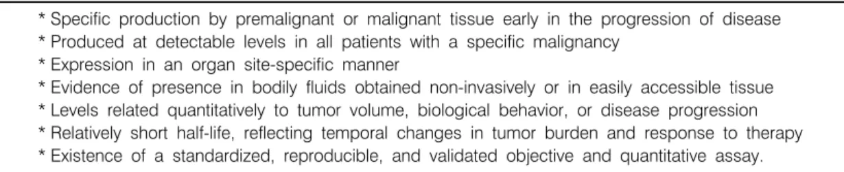
Table 2. Characteristics of the Ideal Tumor Marker
* Specific production by premalignant or malignant tissue early in the progression of disease
* Produced at detectable levels in all patients with a specific malignancy
* Expression in an organ site-specific manner
* Evidence of presence in bodily fluids obtained non-invasively or in easily accessible tissue
* Levels related quantitatively to tumor volume, biological behavior, or disease progression
* Relatively short half-life, reflecting temporal changes in tumor burden and response to therapy
* Existence of a standardized, reproducible, and validated objective and quantitative assay.
자의 발굴에 도움이 될 수 있다.
이와 같이 이상적인 암 바이오마커(Table 2)의 개발을 위 하여 암의 생물학적 이소성(heterogeneity), 인체/암의 이질 성, 분석의 감도와 저해 등 분석학적인 요인, 임상적, 병리 학적 요인, 보건의료서비스와 시장 요인 등에 대한 보다 정 확한 이해가 필요하다.
발암과정의 생물학적 의의
기관지 상피의 전암성 병변으로 알려진 편평상피 이형성 증(squamous dysplasia)과 상피 내암종(carcinoma in situ)은 편평상피세포암으로, atypical adenomatous hyperplasia는 선 암으로, diffuse idiopathic pulmonary neuroendocrine cell hype- rplasia는 carcinoid로 진행한다고 알려져 있다. 그러나 소세 포 폐암에 대한 전암성 병변은 아직 알려지지 않았다(7,8).
유전자 발현과 염색체 구조의 변화는 전암성병변에서 순 차적으로 일어나며, 이런 변화의 발생 빈도 증가는 이형성 을 증가시킨다. 전암성병변에서 발견되는 변화로는 과다한 분화(hyperproliferation), 세포 주기조절의 실패, p53 경로 이 상, Ras 유전자와 3p14.2 부위 유전자들의 이상, 유전자 촉 진자(gene promoter)의 과도한 메틸화, 혈관 성장의 증가, 세 포외 기질(extracellular matrix)의 변화, retinoic acid와 reti- noid X receptor 발현의 감소, 여러 가지 단백질 발현의 변화 등이 있다. 이러한 변화들이 바이오마커의 후보물질로 이 용될 수 있다(8-10).
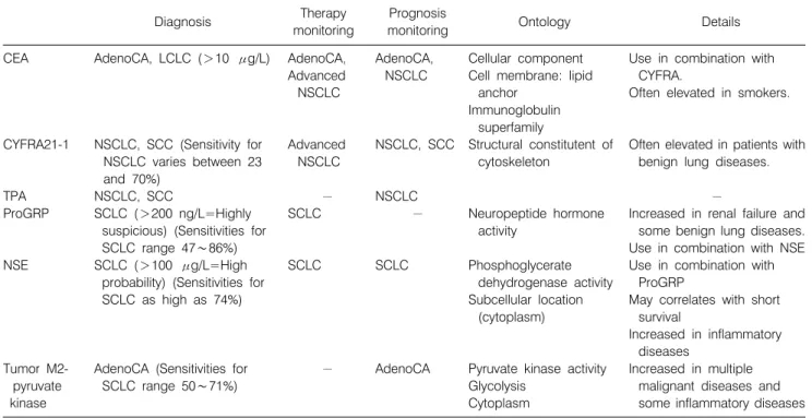
최근에 사용되는 폐암 바이오마커들
다음 표지자들은 폐암 진단과 모니터링에 최근에 권장되 지는 않지만, 일부 영역에서는 임상적으로 사용해 볼 수 있 다(Table 3).
1) Carcinoembryonic antigen
Carcinoembryonic antigen (CEA)는 oncofetal 단백질로 성 인에서는 검출되지 않는다. 암세포에서 증가는 CEA enco-
ding 유전자들 억제에 의해 야기 된다. 폐암(특히 선암, 대세 포 폐암)에서 CEA가 상승하는데, 진단에 CEA만으로는 한 계가 있고 CYFRA 함께 측정이 선호된다. 또한 진행된 비소 세포폐암의 치료 반응 평가를 위해 이용할 수 있다(11). 하 지만 CEA는 종종 흡연자에서도 상승하여 민감하지도 특이 하지도 않다. CEA는 폐암 재발을 스크리닝하는데 다른 방 법과 함께 사용될 수 있으며, 높은 수치는 불량한 생존율을 예측하는데 도움을 준다(12).
2) CYFRA 21-1
CYFRA 21-1는 상피세포에 존재하는 cytokeratin 19 frag- ments를 측정한 것이다. 이는 비소세포폐암 특히 편평상피 세포암을 포함하는 상피세포암과 연관성이 있다. CYFRA 21-1는 비소세포폐암에서 민감도는 23∼70%이고(13), 양성 폐질환에서도 상승한다. CYFRA 21-1는 질병의 치료 평가 와 예후에 연관성이 있다는 보고가 있어, 다른 검사와 함께 예후와 재발의 모니터링에 사용될 수도 있다. Haam 등(14) 은 수술적으로 완전히 절제한 비소세포폐암 환자에서 Cyfra21-1은 생존기간, 재발, 전이를 예측할 수 있는 예후인 자로 사용할 수 있다고 하였다. CEA와 CYFRA는 대장암같 은 다른 종류의 종양과도 관련성을 보인다. 그래서 CEA, CYFRA는 폐암 특이적 바이오마커보다는 일반적인 암 바 이오마커로 여겨지고 있다(15).
3) Squamous cell carcinoma antigen
Squamous cell carcinoma (SCC) 항원은 세포질 단백의 구 조물로, 편평상피암에서 혈액 내 농도가 증가될 수 있고, 전 이 가능성과도 연관성이 있다. 그러나 피부 질환과 염증성 폐 질환에서도 상승할 수 있다. SCC 항원은 편평상피세포 암에서 가장 높은 민감도를 보이며, 연구에 따르면 비소세 포 암에서 민감도는 15∼55%이다(13).
4) Neuron-specific enolase
Neuron-specific enolase (NSE)는 중추 및 말초 신경, 신경 외배엽에서 기원하는 악성 종양에서 생산되는 해당작용
Table 3. Protein-Based Biomarkers in Detection of Lung Cancer: Currently Available
Diagnosis Therapy
monitoring
Prognosis
monitoring Ontology Details
CEA
CYFRA21-1
TPA ProGRP
NSE
Tumor M2- pyruvate kinase
AdenoCA, LCLC (>10 μg/L)
NSCLC, SCC (Sensitivity for NSCLC varies between 23 and 70%)
NSCLC, SCC
SCLC (>200 ng/L=Highly suspicious) (Sensitivities for SCLC range 47∼86%) SCLC (>100 μg/L=High
probability) (Sensitivities for SCLC as high as 74%)
AdenoCA (Sensitivities for SCLC range 50∼71%)
AdenoCA, Advanced NSCLC
Advanced NSCLC
− SCLC
SCLC
−
AdenoCA, NSCLC
NSCLC, SCC
NSCLC
−
SCLC
AdenoCA
Cellular component Cell membrane: lipid
anchor Immunoglobulin
superfamily
Structural constitutent of cytoskeleton
Neuropeptide hormone activity
Phosphoglycerate dehydrogenase activity Subcellular location
(cytoplasm)
Pyruvate kinase activity Glycolysis
Cytoplasm
Use in combination with CYFRA.
Often elevated in smokers.
Often elevated in patients with benign lung diseases.
−
Increased in renal failure and some benign lung diseases.
Use in combination with NSE Use in combination with
ProGRP
May correlates with short survival
Increased in inflammatory diseases
Increased in multiple malignant diseases and some inflammatory diseases CEA: carcinoembryonic antigen, CYFRA 21-1: cytokeratin 19 fragment, TPA: tissue polypeptide antigen, ProGRP: progastrin- releasing peptide, NSE: neuron-specific enolase, AdenoCA: adenocarcinoma, SCC: squamous cell carcinoma, SCLC: small cell lung cancer, NSCLC: non-small cell lung cancer
(glycolysis) 효소이다. 이는 갑상선 수질암과 소세포암에서 증가한다. 그러나 이것은 혈액 내의 세포들에서 발견되고, 염증성 질환에서 상승한다. 소세포암에서 검출되는 경우 민감도는 74% 정도로 높으며, 상승되어 있는 경우 짧은 생 존기간과 연관성이 있다(16,17). CEA, CTFRA 21-1, SCC Ag, NSE 조합은 때때로 폐암 진단의 민감도를 증가시킬 수 있으나, 이러한 표지자들은 특이도가 떨어져서 일차 폐암 의 조사에서는 이용되지 않는다. 그러나 이들은 치료 후 재 발 모니터링에 사용될 수 있다는 보고들이 있다.
5) Progastrin-releasing peptide
Progastrin-releasing peptide (ProGRP)는 gastrin-releasing peptide (GRP)의 안정된 생화학적 전구체이다. GRP는 위 장 관과 호흡기의 신경내분비조직에서 생산되어 호르몬 분비 를 자극한다. 이 펩티드는 신부전과 양성 폐질환에서도 증 가하지만, 때때로 소세포암에서 바이오마커로 사용된다. 소 세포암에서 만감도는 47∼86%이다(12). 그러나 ProGRP의 상승으로 예후를 예측할 수는 없다. ProGRP과 NSE는 소세 포암에서 치료에 대한 반응을 모니터링 하기 위해 함께 사 용된다(13,18).
6) Tumor M2-pyruvate kinase
Tumor M2-Pyruvate Kinase는 pyruvate kinase의 2분자체형 태로 암세포에서 과다이상 발현된다. 이 효소는 여러 염증 상태뿐만 아니라 다발성 악성질병에서도 상승한다. 특히 폐암의 민감도는 50∼71%이고(13), 선암에서 가장 높은 민 감도를 보인다. 이 표지자는 아마도 치료 후 재발의 모니터 링에 사용될 수도 있다.
7) C-reactive protein
C-reactive protein (CRP)는 암을 포함한 많은 염증성 질환 에서 상승한다. 비록 폐암에는 특이적이지는 않지만, CRP 수치의 상승은 예후를 예측하는데 유용하다. 최근 연구에 따르면 평균 10년 이상 7,017환자를 대상으로 높은 농도 CRP는 암의 발생 위험성이 증가하고, 폐암과 가장 높은 연 관성을 보였다(19). CRP 수치 감소와 연관이 있는 CRP 유 전자 변이는 암의 위험성을 증가와 연관되어 있다. 일부 연 구에서는 만성염증과 이것에 의한 방어기전 장애가 만성염 증이 폐암의 유발인자 가능성이 있다고 보고하였다.
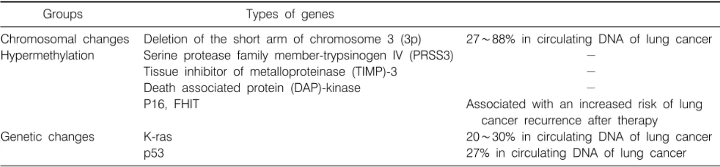
Table 4. Gene-Based Biomarkers in Detection of Lung Cancer: Potential
Groups Types of genes
Chromosomal changes Deletion of the short arm of chromosome 3 (3p) 27∼88% in circulating DNA of lung cancer Hypermethylation Serine protease family member-trypsinogen IV (PRSS3) −
Tissue inhibitor of metalloproteinase (TIMP)-3 −
Death associated protein (DAP)-kinase −
P16, FHIT Associated with an increased risk of lung
cancer recurrence after therapy
Genetic changes K-ras 20∼30% in circulating DNA of lung cancer
p53 27% in circulating DNA of lung cancer
8) Epidermal growth factor receptor
비소세포폐암의 생물적인 측면에서 Epidermal growth factor receptor (EGFR)과 세포 내 tyrosine kinase의 역할에 대하여 많은 연구가 이루어졌고, 이와 관련된 표적제로는 tyrosine kinase를 억제하는 gefitinib, erlotinib과 같은 작은 분 자들과 extracellular EGFR에 대한 monoclonal antibodies로 작용하는 cetuximab과 panitumumab이 있다. 돌연변이 EGFR 은 tyrosine kinase inhibitors에 대한 반응과 연관성이 있고, 최근 연구에서 EGFR copy 수와 mRNA 발현은 다른 중요한 매개변수로 소개되었다(20). 그래서 돌연변이 EGFR와 EGFR copy 수를 조직검사에서 측정하고, 임상의사가 치료 약제를 선택하는데 이용될 수 있다.
9) Other markers
Lactate dehydrogenase (LDH)는 비소세포폐암의 침범과 연관이 있고(12), Carbohydrate antigen 125 (CA-125)는 태아 조직의 당단백질로 주로 난소암의 진단에 사용된다. 그러 나 폐 선암과 대 세포폐암에서도 상승한다. 신경세포 유착 분자(neural cell adhesion molecule)는 전이과정에서 유착과 융합에 관여하는 것으로 알려져 있으며, 소세포폐암에서 높은 농도로 발견된다(15). Tissue polypeptide antigen (TPA) 은 cytokeratin 8, 18, 19을 의미하며 비소세포폐암에서 발견 된다(21).
잠재성 폐암 바이오마커
폐암의 악성화 과정에 유전학적, 후성유전학적(epigene- tics)의 수없이 많은 변화가 잠재적 바이오마커로 존재할 수 있다. 폐의 발암 과정의 생물학적 이해와 바이오마커 발굴 을 위한 high-throughput 기술의 발전, 폐암의 조기진단에 대 한 관심이 폐암 바이오마커 영역을 확장시켰다. 아직까지 폐암의 조기발견을 위한 바이오마커로서 적당한 민감도와
특이도, 입증할 만한 재현성을 가지는 바이오마커를 찾지 는 못하고 있다. 하지만 최근에 폐암의 바이오마커 연구에 대한 전반적인 것을 알아보고자 한다.
1) 혈액 DNA
혈장과 혈청에서 cell-free DNA 양은 일반적으로 건강한 성인에서보다 암을 가진 환자에서 더 높게 측정된다고 보 고하고 있다(22). 혈액 DNA연구방법은 암의 종류, 병기와 DNA 측정 기술에 따라 농도가 다양하게 나타나서, 그 자체 가 바이오마커로 유용성은 없다. 폐암에서 혈중 내 증가된 DNA의 근원은 괴사와 세포고사로 인한 암세포 DNA가 혈 액 내로 유출되어 증가한다고 생각되며(23), 이러한 혈액 내 DNA를 사용하여 보다 더 특이적인 표지자를 찾아내는 데 관심이 높아지고 있다. 폐암환자에서 혈액 내 DNA는 암 의 유전학적, 후생유전학적의 전형적인 변화를 표현한다 (chromosome loss, oncogene activation, tumor suppressor gene inactivation by methylation) (24,25) (Table 4).
2) DNA을 대상으로 하는 폐암 바이오마커
암은 유전적 변이와 환경적 요소들의 상호작용에 의하여 발생한다. 암의 경우 10% 미만에서만 Mendelian 유전법칙 을 따르지만, 유전학과 밀접한 관련이 있다(26). 유전학의 발달에 따라 DNA 또는 RNA에 기반을 둔 암 마이오마커는 임상적 사용에 중요한 진전을 보였다.
다른 암과 유사하게 폐암 암화과정 역시 cell cycle, sene- scence, apoptosis, repair, differentiation, cell migration controls 에서 유전자의 유전적, 후생유전학적인 이상으로 인한 변 화된 분자축척에 의해 다단계 과정에 의한다(27-29). 이와 같은 유전적 변화와 폐암의 악성 변화 사이에는 밀접한 관 련이 있어 유전학은 폐암 바이오마커로 가능성을 보인다.
최근에 single nucleotide polymorphism (SNP)는 여러 질환에 서 좋은 바이오마커로 활용되고 있다. 특히 genome-wide association study (GWAS)를 통해 종양 감수성에 대해 위험
성 평가(risk assessment) 수준에서 많은 연구결과가 이루어 지고 있다. 또한 DNA 수준의 deletion 및 amplification 등과 같은 copy number variation (CNV) 분석도 강력한 association study의 표지자로 사용되고 있다. 두 가지 표지자의 경우 DNA를 활용하기 때문에 시료를 확보하는 것이 용이하다고 할 수 있다. 최근에 보고되기 시작하는 microRNA는 mRNA 와 함께 유전자 발현 수준에서 변화를 분석하는 것으로 세 포의 상태를 반영하는 좋은 표지자이다. 아직 많은 논문은 아니지만, 기존의 mRNA보다 종양의 분류에서 매우 우수한 표지자로 활용이 기대되고 있다.
3) 유전학적 변화
암세포 분열 동안 암 억제유전자의 비활성화는 암세포의 조절되지 않는 성장, 이동, 전이가 되게 하는 주 요인 중 하 나이다. 대부분의 경우 비활성화는 DNA 소실이나 세포 분 열 동안 우연히 발생하는 염색체 재배열(chromosomal rear- rangement)에 의한다. 가장 잘 알려진 이상 소견은 일부 암 억제 유전자가 있는 3번 염색체의 단완 소실(short arm deletion)이다.
염색체 3p의 유전적 물질 소실은 기관지 발암 과정 중 가 장 초기에 일어나는 유전적 변화의 하나이다. 여러 연구를 통해 혈액 내 DNA에서 발견되는 microsatellite instability (MSI), loss of hetero-zygosity (LOH)와 같은 변화가 알려졌 고, 이런 유전자 변화는 폐암환자의 27∼88%에서 혈액 내 DNA에서 발견된다(22). 그래서 위의 같은 여러 가지 변화 의 마커를 조합하면 민감도가 증가할 수 있으나, 비용적인 부분이 문제가 된다.
Gene silencing과는 반대로 성장인자 및 성장인자 수용체, 이와 연결된 매개체와 관련된 유전자의 활성, 돌연변이에 의해 세포주기 활성제는 암화 과정에 중요한 역할을 한다.
폐암환자의 바이오마커로 가능성이 있는 가장 주된 유전자 변이는 K-ras, p53이다. 왜냐하면 이것들은 폐암과 전암성 병변에서 가장 잘 알려진 변이이기 때문이다. 대부분 K-ras mutation은 codon 12에서 발생하며, 폐암의 전암상태와 관 련이 있다(30,31). p53-Retinoblastoma gene (Rb) mutation은 Rb phosphorylation을 조절하지 못하고, G1 arrest를 야기하 여 종양세포가 무분별하게 성장하게 한다(32-34). 혈액 내 DNA에서 K-ras 돌연변이는 폐암환자 20∼30%에서 발견되 고, p53 돌연변이는 27%에서 발견된다(35). 여러 보고에서 이 러한 돌연변이가 항암화학치료 시 내성을 예측할 수 있는지 에 대하여는 논란의 여지가 있다. 또한 Proto-oncogene 돌연변 이이나 변이는 폐암의 좋은 바이오마커가 될 수 있다.
4) 유전자 과메틸화
폐암 발암과정 초기에 다양한 암 억제 유전자 촉진자 (promoter)에 메틸화(methylation)가 일어난다. 유전자 촉진 자 부위의 과메틸화는 전사가 일어나지 않아 과메틸화에 의한 유전자 비 활동상태는 정상 세포의 모든 기능에 관여 하여, 악성으로의 변화와 진행에 치명적인 촉매 역할을 한 다. 즉, 과메틸화(Hypermethylation) 변화, 다양한 촉진자 부 위 CpG islands의 메틸화는 세포의 대표적인 후생유전학적 변화로 gene silencing을 야기한다. 비활성 암 억제 유전자들 의 alternative mechanism으로 과메틸화는 폐암을 포함하여 대부분의 종양에서 발견되었다(36-42).
메틸화는 methylation specific PCR로 분석측정하는데, bisulfites가 모든 unmethylated cytosines을 uracil로 변화시키 고, 증폭이 unmethylated DNA에 비해 methylated에 특이적 인 primer에 의해 일어난다. 많은 폐암연구에서 p16INK4a, APC, TMS1, RASSF1, DAPK의 비정상적인 메틸화가 보고 되었고(22,38,43-45) 최근 Belinsky 등(46)의 보고에 따르면 혈액과 객담에서 여러 다른 유전자들의 촉진자 부위 과메 틸화는 폐암과 연관성이 있으며 폐암 진단에 임상적으로 사용 가능할 것이라 보고하였다. 최근 다른 연구에 따르면 PRSS3 (serine protease family member-trypsinogen IV - a putative tumor suppressor gene) (47), human DAB2 interactive protein gene (48), CARD (49)을 포함한 apoptosis-associated speck-lick protein의 촉진자 부위의 비정상 메틸화가 폐암에 서 보고되어졌다.
p16과 FHIT 유전자의 과메틸화는 치료 후 재발 위험성 증가와 연관이 있으며(50,51), 폐포 세척액에서 p16, RAS- SF1A, H-cadherin, RARβ의 메틸화는 폐암 존재와 연관이 있고, FHIT 메틸화는 담배 노출과 연관성이 있다는 보고가 있다. 기관지 흡인액에서 RASSF1A 촉진자의 과메틸화는 소세포폐암 88%, 비소세포폐암의 28%에서 발견되지만, 양 성 폐질환 환자에서는 과메틸화가 발견되지 않는다(52). 다 른 연구에서는 폐암 환자의 기관지 흡입액에서 APC, p16, RARB2 촉진자의 비정상 메틸화가 발견되었다. 이러한 유 전자 조합을 사용하여 폐암진단에서 민감도 69%와 특이도 87%가 있다고 보고하였다. High-throughput global expression profiling approach을 통한 새로운 암 특이적 메틸화 마커를 찾기 위한 노력이 이루어지고 있다. 여러 폐암 세포주에서 이러한 기술을 이용하여 132 유전자가 메틸화에 의해 억제 됨을 확인하였고, 연구자들은 정상 폐에서 발현되는 유전 자들이 폐암 환자에서는 발현되지 않음도 확인하였다. 또 한 96명의 폐암환자로부터 폐암조직과 인접한 정상조직 간
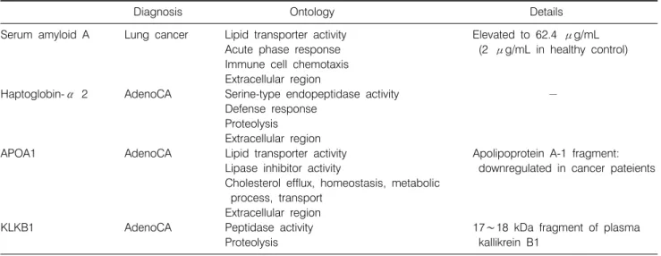
Table 5. Protein-Based Biomarkers for the Detection of Lung Cancer: Potential
Diagnosis Ontology Details
Serum amyloid A Lung cancer Lipid transporter activity Elevated to 62.4 μg/mL Acute phase response (2 μg/mL in healthy control) Immune cell chemotaxis
Extracellular region
Haptoglobin-α 2 AdenoCA Serine-type endopeptidase activity −
Defense response Proteolysis Extracellular region
APOA1 AdenoCA Lipid transporter activity Apolipoprotein A-1 fragment:
Lipase inhibitor activity downregulated in cancer pateients Cholesterol efflux, homeostasis, metabolic
process, transport Extracellular region
KLKB1 AdenoCA Peptidase activity 17∼18 kDa fragment of plasma
Proteolysis kallikrein B1
에 서로 다른 47개 유전자의 촉진자 부위가 메틸화되었음 을 확인하였다(53). Lee 등(54)은 완전히 절제한 비소세포폐 암 환자에서 RARβ유전자의 P2 hypermethylation과 DAPK 의 unmethylation은 조기 암 재발의 중요한 예측인자라고 하 였다.
또한 최근에 우리나라에서 시행한 연구에서 histone meth- yltransferase의 SNP는 폐암의 위험성을 의미 있게 증가시키 는 것과 연관이 있다고 보고하였다(55).
5) 단백질 마커들
유전학에 기반을 둔 바이오마커의 현저한 발전에도 불구 하고, 여전히 임상적으로 사용되는 새로운 암 바이오마커 는 없는 실정이다. DNA에 근간을 둔 바이오마커는 폐암 바 이오마커의 가능성을 제시하지만, 민감도와 특이도가 낮고, 재현성이 매우 적다. 이는 mRNA가 실제 생리학적 기능을 갖는 단백질과 직접 연관되지 않기 때문이다. 현재 인체 게 놈은 약 20,488 유전자를 포함하고 있는 것으로 알려져 있 다. 그러나 단백질은 alternative splice variants, protease cleavages, post-translational modifications (glycosylation, phos- phorylation, ubiquitination, methylation, acetylation 등) 때문에 매우 다양함을 보인다. 이러한 이유로 단백질 바이오마커 는 암 유형, 암 상태에 따라 보다 더 특이적이어야 한다.
폐암 단백질 바이오마커는 단백질 얻는 곳에 따라 serum biomarker, tissue biomarker, sputum biomarker로 분류할 수 있다. 객담은 암 부위에서 분리된 암 세포가 주요 단백질의 근원이 된다. 조직검사를 통해 얻은 폐암 조직은 암 세포뿐 만 아니라 인체 자가방어와 관련된 분자들(immune cell, cytokines, immune or inflammatory response에 의한 유도물
질)이 포함된다. 반면 혈액에서는 더 강력한 바이오마커가 존재하며, 이는 조직검사을 통해 얻은 암 조직에서 발견된 바이오마커와 diseased tissue microenvironment에서 형성된 circulating protein fragment와 diseased tissue에서 유도된 순 환하는 단백질과 세포가 포함한다. 바이오마커의 궁극적 목적은 조기진단, 비침습적 진단, 치료 반응 평가이기 때문 에 혈액이 가장 적절한 생물학적 재료로 생각 된다. 그래서 많은 바이오마커 연구는 혈액을 기본으로 하여 이루어지 며, 이미 많은 혈청 바이오마커가 연구되고 있다(56,57).
아직 임상적으로 사용되지 않으나, 가능성이 있는 많은 폐암 바이오마커 분자들이 있다(Table 5).
(1) hnRNPB1: hnRNPB1, RNA-binding protein은 편평상피 세포암에서 흔하게 mRNA 전달과 RNA 돌연변이에 관여한 다. 이 단백질이 객담에서 발현되는 경우 특정 집단에서는 폐암 발생의 위험성과 연관이 있다. 그리고 이 단백질은 조 기 폐암에서 과도하게 발현된다. 최근 연구에 의하면 hnRNPB1 수치는 대조군 12%와 비교할 때 폐암환자에서는 46%로 증가하였다.
(2) Telomerase: Telomeres의 기능은 염색체의 끝을 보호 하는 구조로서 작용한다. Telomeres의 기능이 소실되면 암 의 시작과 진행에 중요한 역할을 한다. Human telomerase catalytic component (hTERT)는 여러 암에서 증가하고, EGFR은 폐암에서 종종 증가한다. Miura 등(58)의 보고에 의 하면 112명 폐암환자와 80명 대조군에서 quantitative one- step real-time RT PCR (reverse transcription polymerase chain reaction)을 이용하여 hTERT와 EGFR mRNA을 측정하였다.
혈청 hTERT mRNA copy number는 암 병기, 전이, 재발과 관계가 있고, copy number는 치료 후 감소하였다. hTERT는
민감도와 특이도가 89%, 73%, EGFR은 71%, 80%로 보고하 였다. Telomere 안정성을 조절하는데 도움을 주는 유전자인 RAP1 발현은 폐암 환자의 생존율를 향상시킨다(51 vs 15 months) (59).
(3) Survivin: Survivin는 apoptosis을 억제하고 mitosis를 촉 진하는 단백질이다. 암 조직 핵내 survivin 양은 수술한 비소 세포폐암 환자에서 나쁜 예후와 재발을 예측할 수 있게 하 고, 핵내 survivin를 가진 환자는 나쁜 생존율을 가진다.
(4) Others: Osteopontin과 발현은 폐암 환자 조직과 혈청 에서 발견되고, 병의 악화와 연관이 있다. Fas-associated death domain은 NF-kB를 비활성화 시키고, cyclins D1과 B1 과발현과 연관되어 세포주기에 영향을 미친다. 폐암에서 phosphorylated Fas-associated death domain mRNA와 이 단백 질의 증가는 예후에 나쁜 영향을 준다. Functional poly- morphisms of matrix metalloproteinase-9는 폐암 발생의 위험 성 및 재발과 연관이 있다. 또한 폐암 환자에서 soluble E-cadherin이 증가한다. Plasma kallikrein B1 (KLKB1) frag- ment가 선암 진단에 유용한 바이오마커로서 가능성을 제시 하였고, KLKB1의 18 kDa fragment (H4 domain 포함)은 폐 선암에서 높게 측정되었다.
(5) 단백질체와 유전체: 2007년 Chen 등(24)은 폐암 예후 에 대한 유전자 연구결과를 보고 하였다. Chen 등은 수술적 치료를 받은 조기 폐암 조직에서 microarray 분석을 수행하 여 생존율 향상과 관련된 16개 유전자를 발견하였다. 이 중 RT-PCR 방법을 통해 5가지 유전자, dual specificity phos- phatase 6, monocyte to macrophage differentiation associated protein, signal transducer, activator of transcription 1, v-erb-b2 avian erythroblastic leukemia viral oncogene homolog 3, lymphocyte-specific protein tyrosine kinase을 확인하였다. 위 험성 높은 유전자를 가진 환자는 수술 후 5년 이내에 어떠 한 원인이든 사망하는 경우가 3∼4배 높았다. 또한 향후 randomized clinical trials에서 genetic profiling을 토대로 특별 한 약제에 대한 반응을 예측할 수 있을 것이다.
Human genome project 종결 이후 단백질 체(proteomics)라 는 새로운 학문분야로 나타나게 되었다. 단백질 체는 매우 복잡한 특징들을(isoforms, modifications, interactions, func- tional structures 등) 포함한 단백질 large scale characterization 을 의미한다. Protein separation, quantification, identification 같은 protemic technologis의 획기적인 발전은 집중적으로 단 백질 연구를 가능케 하였으며, 단백질 기능을 심도 있게 이해 할 수 있게 해 주었다. 그로 인해 전체적으로 발현된 단백질 (whole expressed proteins)을 전반적으로 검토할 수 있어, 진단, 치료, 예후을 평가하여 암 치료의 발전을 제시하고 있다.
폐암 환자에서 단백질체 연구(proteomic study)는 두 가지 측면에서 암 바이오마커 분야에 접근할 수 있다. 첫째는 Protein profiling으로, 전체적 단백질 발현 양상을 보고 실제 적으로 이 단백질이 관여한 것인지, 폐암의 마커로 사용할 수 있는 단백질인지 확인하여 폐암 진단과 치료에 도움을 줄 수 있다. 둘째는 matrix assisted laser desorption/ionization time-of-flight mass spectrometry (MALDI-TOF MS)을 이용하 여 정확한 크기와 극성을 결정하여 정확한 단백질을 확인 할 수 있어 새로운 암 바이오마커를 발견할 수 있다. 이 방 법을 통해 폐암에서 혈청 amyloid A와 macrophage migration inhibitory factor가 증가함을 확인하였다. 추적 연구로 enzy- me-linked immunosorbent assay (ELISA)을 통해 amyloid A가 폐암 환자에서 증가하고, macrophage-inhibitory factor는 폐 암뿐만 아니라 다른 질병도 증가하여 구분할 수 없음을 확 인하였다. 여러 다른 연구에서도 surface-enhanced laser des- orption/ionization time-of-flight mass spectrometry (SELDI- TOF MS)을 이용하여 혈액 내 단백질 peak pattern을 보고, 대조군과 암 환자를 구분할 수 있다. 최근 연구에서는 158 명 폐암 환자와 50명 대조군의 혈액을 분석하여 5개 단백질 peak pattern으로 비소세포폐암을 진단 할 수 있고, 민감도는 91.4%로 보고하였다.
(6) 다른 분자학적 마커들: Cytokinesis-block micronucleus assay는 게놈 불안정성에 대한 바이오마커로서, NNK (toba- cco carcinogen)에 의해 유도되는 유전적 손상을 이 방법으 로 매우 민감하게 찾을 수 있고, 폐암 위험성의 예측인자로 사용될 수 있다. 대사적으로 활성화된 중개물질을 통하여 DNA adduct을 형성한 많은 발암물질이 영향을 미칠 수 있 는데, 조직에서 adduct을 확인하는 것이 암 위험성 연구의 도구로 사용할 수 있다(60).
또한 Mitochondrial DNA는 암 바이오마커로 사용될 수 있 는데, Jakupciak 등(61)은 최근에 rapid and high-throughput sequencing 방법으로 mitochondrial DNA에서 sequence vari- ants를 확인하였다. 폐암 진단을 위해 호기 시 공기를 표지 자로 사용하는 것은 흥미로운 생각이다. 한 연구에서는 개 가 폐암 환자와 건강한 대조군을 호흡샘플로 정확하게 구 별할 수 있다고 보고 하였다(62). 폐암 환자에서는 volatile organic compounds, 주로 alkanes과 aromatic compounds가 만 들어지고 호기 때 공기로 배출된다. Poli 등(63)은 호기 시 나온 공기에서 13가지 혼합 volatile organic compounds를 측 정하여 80% 환자에서 폐암을 구별할 수 있다고 하였다.
Wang 등(64)은 민감도와 특이도를 증가시키기 위하여 혈 장과 객담에서 여러 가지 DNA 표지 자를 혼합하여, meth- ylation-specific PCR 방법으로 세가지 암 억제 유전자의 촉
진자 과메틸화를 확인하였고, 79명의 폐암환자 객담에서 LOH와 microsatellite instability (MSI)을 이용하여, 8개 mic- rosatellite markers의 불안정성을 확인하였다. 또한 Greeng- berg와 Lee (65)는 폐암진단을 위한 최대의 민감도와 특이 도를 얻기 위해 표지자들을 혼합하는 방법으로 객담을 분 석하여 LOH 부위 D9S286, D9S942, GATA49D12, D13S170, MSI 부위 D9S942, p16INK4a와 RARb의 methylation 등 7개 의 바이오마커를 찾아냈다.
결 론
비침습적인 방법을 통한 폐암의 바이오마커의 개발은 폐 암의 치료와 진단에 새로운 장을 제시 할 수 있다. 이러한 바이오마커는 CT에서 발견된 양성 폐 결절과 조기 암을 구 별할 수 있고, 암 특성을 기반으로 한 개인 특성에 맞는 폐 암 치료를 가능하게 할 수 있다. 그러나 현재 이용할 수 있 거나 잠재력있는 폐암 바이오마커는 진단, 분류, 예후, 약물 반응을 평가할 수 있을 정도의 높은 민감도와 특이도를 보 이지 않으므로, 유용한 폐암 바이오마커를 찾기 위해서는 고려되어야 할 점이 있다. 먼저 현재 사용가능하거나 가능 성이 있는 바이오마커를 다른 암 조직형 및 염증성 질환 같은 다른 질병을 포함하여 다양한 질병에서 분석해 보아 야 한다. 둘째는 기술의 발전에 힘입어 폐암의 특이한 아류 형(specific subtype)에 초점을 맞춘 매우 특이적이고 적은 양 의 폐암 바이오마커 연구에 집중해야 한다. 셋째는 하나의 특징적인 바이오마커만으로는 폐암을 예측하거나 평가하 기 어려워, 몇몇의 good biomarkers를 함께 사용하여 실제 임상에 필요한 정보를 얻어야 한다.
암화 과정과 관련된 분자생물학적, 유전학적 변화에 대 한 괄목할만한 연구 발전은 폐암 바이오마커의 발굴 가능 성의 기반을 제공한다. 새롭게 향상된 high-throughput tech- nology의 발전이 바이오마커 발굴에 지대한 영향을 주었으 며, 또한 지난 몇 년간 폐암 바이오마커에 대한 연구도 폭발 적으로 이루어져서, 많은 연구를 통해 높은 민감도와 특이 도를 보고 하였다.
그러나 이러한 연구는 작은 규모의 연구, 재현성 부족, 연 구 간의 불일치 등의 문제가 있어, 모두 바이오마커 확인은 larger clinical cohorts에서 검증되어야 한다. 예후와 진단 후 치료의 개인화에 대한 바이오마커는 곧 발굴될 것이며, 빠 른 시일 내에 폐암의 조기진단을 위한 실용적 바이오 마커 의 발굴도 기대해 본다.
REFERENCES
1. Kim YC, Kwon YS, Oh IJ, et al. National survey of lung cancer in Korea, 2005. J Lung Cancer 2007;6:67-73.
2. Mulshine JL, Sullivan DC. Clinical practice. Lung cancer screening. N Engl J Med 2005;352:2714-2720.
3. Dalton WS, Friend SH. Cancer biomarkers: an invitation to the table. Science 2006;312:1165-1168.
4. Biomarker Definitions Working Group. Biomarkers and surro- gate endpoints: preferred definitions and conceptual frame- work. Clin Pharmacol Ther 2001;69:89-95.
5. Oh P, Li Y, Yu J, et al. Subtractive proteomic mapping of the endothelial surface in lung and solid tumours for tissue- specific therapy. Nature 2004;429:629-635.
6. Park HJ, Kim BG, Lee SJ, et al. Proteomic profiling of endothelial cells in human lung cancer. J Proteome Res 2008;
7:1138-1150.
7. Travis WD, Colby TV, Corrin B, Shimosato Y, Brambilla E, Collaborators from 14 Countries; World Health Organization.
International histological classification of tumors. Histological typing of lung and pleural tumors. 3rd ed. New York: Sprin- ger-Verlag; 1999.
8. Greenberg AK, Yee H, Rom WN. Preneoplastic lesions of the lung. Respir Res 2002;3:20.
9. Belinsky SA. Gene-promoter hypermethylation as a biomarker in lung cancer. Nat Rev Cancer 2004;4:707-717.
10. Brabender J, Metzger R, Salonga D, et al. Comprehensive expression analysis of retinoic acid receptors and retinoid X receptors in non-small cell lung cancer: implications for tumor development and prognosis. Carcinogenesis 2005;26:525-530.
11. Ardizzoni A, Cafferata MA, Tiseo M, et al. Decline in serum carcinoembryonic antigen and cytokeratin 19 fragment during chemotherapy predicts objective response and survival in patients with advanced nonsmall cell lung cancer. Cancer 2006;107:2842-2849.
12. Okada M, Nishio W, Sakamoto T, et al. Prognostic signi- ficance of perioperative serum carcinoembryonic antigen in non-small cell lung cancer: analysis of 1,000 consecutive resections for clinical stage I disease. Ann Thorac Surg 2004;
78:216-221.
13. Schneider J. Tumor markers in detection of lung cancer. Adv Clin Chem 2006;42:1-41.
14. Haam SJ, Kim GD, Cho SH, Lee DY. Clinical effectiveness of tumor markers (CEA, NSE, Cyfra 21-1) in completely resected non-small cell lung cancer. J Lung Cancer 2006;5:
75-83.
15. Kulpa J, Wojcik E, Reinfuss M, Kolodziejski L. Carcinoem- bryonic antigen, squamous cell carcinoma antigen, CYFRA 21-1, and neuron-specific enolase in squamous cell lung cancer patients. Clin Chem 2002;48:1931-1937.
16. Ferrigno D, Buccheri G, Giordano C. Neuron-specific enolase is an effective tumour marker in non-small cell lung cancer (NSCLC). Lung Cancer 2003;41:311-320.
17. Pujol JL, Quantin X, Jacot W, Boher JM, Grenier J, Lamy PJ. Neuroendocrine and cytokeratin serum markers as pro- gnostic determinants of small cell lung cancer. Lung Cancer 2003;39:131-138.
18. Molina R, Filella X, Auge JM. ProGRP: a new biomarker for small cell lung cancer. Clin Biochem 2004;37:505-511.
19. Siemes C, Visser LE, Coebergh JW, et al. C-reactive protein levels, variation in the C-reactive protein gene, and cancer risk:
the Rotterdam Study. J Clin Oncol 2006;24:5216-5222.
20. Dziadziuszko R, Witta SE, Cappuzzo F, et al. Epidermal growth factor receptor messenger RNA expression, gene dosage, and gefitinib sensitivity in non-small cell lung cancer.
Clin Cancer Res 2006;12:3078-3084.
21. Barak V, Goike H, Panaretakis KW, Einarsson R. Clinical utility of cytokeratins as tumor markers. Clin Biochem 2004;37:529-540.
22. Xue X, Zhu YM, Woll PJ. Circulating DNA and lung cancer.
Ann N Y Acad Sci 2006;1075:154-164.
23. Jahr S, Hentze H, Englisch S, et al. DNA fragments in the blood plasma of cancer patients: quantitations and evidence for their origin from apoptotic and necrotic cells. Cancer Res 2001;61:1659-1665.
24. Chen HY, Yu SL, Chen CH, et al. A five-gene signature and clinical outcome in non-small-cell lung cancer. N Engl J Med 2007;356:11-20.
25. Sozzi G, Musso K, Ratcliffe C, Goldstraw P, Pierotti MA, Pastorino U. Detection of microsatellite alterations in plasma DNA of non-small cell lung cancer patients: a prospect for early diagnosis. Clin Cancer Res 1999;5:2689-2692.
26. Ludwig JA, Weinstein JN. Biomarkers in cancer staging, prognosis and treatment selection. Nat Rev Cancer 2005;5:
845-856.
27. Brambilla C, Fievet F, Jeanmart M, et al. Early detection of lung cancer: role of biomarkers. Eur Respir J Suppl 2003;39:
36s-44s.
28. Chung GT, Sundaresan V, Hasleton P, Rudd R, Taylor R, Rabbitts PH. Sequential molecular genetic changes in lung cancer development. Oncogene 1995;11:2591-2598.
29. Kishimoto Y, Sugio K, Hung JY, et al. Allele-specific loss in chromosome 9p loci in preneoplastic lesions accompanying non-small-cell lung cancers. J Natl Cancer Inst 1995;87:1224- 1229.
30. Rodenhuis S, Slebos RJ. Clinical significance of ras oncogene activation in human lung cancer. Cancer Res 1992;52:2665S- 2669S.
31. Sugio K, Ishida T, Yokoyama H, Inoue T, Sugimachi K, Sasazuki T. Ras gene mutations as a prognostic marker in adenocarcinoma of the human lung without lymph node metastasis. Cancer Res 1992;52:2903-2906.
32. Brambilla E, Gazzeri S, Lantuejoul S, et al. p53 mutant immunophenotype and deregulation of p53 transcription pathway (Bcl2, Bax, and Waf1) in precursor bronchial lesions of lung cancer. Clin Cancer Res 1998;4:1609-1618.
33. Gazzeri S, Brambilla E, Caron de Fromentel C, et al. p53 genetic abnormalities and myc activation in human lung
carcinoma. Int J Cancer 1994;58:24-32.
34. Levine AJ. p53, the cellular gatekeeper for growth and division. Cell 1997;88:323-331.
35. Aviel-Ronen S, Blackhall FH, Shepherd FA, Tsao MS. K-ras mutations in non-small-cell lung carcinoma: a review. Clin Lung Cancer 2006;8:30-38.
36. Belinsky SA, Nikula KJ, Palmisano WA, et al. Aberrant methylation of p16 (INK4a) is an early event in lung cancer and a potential biomarker for early diagnosis. Proc Natl Acad Sci U S A 1998;95:11891-11896.
37. Chaussade L, Eymin B, Brambilla E, Gazzeri S. Expression of p15 and p15.5 products in neuroendocrine lung tumours:
relationship with p15 (INK4b) methylation status. Oncogene 2001;20:6587-6596.
38. Esteller M, Sanchez-Cespedes M, Rosell R, Sidransky D, Baylin SB, Herman JG. Detection of aberrant promoter hypermethylation of tumor suppressor genes in serum DNA from non-small cell lung cancer patients. Cancer Res 1999;59:
67-70.
39. Kurakawa E, Shimamoto T, Utsumi K, Hirano T, Kato H, Ohyashiki K. Hypermethylation of p16 (INK4a) and p15 (INK4b) genes in non-small cell lung cancer. Int J Oncol 2001;19:277-281.
40. Palmisano WA, Divine KK, Saccomanno G, et al. Predicting lung cancer by detecting aberrant promoter methylation in sputum. Cancer Res 2000;60:5954-5958.
41. Virmani AK, Rathi A, Zochbauer-Muller S, et al. Promoter methylation and silencing of the retinoic acid receptor-beta gene in lung carcinomas. J Natl Cancer Inst 2000;92:1303- 1307.
42. Zochbauer-Muller S, Fong KM, Virmani AK, Geradts J, Gazdar AF, Minna JD. Aberrant promoter methylation of multiple genes in non-small cell lung cancers. Cancer Res 2001;61:249-255.
43. Usadel H, Brabender J, Danenberg KD, et al. Quantitative adenomatous polyposis coli promoter methylation analysis in tumor tissue, serum, and plasma DNA of patients with lung cancer. Cancer Res 2002;62:371-375.
44. Ramirez JL, Sarries C, de Castro PL, et al. Methylation patterns and K-ras mutations in tumor and paired serum of resected non-small-cell lung cancer patients. Cancer Lett 2003;193:207-216.
45. Belinsky SA, Klinge DM, Dekker JD, et al. Gene promoter methylation in plasma and sputum increases with lung cancer risk. Clin Cancer Res 2005;11:6505-6511.
46. Belinsky SA, Liechty KC, Gentry FD, et al. Promoter hyper- methylation of multiple genes in sputum precedes lung cancer incidence in a high-risk cohort. Cancer Res 2006;66:3338- 3344.
47. Marsit CJ, Okpukpara C, Danaee H, Kelsey KT. Epigenetic silencing of the PRSS3 putative tumor suppressor gene in non-small cell lung cancer. Mol Carcinog 2005;44:146-150.
48. Yano M, Toyooka S, Tsukuda K, et al. Aberrant promoter methylation of human DAB2 interactive protein (hDAB2IP) gene in lung cancers. Int J Cancer 2005;113:59-66.
49. Zhang Z, Tan S, Zhang L. Prognostic value of apoptosis- associated speck-like protein containing a CARD gene pro- moter methylation in resectable non-small-cell lung cancer.
Clin Lung Cancer 2006;8:62-65.
50. Maruyama R, Sugio K, Yoshino I, Maehara Y, Gazdar AF.
Hypermethylation of FHIT as a prognostic marker in nonsmall cell lung carcinoma. Cancer 2004;100:1472-1477.
51. Kim JS, Kim JW, Han J, Shim YM, Park J, Kim DH.
Cohypermethylation of p16 and FHIT promoters as a prog- nostic factor of recurrence in surgically resected stage I non- small cell lung cancer. Cancer Res 2006;66:4049-4054.
52. Grote HJ, Schmiemann V, Geddert H, et al. Methylation of RAS association domain family protein 1A as a biomarker of lung cancer. Cancer 2006;108:129-134.
53. Ehrich M, Field JK, Liloglou T, et al. Cytosine methylation profiles as a molecular marker in non-small cell lung cancer.
Cancer Res 2006;66:10911-10918.
54. Lee SH, Kim YT, Sung SW, Kim JH. Correlation between aberrant promoter hypermethylation of CpG islands and the clinical outcome of non-small cell lung cancer after curative resection. J Lung Cancer 2004;3:77-85.
55. Yoon KA, Hwangbo B, Kim IJ, et al. Novel polymorphisms in the SUV39H2 histone methyltransferase and the risk of lung cancer. Carcinogenesis 2006;27:2217-2222.
56. Omenn GS. Strategies for plasma proteomic profiling of cancers. Proteomics 2006;6:5662-5673.
57. Rifai N, Gillette MA, Carr SA. Protein biomarker discovery and validation: the long and uncertain path to clinical utility.
Nat Biotechnol 2006;24:971-983.
58. Miura N, Nakamura H, Sato R, et al. Clinical usefulness of serum telomerase reverse transcriptase (hTERT) mRNA and epidermal growth factor receptor (EGFR) mRNA as a novel tumor marker for lung cancer. Cancer Sci 2006;97:1366-1373.
59. Lin X, Gu J, Lu C, Spitz MR, Wu X. Expression of telomere-associated genes as prognostic markers for overall survival in patients with non-small cell lung cancer. Clin Cancer Res 2006;12:5720-5725.
60. El-Zein RA, Schabath MB, Etzel CJ, Lopez MS, Franklin JD, Spitz MR. Cytokinesis-blocked micronucleus assay as a novel biomarker for lung cancer risk. Cancer Res 2006;66:6449- 6456.
61. Jakupciak JP, Wang W, Markowitz ME, et al. Mitochondrial DNA as a cancer biomarker. J Mol Diagn 2005;7:258-267.
62. McCulloch M, Jezierski T, Broffman M, Hubbard A, Turner K, Janecki T. Diagnostic accuracy of canine scent detection in early- and late-stage lung and breast cancers. Integr Cancer Ther 2006;5:30-39.
63. Poli D, Carbognani P, Corradi M, et al. Exhaled volatile organic compounds in patients with non-small cell lung cancer:
cross sectional and nested short-term follow-up study. Respir Res 2005;6:71.
64. Wang YC, Hsu HS, Chen TP, Chen JT. Molecular diagnostic markers for lung cancer in sputum and plasma. Ann N Y Acad Sci 2006;1075:179-184.
65. Greenberg AK, Lee MS. Biomarkers for lung cancer: clinical uses. Curr Opin Pulm Med 2007;13:249-255.