This is an open-access article distributed under the terms of the Creative Commons Attribution Non-Commercial License (http://creativecommons.org/
licenses/by-nc/4.0/), which permits unrestricted non-commercial use, distribution, and reproduction in any medium, provided the original work is properly cited.
CC
Cone-beam computed tomography characterization of the intraosseous vascular canal in the lateral wall of the maxillary antrum
Shishir Ram Shetty1, Saad Wahby Al Bayatti1, Hesham Marei2, Raghavendra Shetty3, Hossam Abdelatty Abdelmagyd4, Alexander Maniangat Luke3
1Oral Radiology, Department of Oral and Craniofacial Health Sciences, College of Dental Medicine, University of Sharjah, Sharjah,
2Oral and Maxillofacial Surgery, College of Dentistry, Gulf Medical University, 3Department of Clinical Sciences, College of Dentistry, Ajman University, 4Periodontics, College of Dentistry, Gulf Medical University, Ajman, United Arab Emirates
Abstract(J Korean Assoc Oral Maxillofac Surg 2021;47:34-39)
Objectives: The purpose of the study was to assess the occurrence, location, and dimensions of the intraosseous vascular canal in the lateral wall of the maxillary antrum using cone-beam computed tomography (CBCT).
Materials and Methods: In this retrospective study, we examined 400 CBCT scans from our archive of patients who had earlier reported to a dental teaching hospital in the United Arab Emirates. The prevalence, location, and dimensions of the lateral antral intraosseous canal (LAIC) in the maxillary antrum were evaluated by 2 examiners using standardised methods. A third examiner was consulted in cases of disagreement.
Results: The prevalence of LAIC was 62.3% (249 maxillary antra) among the study population. The mean distance between the most inferior point of the alveolar bone and the inferior border of the LAIC in the posterior maxillary region was 19.83±3.12 mm. There was a significant difference (P=0.05) between the maxillary molar and premolar regions in mean distance from the most inferior point of the alveolar bone and the inferior border of the LAIC. There was no statistically significant difference in mean distance between the most inferior point of the alveolar bone and the inferior border of the LAIC between dentulous and edentulous areas (P=0.1). The G3-intrasinusal type canal less than 1mm in diameter was the most common type of LAIC.
Conclusion: This study established the approximate location of the LAIC in a United Arab Emirates cohort, which will assist the oral surgeon in se- lecting the appropriate site for sinus lift procedures with reduced risk of surgical hemorrhage.
Key words: Maxillary sinus, Cone-beam computed tomography, Sinus floor augmentation, Hemorrhage, Alveolar bone loss
[paper submitted 2020. 9. 16 / revised 2020. 11. 3 / accepted 2020. 11. 10]
Copyright © 2021 The Korean Association of Oral and Maxillofacial Surgeons. All rights reserved.
I. Introduction
Dental implants are gaining popularity in the Middle East. According to recent surveys in the Middle East, 76%
to 80% of respondents with tooth loss were willing to un- dergo implant therapy1. Newer surgical techniques have allowed placement of dental implants in clinical conditions
which were previously contraindicated2. The lateral window technique in sinus lift surgery is sometimes associated with risk of hemorrhage as a result of inadvertent laceration of the lateral intraosseous blood vessels located in the lateral wall of the maxillary antrum3,4. Due to their intraosseous location in the lateral antral wall, these vessels have an es- timated 20% chance of sustaining injury during direct sinus lift procedures5,6. Recently published studies have revealed variations in occurrence and location of the lateral antral in- traosseous canals (LAIC)4,7-11. Currently, no studies regarding the incidence and location of the lateral antral intraosseous blood vessels in the Middle Eastern population have been published. We conducted a study to evaluate the occurrence and location of lateral antral intraosseous vascular canals in a United Arab Emirates (UAE) population.
Shishir Ram Shetty
Oral Radiology, Department of Oral and Craniofacial Health Sciences, College of Dental Medicine, University of Sharjah, P.O. Box 27272 Sharjah, United Arab Emirates
TEL: +971-067435772
E-mail: [email protected]
ORCID: https://orcid.org/0000-0002-8097-6024
II. Materials and Methods
The study was conducted between January to August 2020.
Cone-beam computed tomography (CBCT) images from 200 patients (400 maxillary sinuses) at Thumbay Dental Hospital, Gulf Medical University, Ajman, UAE, were evaluated in this retrospective study. Prior to commencement of the study, ethical clearance was obtained from the Institutional Review Board of Gulf Medical University (No. INT/COD/FR/006- 2020), and the written informed consent was obtained from all patients. The CBCT examinations of the sino-nasal vari- ants were performed using a ProMax 3D Mid (Planmeca, Helsinki, Finland), operated at 90 kVp and 10 mA with a 9×16 cm field of view. Assessment of CBCT scans was di- rectly performed on a 1,920×1,080 pixel and 23-inch DELL monitor (DELL, Round Rock, TX, USA). Voxel edge length was 0.2 mm, and slice thickness was 1 mm. The CBCT scans of patients with a history of trauma or surgery involving the maxillofacial region or those with clinical or radiographic in- dication of developmental anomalies affecting the maxillofa- cial region were excluded from the study. The CBCT scans of subjects with first premolars located anterior to the maxillary antrum were excluded from the study, following research by Mung San Aung et al.4. The CBCT scans of the subjects were evaluated by an oral radiologist and an oral surgeon with 10 years of clinical experience. In cases of inter-observer dis-
agreement, a 3rd examiner (an oral surgeon with 10 years of clinical experience) was consulted for the final decision.
To determine intra-observer variance, 50 CBCT scans (100 maxillary antra) were examined twice with one-month by the same examiners (r=0.92).
The criteria used for identification and dimensional measurement of LAIC were from the study conducted by Mardinger et al.12. According to these criteria, four reference CBCT landmarks were considered for locating the position of the LAIC.(Fig. 1) The reference points were the centres of the crowns of the first premolar, second premolar, first molar, and second molar mesiodistally and bucco-lingually at the level of the alveolar crest in the axial view, with an equiva- lent distance from the adjacent tooth of the dentate side13. The diameters of the LAIC were divided into four categories based on the criteria employed by Mardinger et al.12 in their computed tomography (CT)-based study. To locate the refer- ence points in the sub-sinusal edentulous maxilla (Fig. 2) and determine the horizontal dimensions of LAIC, the technique devised by Lee et al.13 was applied. The data were analysed using IBM SPSS Statistics (ver. 26; IBM, Armonk, NY, USA).
A P-value <0.05 was considered statistically significant.
III. Results
The prevalence of LAIC was 62.3% (249 maxillary antra)
A
B C
Fig. 1. Procedure followed for the measurement of lateral antral intraosseous canal (LAIC) location. Lower border of the LAIC (A), lowest point of the alveolar crest (B) used for measurement of dis- tance (C).
Shishir Ram Shetty et al: Cone-beam computed tomography characterization of the intraosseous vascular canal in the lateral wall of the maxillary antrum. J Korean Assoc Oral Maxillofac Surg 2021
A
C
B
Fig. 2. Procedure followed for the measurement of lateral antral intraosseous canal (LAIC) location in edentulous patient. Lower border of the LAIC (A), lowest point of the residual alveolar ridge (B) used for measurement of distance (C).
Shishir Ram Shetty et al: Cone-beam computed tomography characterization of the intraosseous vascular canal in the lateral wall of the maxillary antrum. J Korean Assoc Oral Maxillofac Surg 2021
among the study population (200 subjects, 400 maxillary an- tra). Among the 200 CBCT scans, 137 scans (274 maxillary antra) were of male subjects, and 63 scans (126 maxillary an- tra) were of female subjects. The prevalence of LAIC in the maxillary antra of male subjects was 63.1% (173 maxillary sinuses). The prevalence of LAIC in the maxillary antra of female subjects was 60.3% (76 maxillary antra). There was no statistically significant difference in prevalence of LAIC between male and female subjects (P=0.542). The mean distance between the LAIC and the alveolar crest in the pos- terior maxillary teeth is stated in Table 1. The mean distance between the most inferior point of the alveolar bone and the inferior border of the LAIC in the posterior maxillary region was 19.83±3.12 mm.
There was a significant difference between maxillary molar and premolar regions in mean distance from the most infe- rior point of the alveolar bone and the inferior border of the LAIC.(Table 2)
Fifty-three subjects had an edentulous area in the maxillary or mandibular region. Among them, 32 subjects had a sub- sinusal edentulous area (edentulous area extending anteriorly from the mesial aspect of the maxillary first premolar pos- teriorly to the distal aspect of the maxillary second molar).
Among these 32 subjects, 12 had bilateral sub-sinusal edentu- lous areas and 20 had a unilateral edentulous area. There was no statistically significant difference in mean distance from the most inferior point of the alveolar bone and the inferior border of the LAIC between dentulous and edentulous areas
(P=0.1).(Table 3)
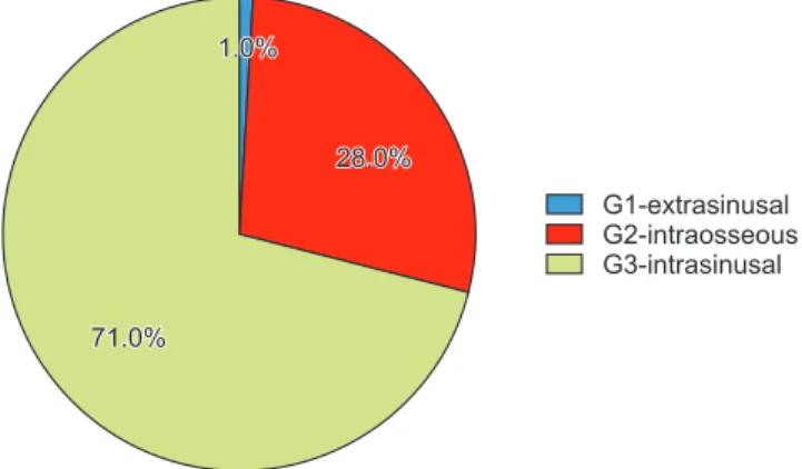
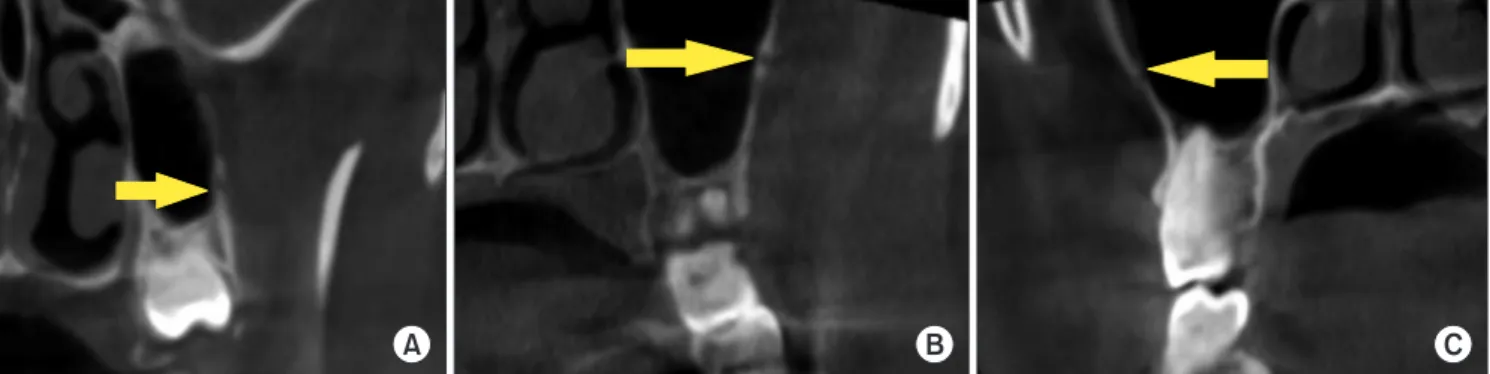
The LAICs were categorised according to medio-lateral location within the antral wall based on the criteria by Lee et al.13.(Fig. 3) In only 3 cases, the canal bulged towards the outer side of the wall (G1-extrasinusal).(Fig. 4. A) Canals were embedded within the wall (G2-intraosseous) (Fig. 4. B) in 70 sinus walls, whereas canals bulged towards the inner side of the wall in 176 sinus walls (G3-intrasinusal).(Fig. 4.
C) The diameter of the antral canal was classified based on the criteria devised by Mardinger et al.12. The diameter was less than 1 mm in the majority of cases (n=113). Eighty-three LAICs had a diameter of 1-2 mm, and 53 LAICS had a diam- eter of 2-3 mm.(Fig. 5)
IV. Discussion
In the present study, LAIC was detected in 62.3% of 200 CBCT scans. A similar percentage (67.1%) was reported by Rahpeyma et al.7, although their study only evaluated 35 CBCT scans. In a Thai study of 184 CBCT scans, the LAIC was detected in 60.3% of the samples4. Higher percentage was reported in recently conducted studies in a Chinese
Table 1. Distance between the most inferior point of the alveolar bone and the inferior border of the lateral antral intraosseous canal
Area of dentition Distance (mm)
Maxillary first molar 16.24±2.59
Maxillary second molar 16.53±2.71
Maxillary first premolar 25.24±3.65 Maxillary second premolar 21.33±3.54 Values are presented as mean±standard deviation.
Shishir Ram Shetty et al: Cone-beam computed tomography characterization of the intraosseous vascular canal in the lateral wall of the maxillary antrum. J Korean Assoc Oral Maxillofac Surg 2021
Table 2. Comparison of mean distances between maxillary molar and premolar areas
Area of dentition Distance (mm) P-value
Maxillary molar area 16.38±2.65 0.05
Maxillary premolar area 23.28±3.59 Values are presented as mean±standard deviation.
P-value by t-test.
Shishir Ram Shetty et al: Cone-beam computed tomography characterization of the intraosseous vascular canal in the lateral wall of the maxillary antrum. J Korean Assoc Oral Maxillofac Surg 2021
Table 3. Comparison of mean distances between dentulous and edentulous areas
Dentulous area vs
edentulous area Distance (mm) P-value Dentulous sub-sinusal area 19.86±3.10 0.1 Edentulous sub-sinusal area 19.80±3.15
Values are presented as mean±standard deviation.
P-value by independent sample t-test.
Shishir Ram Shetty et al: Cone-beam computed tomography characterization of the intraosseous vascular canal in the lateral wall of the maxillary antrum. J Korean Assoc Oral Maxillofac Surg 2021
G1-extrasinusal G2-intraosseous G3-intrasinusal 1.0%
1.0%
28.0%
28.0%
71.0%
71.0%
Fig. 3. Categorization of the canals based on the medio-lateral location within the antral wall.
Shishir Ram Shetty et al: Cone-beam computed tomography characterization of the intraosseous vascular canal in the lateral wall of the maxillary antrum. J Korean Assoc Oral Maxillofac Surg 2021
population14 (87.6% of 484 CBCT scans) and a Mexican population15 (90% of 1,005 CBCT scans). One of the earliest reported studies12 in 2007 demonstrated lateral intraosseous antral canals in 55% of 208 scans. However, the study was conducted using CT. Another CT based study conducted in 2016 reported presence of intraosseus antral canal in 32%
of the 284 scans that were examined13. Generally, CBCT- based studies show a higher percentage and better detection of LAIC than CT-based studies, perhaps because CBCT tends to display a superior spatial resolution compared to fan beam computed tomography16. In addition, some researchers have suggested that the higher spatial resolution observed in CBCT is due to the smaller voxel size of the scan17.
On the other hand, the differences in percentages of LAIC could be attributed to racial variations, since these studies were carried out in different parts of the world.
The present study demonstrated no significant difference in prevalence of LAIC between male and female subjects. Simi-
lar outcomes were reported by Lee et al.13 and Rahpeyma et al.7. In one recent CBCT-based study, the occurrence of lateral antral canal walls was higher in males compared to females6. The researchers stated that the probability of detec- tion of antral canals increases significantly as thickness of the antral wall increases, which is the probable reason for the increased radiographic detection of antral canals in male sub- jects in their study. When the location of the canal in the me- diolateral aspect of the antral wall was evaluated, we found that 78% of the canals had an intrasinusal position. Similar radiographic features were reported by Lee et al.13. Extra- sinusal is the least common location of the canal within the antral wall. However, one Korean study18 conducted on cadav- ers reported that the extrasinusal variety was most common.
When the diameter of LAIC was evaluated, most were less than 1 mm. The diameter of the canals plays a very important role in the radiographic detection of antral canals. LAICs with a diameter less than 0.5 mm are difficult to detect on routine CBCT scans19. Furthermore, the diameter of the LAIC has clinical significance in surgical interventions in the posterior maxillary sinus antrum area as its alerts the surgeon to possible risk of hemorrhage during surgery20. The surgeon can adjust the location of the osteotomy window according to pre-surgical CBCT evaluation to avoid complications9. De- spite a detailed radiographic examination of the surgical site, however, accidental exposure of the antral vessels, which might lead to bleeding, can occur20,21. This can be attributed to the heterogeneous pathways of the blood vessels, which can be partially intraosseous or completely extraosseous22. Researchers have advised the use of electrocautery or bone wax for management of bleeding subsequent to accidental rupture of these blood vessels22. Future CBCT-based research should focus on not only determining the point position of
A B C
Fig. 4. Tomographic appearance of lateral antral intraosseous canals according to classification of Lee et al.13. A. G1-extrasinusal type. B.
G2-intraosseous type. C. G3-intrasinusal type.
Shishir Ram Shetty et al: Cone-beam computed tomography characterization of the intraosseous vascular canal in the lateral wall of the maxillary antrum. J Korean Assoc Oral Maxillofac Surg 2021
<1 mm 1-2 mm 2 3 mm- 45.4%
33.3%
21.3%
Fig. 5. Categorization of lateral antral intraosseous canals accord- ing to diameter of the canals.
Shishir Ram Shetty et al: Cone-beam computed tomography characterization of the intraosseous vascular canal in the lateral wall of the maxillary antrum. J Korean Assoc Oral Maxillofac Surg 2021
the LAIC, but also on tracing its pathways.
V. Conclusion
This study established the average measurement for loca- tion of the LAIC with reference to maxillary alveolar bone in the UAE, which has not been previously reported. This data will aid the surgeon in avoiding the LAIC during sinus lift procedures and reduce the risk of hemorrhage.
ORCID
Shishir Ram Shetty, https://orcid.org/0000-0002-8097-6024 Saad Wahby Al Bayatti, https://orcid.org/0000-0002-5815- 2221
Hesham Marei, https://orcid.org/0000-0002-5967-6473 Raghavendra Shetty, https://orcid.org/0000-0003-1209- 7295
Hossam Abdelatty Abdelmagyd, https://orcid.org/0000- 0002-3958-4563
Alexander Maniangat Luke, https://orcid.org/0000-0002- 5885-7694
Authors’ Contributions
S.R.S. participated in data collection and wrote the manu- script. S.W.A.B., H.M., R.S., and H.A.A. participated in the study design and performed the statistical analysis. A.M.L.
participated in the study design and coordination and helped to draft the manuscript. All authors read and approved the final manuscript.
Ethics Approval and Consent to Participate
The study was approved by the Institutional Review Board of Gulf Medical University (No. INT/COD/FR/006-2020), and the written informed consent was obtained from all pa- tients.
Conflict of Interest
No potential conflict of interest relevant to this article was reported.
References
1. Albugami RA, Smith S, Hassan MA, Almas K. Trends in implant dentistry: implant systems, complications and barriers in Ri- yadh, Saudi Arabia. Dent Med Probl 2019;56:223-30. https://doi.
org/10.17219/dmp/108659
2. Akeel R. Attitudes of Saudi male patients toward the replacement of teeth. J Prosthet Dent 2003;90:571-7. https://doi.org/10.1016/
j.prosdent.2003.09.007
3. Zijderveld SA, van den Bergh JP, Schulten EA, ten Bruggen- kate CM. Anatomical and surgical findings and complications in 100 consecutive maxillary sinus floor elevation procedures. J Oral Maxillofac Surg 2008;66:1426-38. https://doi.org/10.1016/
j.joms.2008.01.027
4. Mung San Aung C, Panmekiat S, Pimkhaokham A. The study of the alveolar antral artery canal in using cone beam computed to- mography. Mahidol Dent J 2017;37:63-9.
5. Elian N, Wallace S, Cho SC, Jalbout ZN, Froum S. Distribution of the maxillary artery as it relates to sinus floor augmentation. Int J Oral Maxillofac Implants 2005;20:784-7.
6. Varela-Centelles P, Loira M, González-Mosquera A, Romero-Men- dez A, Seoane J, García-Pola MJ, et al. Study of factors influencing preoperative detection of alveolar antral artery by CBCT in sinus floor elevation. Sci Rep 2020;10:10820. https://doi.org/10.1038/
s41598-020-67644-9
7. Rahpeyma A, Khajehahmadi S, Amini P. Alveolar antral artery:
does its diameter correlate with maxillary lateral wall thickness in dentate patients? Iran J Otorhinolaryngol 2014;26:163-7.
8. Park WH, Choi SY, Kim CS. Study on the position of the posterior superior alveolar artery in relation to the performance of the maxil- lary sinus bone graft procedure in a Korean population. J Korean Assoc Oral Maxillofac Surg 2012;38:71-7. https://doi.org/10.5125/
jkaoms.2012.38.2.71
9. Kim JH, Ryu JS, Kim KD, Hwang SH, Moon HS. A radiographic study of the posterior superior alveolar artery. Implant Dent 2011;20:306-10. https://doi.org/10.1097/ID.0b013e31822634bd 10. Güncü GN, Yildirim YD, Wang HL, Tözüm TF. Location of pos-
terior superior alveolar artery and evaluation of maxillary sinus anatomy with computerized tomography: a clinical study. Clin Oral Implants Res 2011;22:1164-7. https://doi.org/10.1111/j.1600- 0501.2010.02071.x
11. Ilgüy D, Ilgüy M, Dolekoglu S, Fisekcioglu E. Evaluation of the posterior superior alveolar artery and the maxillary sinus with CBCT. Braz Oral Res 2013;27:431-7. https://doi.org/10.1590/
S1806-83242013000500007
12. Mardinger O, Abba M, Hirshberg A, Schwartz-Arad D. Preva- lence, diameter and course of the maxillary intraosseous vascular canal with relation to sinus augmentation procedure: a radiographic study. Int J Oral Maxillofac Surg 2007;36:735-8. https://doi.
org/10.1016/j.ijom.2007.05.005
13. Lee J, Kang N, Moon YM, Pang EK. Radiographic study of the distribution of maxillary intraosseous vascular canal in Koreans.
Maxillofac Plast Reconstr Surg 2016;38:1. https://doi.org/10.1186/
s40902-015-0045-x
14. Sun W, Liu A, Gong Y, Shu R, Xie Y. Evaluation of the anastomo- sis canal in lateral maxillary sinus wall with cone beam computer- ized tomography: a clinical study. J Oral Implantol 2018;44:5-13.
https://doi.org/10.1563/aaid-joi-D-17-00129
15. Cruz ILA, Palacios VDE, Miranda VJE, Cazar AM, Martínez OPA.
[CBCT evaluation of the alveolar antral artery in a Mexican cohort and its relationship to maxillary sinus floor lifting]. Rev ADM 2016;73:286-90. Spanish.
16. Lechuga L, Weidlich GA. Cone beam CT vs. fan beam CT: a comparison of image quality and dose delivered between two dif- fering CT imaging modalities. Cureus 2016;8:e778. https://doi.
org/10.7759/cureus.778
17. Watanabe H, Honda E, Tetsumura A, Kurabayashi T. A compara- tive study for spatial resolution and subjective image characteristics of a multi-slice CT and a cone-beam CT for dental use. Eur J Ra- diol 2011;77:397-402. https://doi.org/10.1016/j.ejrad.2009.09.023 18. Hur MS, Kim JK, Hu KS, Bae HE, Park HS, Kim HJ. Clinical
implications of the topography and distribution of the posterior su- perior alveolar artery. J Craniofac Surg 2009;20:551-4. https://doi.
org/10.1097/SCS.0b013e31819ba1c1
19. Yoshida S, Kawai T, Asaumi R, Miwa Y, Imura K, Koseki H, et al. Evaluation of the blood and nerve supply patterns in the molar region of the maxillary sinus in Japanese cadavers. Okajimas Folia Anat Jpn 2010;87:129-33. https://doi.org/10.2535/ofaj.87.129 20. Kim J, Jang H. A review of complications of maxillary sinus
augmentation and available treatment methods. J Korean Assoc Oral Maxillofac Surg 2019;45:220-4. https://doi.org/10.5125/jka- oms.2019.45.4.220
21. Kim YK, Ku JK. Sinus membrane elevation and implant place-
ment. J Korean Assoc Oral Maxillofac Surg 2020;46:292-8. https://
doi.org/10.5125/jkaoms.2020.46.4.292
22. Maridati P, Stoffella E, Speroni S, Cicciu M, Maiorana C. Al- veolar antral artery isolation during sinus lift procedure with the double window technique. Open Dent J 2014;8:95-103. https://doi.
org/10.2174/1874210601408010095
How to cite this article: Shetty SR, Al Bayatti SW, Marei H, Shet- ty R, Abdelmagyd HA, Luke AM. Cone-beam computed tomog- raphy characterization of the intraosseous vascular canal in the lat- eral wall of the maxillary antrum. J Korean Assoc Oral Maxillofac Surg 2021;47:34-39. https://doi.org/10.5125/jkaoms.2021.47.1.34