Secondary Dental Pain and Facial Pain Due to Pansinusitis : A Case Report
Nam-Koo Kim, D.M.D.
1, Q-Schick Auh, D.M.D.,M.S.D.,Ph.D.
1,
Yang-Hyun Chun, D.M.D.,M.S.D.,Ph.D.
1, Jung-Pyo Hong, D.M.D.,M.S.D.,Ph.D.
1,2Department of Oral Medicine, School of Dentistry, Kyung Hee University1 Institute of Oral biology, School of Dentistry, Kyung Hee University2
Toothache is a common complaint in the dental office. Most toothaches have their origin in the pulpal tissues or periodontal structures1).
These odontogenic pains are managed well and predictably by dental therapies. One of the most frequent encounters and most confusing phenomena with which the dental diagnostician must deal is the problem of referred pain. The most important step toward proper management of a toothache is to consider that the pain may not be of dental origin.
And Patients with orofacial pain, especially those in chronic pain, present a diagnostic and management challenge for the practitioners. There are many structures in the head and neck that can produce heterotopic pains felt in the teeth and other structures. Once referred pain is suspected, the true source of the pain must be located in order to render effective therapy.
With increased interest in temporomandibular disorders and orofacial pain, many studies of accurate diagnosis and differential diagnosis about orofacial pain have been established.
The purpose of this paper is to present a case for pansinusitis which produced pain referral in teeth and mimicked the symptoms of migraine.
Key words : Secondary dental pain, Referred pain, Facial pain, Pansinusitis, MRI
1)
I. INTRODUCTION
Toothache is a common complaint in the dental office. Most toothaches have their origin in the pulpal tissues or periodontal structures.
1)Corresponding Author : Jung-Pyo Hong
Department of Oral Medicine, School of Dentistry, Kyung Hee University,
Hoegi-dong 1, Dongdaemun-ku, Seoul 130-701, Korea Tel: 82-2-958-9355
Fax: 82-2-968-2043
E-mail: [email protected] Received: 2007-06-22
Accepted: 2007-09-02
These odontogenic pains are managed well and predictably by dental therapies. One of the most frequent encounters and most confusing phenomena with which the dental diagnostician must deal is the problem of referred pain. The most important step toward proper management of a toothache is to consider that the pain may not be of dental origin.
And Patients with orofacial pain, especially those
in chronic pain, present a diagnostic and
management challenge for the practitioners. There
are many structures in the head and neck that can
produce heterotopic pains felt in the teeth and other
structures. Once referred pain is suspected, the true
source of the pain must be located in order to render
effective therapy.
With increased interest in temporomandibular disorders and orofacial pain, many studies of accurate diagnosis and differential diagnosis about orofacial pain have been established.
The purpose of this paper is to present a case for pansinusitis which produced pain referral in teeth and mimicked the symptoms of migraine.
Ⅱ. CLINICAL CASE
A 28-year-old man, a salaryman at an electric gift producing company, was referred from the department of conservative dentistry for the differential diagnosis of nonodontogenic pain in the left upper molar and facial area. The intermittent severe throbbing pain of the left anterior temporal, eye area, left upper gingival area, left masseter, and the left sternocleidomastoid muscle area spontaneously happened approximately 10 days earlier.
Since that time, episodic severe pulsating pain had been generated for about 1 hour once or twice a day, and dramatically removed according to the patient.
The patient said that he had suffered from this pain, especially in the summer for up to a month at a time every year for about 7 years. The left maxillary second premolar had been extracted earlier as a result of the pain in this area.
The left maxillary first molar was filled with ZOE. The adjacent second molar was filled with amalgam on buccal pit. The first molar was sensitive to percussion slightly. As the history indicated that the patient had felt stiffness on neck area before the symptom. With the symptom, especially for swelling and heating sensation on the temporal muscle area and redness of the left eye with pain were presented.
He complained pain on the TMJ area and nocturnal bruxism. His 'range of motion' is within normal limit. The patient's right TMJ is revealed click sound during mouth opening but without pain.
The temporomandibular ligaments on both sides, masseter muscles on both sides, left anterior temporal muscle and the left sternocleidomastoid
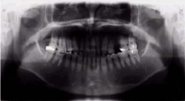
Fig. 1. Panoramic radiograph of the head.
muscle was sensitive to the palpation test slightly.
The panoramic radiograph showed that the left maxillary secondary premolar had been extracted and the lining mucosa on the left maxillary sinus wall was thickened slightly(Fig. 1).
The tongue ridging and linear alba, 3mm midline deviation to the left and bruxofacet on #13,23,33,34 teeth were observed. The diagnosis was myofascial pain in the left temporal, masseter and sternocleidomastoid muscle. The treatment plan was physical therapy which included Fluoromethane vapor coolant therapy and stretch under ultra sound therapy, dry needling with oriental acupuncture and EAST(electrical acupuncture stimulating therapy).
Judging from the pattern and nature of the pain, possible diagnosis of migraine was suspected.
During the first three days only physical therapy treatment was given, and pain in the masseter and sternocleidomastoid muscle areas was decreased.
But the pulsating pain symptoms remained in the anterior temporal area.
With a tentative diagnosis of migraine, Cafergot, a vasoconstrictor ergotamine tartrate, was prescribed to relieve the pain and to diagnose accurately.
But the patient reported severe a stomachache after taking Cafergot, and Sumatriptan was prescribed as a replacement. The patient reported that the duration of pain had been shortened (one hour to half hour) but, not completely eliminated.
Therefore the patient referred to a neurosugery
specialist for a thorough examination and treatment
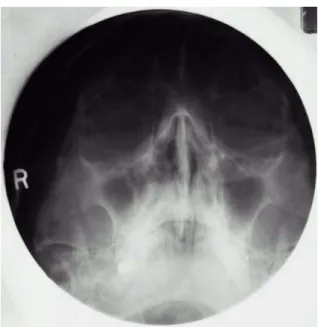
Fig. 2. Waters' radiograph of the paranasal sinus.
Fig. 3. The coronal magnetic resonance imaging of the head.
of tentative diagnosed facial migraine.
After physical examination, Brain magnetic resonance imaging (MRI) was performed. The MRI revealed the lining mucosae on both maxillary sinuses, ethmoidal sinuses and left sphenoidal sinus presented high signal on the T2 weighted image(Fig. 3, 4). And Waters' projection radio- graphic imaging revealed thickening of the lateral wall of the left maxillary sinus(Fig. 2).
Accordingly, pansinusitis was diagnosed. Under
Fig. 4. The horizontal magnetic resonance imaging of the head.
general anesthesia, endoscopic surgery and submucosal resection was performed by the ear-nose-throat specialist. Since surgery, the patient has been free from pain for a period of about 7 months.
Ⅲ. DISCUSSION
By the Subcommittee on Taxonomy of the International Association for the Study of Pain, Pain is defined as " An unpleasant sensory and emotional experience associated with actual or potential tissue damage, or described in terms of such damage".
2)Pain especially chronic pain, is a major health problem. According to the Nuprin Report (Bristol -Myer Co, 1985), of the 1,254 persons questioned, 27% reported having experienced dental pain, and 73% reported headaches during the previous 12 months. This report indicates that the task of managing head and neck pain is a very real one indeed. The problem crosses the lines that demarcate the professions; it should be the concern of all health care professionals.
For the clinician to begin classifying orofacial pain
disorders, he or she must first be able to
differentiate the signs and symptoms associated
with each category. For the successful management
of pain, first of all physical and psychologic factors
must be separated. In this paper I used the pain
classification based on the symptoms by Jeffry P Okeson in Bell's Orofacial Pain.
3)Periodontal pain is deep somatic pain of the musculoskeletal type. The proprioceptors of the periodontal ligament are capable of rather precise localization of the stimulus. Therefore, periodontal pain of all types presents little diagnostic problem because the offending tooth is readily identified.
The dental pulp pain is a visceral organ, and thus pulpal pain possesses characteristics of deep pain of visceral origin. Visceral pain tends to be deep, dull, aching pain that is of a threshold nature and is often difficult to localize.
Migraine is the most common type of neurovascular pain. Neurovascular pain has common mechanisms involving the trigeminovascular system.
4)The distinctive feature of vascular pain is its primary pulsatile or throbbing quality.
Neurovascular pains are a group of visceral pain disorders that are generally characterized by episodic pains accompanied by neurologic, gastrointestinal, and psychologic changes.
3)The secondary facial pain and dental pain from the sinusitis has been reported from some clinicians.
5,6)Inflammatory conditions of the maxillary antrum and other paranasal sinuses are generally misund- erstood. Most sinusitis occur as a complication of the common cold or other upper respiratory tract infections, with occasional presentations due to extension of a periodontal infection under the maxillary. Radiographic evaluation is relatively sensitive. Magnetic resonance image and ultrasono- graphy have limited but specialized applications.
Sinus aspiration and endoscopic sinuscopy may be necessary to recover organisms or to effect drainage.
Decongestants and antihistamines improve nasal obstruction and may improve sinus drainage. Oral antibiotics are often prescribed. Occasionally, surgical interventions are used when disease is chronic and resistant to empiric therapy.
7)The syndrome of symptoms usually thought to be clinically indicative of sinusitis are chiefly secondary autonomic and sensory effects induced by primary
pain emanating from the nasal mucosa. Inflammatory exudate that accumulates in the antral cavity remains below the opening of the antrum into the middle meatus of the nose as long as the head is erect. Bending forward, however, permits the fluid to wash over the pain-sensitive tissue around the ostium, thus eliciting pain. If the ostium is open, antral inflammation can spread to the nasal mucosa and thereby initiate the symptoms. If the ostium is closed by swollen membrane or mucous polyps, the maxillary sinus may remain essentially nonsym- ptomatic.
Pain that emanates from the nasal mucosa is typically a dull, burning sensation that exhibits the clinical characteristics of visceral mucosa pain.
The mechanism of visceral pain has been described by several authors. Ordinarily, sensory information transmitted by afferent neurons of the visceral nervous system remains well below conscious levels. It serves to monitor the involuntary muscle action of vessels, glands, and organs. It reports unusual sensation occurring in the lining membranes of organs and cavities. It helps regulate the autonomic functioning of the body by supplying the CNS with a constant inflow of information. When such sensation becomes noxious, pain is felt. Visceral afferent fibers of both the sympathetic and parasympathetic type are known to mediate pain.
8)Visceral pain usually has a distinct stinging or burning quality. It is said that protracted visceral pain tends to provoke vasomotor effects, local edema, and trophic changes.
9)When treatment of orofacial pain fails, it is often because the condition has not been adequately diagnosed. Although current knowledge of pain mechanisms has many gaps, understanding of the central and the peripheral mechanisms is expanding.
Meller
10)in a focus article on pain mechanisms in the Journal of the American Pain Society, said "It is crucial to the effective treatment of chronic and persistent pain to have a better understanding of the mechanisms that underlie the different types of hyperalgesia".
The diagnostic differentiation between true
primary pain and symptoms that occur as secondary effects of that pain is essential. Such manifestations are usually referred as central excitatory effects on the presumption that they result from hypere- xcitability of CNS interneurons. The neurologic mechanisms involved in the secondary effects of pain are not fully understood. Some researchers think that reflex activity is involved.
11)There seems to be little doubt that convergence of afferent impulses among CNS interneurons takes place.
12)The neurons thus activated are presumed to be those that respond to input from deep structures of the mouth and face. This is taken as evidence that the reference of pain occurs in conjunction with deep pain input rather than a superficial one.
13)It suggests that under certain conditions trigeminal nociceptive inter neurons are subject to subliminal stimulation from structures other than those located in the normal receptive fields. Such evidence supports the convergence theory of pain reference.
Gross
14)reported that referred pain due to visceral disease followed known dermatomes but referred pain from deep somatic structures did not. Mense
15)theorized that there are convergent connections from deep tissues to the dorsal horn neurons, which are opened when nociceptive input from skeletal muscle occurs. This phenomenon would cause referral to myotomes outside of the original lesion and spread of central sensitization to adjacent spinal segment.
Some information
16)suggested that the second- order neuron can change with respect to its processing of impulses. It appears that when a second-order neuron receives a constant barrage of nociceptive input, specific receptors sites can be activated that increase the sensation of that neuron.
Some of the receptors that are involved in this sensitization are the N-methyl-D-aspartic acid (NMDA) receptors.
17)Stimulation of these receptors by excitatory amino acids, such as aspartate and glutamate, increase the sensitization of these neurons. This increased sensitization can alter neural impulses as they are processed on the way to the higher centers. When this occurs even normal
neural impulses can be misinterpreted as noxious.
This alteration in impulses processing is called neuroplasticity.
Clinically, sensitization and central excitatory effects are induced by more or less continuous barrages of noxious sensation emanating from deep somatic structures. If the input is not continuous, these secondary intensity and duration of the deep pain input enhances the secondary effects. Initially these changes in neural sensitization are usual and reversible. With chronicity, however, changes may occur that alter the neuroprocessing more permanently. The symptom of central sensitization complicate continuous deep somatic pains since they increase with the intensity and duration of the primary pain. Such symptoms can occur in otherwise normal structures; therefore, treatment can be misdirected.
3)If the nociceptive activity from the periphery has been of sufficient intensity, changes occur in the central nervous system that significantly alter the central response of subsequent peripheral stimulation. These central neuroplastic changes cause additional allodynia(pain which is produced by normally non-painful stimulus), secondary hyperalgesia(an increased response to stimulation at the site of pain in the absence of any local cause) and expansion of receptive field in the periphery.
Recently, Merril
18)summarized central neuroplastic changes as the following 4 mechanisms.
The mechanisms of these peripheral symptoms are a result of central physiologic, neurochemical, anatomic, and genetic changes.
19)1. Central Physiologic changes
When nerve damage or intense afferent
discharges occur, the resulting central physiologic
changes involve the WDR(the wide dynamic range)
second-order neurons. Mendell
20)showed that
repetitive activation of the C fibers evoked a
sustained augmentation of the dorsal horn WDR
neurons. This phenomenon is called 'windup'
represents facilitation of pain and sensitization of the
WDR in the trigeminal subnucleus caudalis.
Glutamate release is responsible for this facilitation.
The augmentation of the EDR reaction also causes prolonged after discharge, decreased threshold to mechanical stimulation, and expansion of the peripheral receptive field.
2. Central Neurochemical Changes
The cell membrane of the WDR neuron has many receptors that respond to various neurochemical agents. The first nociceptive event, carried by the faster A-delta fibers to the central nervous system, causes a release of neurokinins such as substance P that act on the AMPA and metabotropic receptors on the WDR neuron or on the segmental interneurons. This causes depolarization of the WDR neuron, sending an ascending message to the thalamus and on to the somatosensory cortex, where it is perceived as sharp pain. If the volley of afferent activity is intense enough, C fibers release excitatory amino acid, glutamate which has agonist activity on the NMDA receptor. Glutamate is the main mediator of chronic pain.
21)The receptor is associated with a calcium channel that is normally blocked by Mg
+. The Mg
+is expelled, and the calcium channel is opened with the resultant influx of Ca
++into the WDR neuron. Nitric oxide is produced in the WDR through the influence of calcium/calmodulin complex on nitric oxide synthetase. Nitric oxide is also thought to be one of the agents that causes the windup because pre treatment with an NMDA antagonist or a nonactive substrate of nitric oxide prevents it. Nitric oxide is a gaseous neurotransmitter that easily diffuses out of the cell to cause action in other areas. It is thought to have presynaptic activity in the terminal bouton of the C-PMN, causing further release of the excitatory amino acids such as glutamate as well as release of substance P and other neurokinins. This would result in further excitation of the WDR and bring about a cycle of pain. The system feeds on itself, and pain becomes continuous until the process is interrupted by stopping this central excitatory
mechanism with centrally or peripherally acting medications. This is thought to be the mechanism for continuous pain in such conditions as atypical odontalgia, traumatic neuralgia, and other chronic pain neuropathic pain conditions.
3. Anatomic Changes
Segmental inhibitory interneurons are an important part of the pain modulatory system in the central nervous system. It is thought that part of the mechanism that leads to chronic ongoing pain states is loss of the segmental inhibition. Interneurons may be particularly susceptible to the neurotoxic effects of nitric oxide and lose their ability to function.
4. Genetic Changes
After painful stimulus and influx of calcium and production of nitric oxide, c-fos & c-jun, immediate early oncogenes, direct the transcription of c-fos &
c-jun proteins. These proteins direct the transcription of other regulatory proteins within the cell. The target proteins of this activity apparently alter the function of the cell, causing it to lose its ability to encode a difference between high-frequency nociceptive information and low-frequency mechanostimulation. This represents an alteration of the genetic control of cellular function and may be long lived if not permanent.
This process may account for the chronicity and recalcitrant response to therapies.
When the examination process has not been able
to determine the source of pain, a somatic sensory
anesthetic block is done to determine whether to
pain is centrally or peripherally driven. Anesthetic
blocks relieve pain arising from peripheral processes
and are only partial or ineffective in relieving
centrally driven pain. If pain is only partial relieved
with profound somatic anesthesia, a neuropathic
pain condition with varying degrees of central and
peripheral involvement is to be suspected. If the
somatic blockade is not effective although there is
profound anesthesia, a central pain process is
suspected.
8)To diagnose orofacial pain , it is necessary to be familiar with its clinical characteristics. And the characteristics of odontogenic pain and nonodon- togenic toothache originating from migraine and sinusitis have been well summarized in other papers.
4)To make an accurate diagnosis of orofacial pain and render treatment, all organ systems need to be considered and evaluated. Central sensitization has suggested that treatment of these disorders be problem oriented, addressing both peripheral and central mechanisms if present. The characteristics of signs and symptoms for each orofacial pain must be well known, and other supportive diagnostic method must be correctly used for differential diagnosis.
Many teeth have been mistakenly extracted, and many pulps extirpated because of incorrected diagnosis of this phenomenon. This patient demonstrated the same pain pattern and insisted that the pain originated from the teeth. According to the patient's dental history, it is possible that the removal of the left maxillary secondary premolar was unnecessary.
REFERENCES
1. Lipton JA, Ship JA, Larach-Robinson D. Estimated prevalence and distribution of reported orofacial pain in the United States. J Am Dent Assoc 1993;124:115.
2. Merskey H. Pain terms: A list with definitions and notes on usage. Pain 1979;6:249-252.
3. Okeson JP. Bell's Orofacial Pains. 5th ed., Chicago, 1995, Quintessence Co., pp.61-81
4. Okeson JP, Falace DA. nonodontogenic toothache.
Dent Clin North Am 1997;41(2):367-383.
5. Emshoff R, Bertram S, Kreczy A. Idiopathic maxillary pain: prevalence of maxillary sinus hypereactivity in relation to allergy, chronic mucosal inflammation and eosinophillia. Oral Surg Oral Med Oral Pathol Oral Radiol Endod 1999;87(6):685-90.
6. Chen YH, Tseng CC, Chao WY et al. Toothache with a multifactorial etiology: a case report. Endod Dent Traumatol 1997;13(5):245-247.
7. Bennett JC, Plum F. Cecil text book of medicine. 20th ed., Philadelphia, 1996, WB Saunders Co., pp450-459.
8. Abram SE. Sympathetic pain. In Raj PP(ed): Practical Management of Pain. Chicago, 1986, Year Book, pp 209-214.
9. Gross D. Pain and autonomic nervous system. In Bonica JJ(ed): Advances in Neurology. vol. 4. New York, 1974, Raven Press, pp93-103.
10. Meller AT. Thermal and mechanical hyperalgesia: A distinctive role for different excitatory aminoacid receptors and signal transduction pathways. J Pain 1994;3:215-231.
11. Procacci P, Zoppi M. Pathophysiology and clinical aspect of visceral and referred pain. In Bonica JJ, Lindblom U, Iggo A (ed): Advances in Pain research and Therapy. New York, 1983, Raven Press, pp 643-658.
12. Milne RJ, Foreman RD, Ciesler GJ et al. Visceroso- matic convergence into primate spinothalamic neurons: An explanation for referral of pelvic visceral pain. In Bonica JJ, Lindblom U, Iggo A (ed): Advances in Pain Research Pain Therapy. vol 5. New York, 1983, Raven Press, pp131-137.
13. Sessle BJ, Hu JW, Amano N et al. Convergence of cutaneous, tooth pulp, visceral, neck and muscle afferents onto nociceptive and nonnociceptive neurons in trigeminal subnucleus caudalis (medullary dorsal horn) and its implications for referred pain. Pain 1986;27:219-235.
14. Gross D. Pain and autonomic nervous system. In Bonica JJ (ed): Advances in Neurology. vol 4. New York, 1974, Raven Press, pp93-103.
15. Mense S. Refferral of muscle pain: New aspects. J Pain 1994;3:1-9.
16. Coderre TJ, Katz J, Vaccarino AL, Melzack R.
Contribution of central neuroplasticity to pathological pain: Review of clinical and experimental evidence.
Pain 1993;52:259-285.
17. Woolf CJ, Thompson SWN. The induction and maintenance of central sensitization is dependent on N-methyl-D-aspartic acid receptor activation:
Implications for the treatment of post-injury pain hypersensitivity. Pain 1991;44:293-300.
18. Merril RL. Orofacial pain mechanisms and their clinical application. Dent Clin North Am 1997;41(2):167-188.
19. Dubner R. Neuropathic pain: New understanding leads to new treatments. J Pain 1993;2:8-11.
20. Mendell LM. Physiological properties of unmyelinated fiber projection to the spinal cord. Exp Neurol 1966;16:316-332.
21. Wilcox GL. Spinal mediators of nociceptive neurotra- nsmission and hyperalgesia. J Pain 1993;2:265-275.
국문요약
범부비동염에 의한 이차성 치통과 안면통: 증례보고
경희대학교 치과대학 구강내과학교실1, 경희대학교 치과대학 구강생물학연구소2