후반고리관 양성돌발성 두위현훈의 진단과 치료
성균관대학교 의과대학 삼성서울병원 이비인후과학교실
정 원 호·박 계 훈
Diagnosis and Management of Benign Paroxysmal Positional Vertigo of Posterior Semicircular Canal
Won-Ho Chung, MD and Kye Hoon Park, MD
Department of Otorhinolaryngology-Head and Neck Surgery, Sungkyunkwan University School of Medicine, Samsung Medical Center, Seoul, Korea
서 론
양성돌발성 두위현훈(이하 BPPV)은 회전성 어지러움 을 일으키는 말초성 내이 질환 중 가장 높은 빈도를 보이 는 단일 질환으로, 어지러움 클리닉 내원 환자 중 18.3%
1)를 차지한다고 보고되고 있다. 흔히 특징적인 임상 증상 과 안진소견으로 진단이 용이하고, 대부분의 경우 쉽게 치 유될 수 있는 질환이다. 본 질환의 빈도는 증상이 자연 소 멸되는 경우가 많기 때문에 정확히 평가하기 어려우나, 대부분의 보고에서 100,000명당 10.7명에서 64명으로 보고되었으며,
2)3)소아보다는 중년이상의 성인에서 많이 발생한다.
BPPV을 일으키는 기전은 난형낭의 평형반에서 기원한 유리 부유물(free floating debris)이 반고리관의 내림프 강 내로 들어가면서 부유물에 작용하는 중력의 영향으로 머리 움직임에 따른 내림프 움직임의 비정상적인 항진효 과를 일으키게 되어 안진이 발생되고 어지러움이 유발되 는 것으로 설명된다. 원발성 원인으로는 미상(idiopathic) 인 경우가 50~70%를 차지 한다. 이차성(secondary)
BPPV의 원인으로는 두부외상에 의한 BPPV의 빈도가 가 장 높으며(7~17%), 이외에 전정신경염, 만성중이염, 메니 에르 질환, 편두통, 중이 수술 등을 원인으로 들 수 있다.
침범되는 반고리관은 주로 후반고리관이 가장 많고 외 반고리관과 상반고리관의 순으로 발생한다. 이는 해부학 적으로 누워있는 자세에서 후반고리관이 가장 아래 부위 에 놓여 있어, 생성된 부유물이 중력의 영향으로 후반고 리관으로 들어가기가 용이하기 때문이다.
후반고리관 BPPV의 진단
병 력(History)
환자는 회전성 어지러움의 증상이 갑자기 발생하면서 오심과 구토를 동반하게 된다. 처음 발생의 시작은 주로 새벽에 잠자리에서 일어나거나 몸을 좌우로 돌아눕는 순 간에 발생하게 된다. 이후 계속되는 어지러움은 보통 특 정한 머리의 위치나 움직임에 의해 유발되는 특징이 있다.
어지러움을 유발하는 가장 흔한 동작으로는 잠자리에서 눕거나 일어날 때, 선반에서 물건을 꺼내려고 올려 볼 때 그리고 몸을 앞으로 구부릴 때 등이다. 환자들은 종종 어 지러움을 유발하는 동작을 기억하는 경우가 있어 환측을 예측하는데 도움이 되기도 한다. 몇 번의 발작 경험을 하 게 되면, 환자 스스로 어지러움이 발생되는 자세와 아닌 자세를 알 수 있기 때문에 어지러움을 일으키지 않도록 행
교신저자:정원호, 135-710 서울 강남구 일원동 50번지성균관대학교 의과대학 삼성서울병원 이비인후과학교실 전화:(02) 3410-3579·전송:(02) 3410-3879 E-mail:[email protected]
동의 변화를 보이게 된다. 어지러움의 지속시간은 전형적 으로 30초에서 1분 이내지만 수분 이상 지속된다고 말 하는 환자도 있다. 이러한 차이는 어지러움 발작 이후에 도 지속적인 비회전성의 어지러움이 동반되는 경우가 많 기 때문이다.
회전성의 어지러움 이외에 많은 환자들이 쓰러질 것 같 은 느낌, 떠다니는 느낌, 구역질나는 느낌, 머리움직임에 대한 공포 등을 호소하기도 한다. 특히 회전성 어지러움 을 처음 경험하는 환자는 뇌에 치명적인 질환이 나타난 것 으로 생각하고 심각한 두려움을 갖게 된다.
진 단(Diagnostic maneuvers)
후반고리관형 BPPV에 대한 Dix-Hallpike 검사는 1952 년 처음 기술되었다.
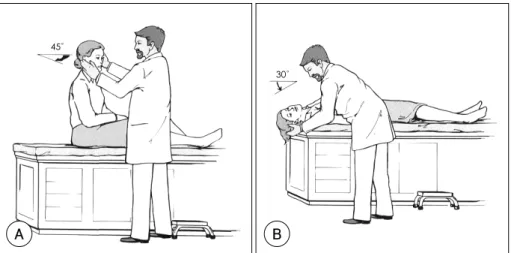
4)정확한 검사를 위하여 다음과 같은 사항을 유의한다. 1) 검사 전에 환자에게 심한 어지러움 이 발생할 수 있음을 설명하고 검사 중에는 눈을 되도록 뜨도록 하고, 중앙의 한 점을 주시하도록 교육한다. 환자 가 한 점을 주시하지 못하는 경우 안진의 방향을 기술하 기 어렵기 때문이다. 2) 검사자의 한 손으로는 최소한 환 자의 한쪽 눈이 크게 떠진 상태로 유지할 수 있도록 한 다. 3) 안진의 잠복기가 긴 환자가 있을 수 있으므로 30 초 정도까지 머리를 낮춘 자세를 유지하고 관찰한다. 앉은 자세에서 환자의 머리를 환측으로 45도 돌리고(A) 환자
를 눕히며 머리를 뒤로 젖혀 검사대보다 30도 정도 머리 가 낮은 위치를 유지한 후(B) 안진을 관찰한다. 특징적 인 안진은 잠복기가 1~5초이며 지속기간은 30초 이내이
Fig. 1. Dix-Hallpike maneuver (right ear). The patient is seated and positioned so that the patient’s head will extend over the top edge of the table when supine. The head is turned 45°toward the ear being tested (position A). The patient is quickly lowered into the supine position with the head extending about 30°below the horizontal (position B).The patient head is held in this position and the examiner observes the patient’s eyes for nystagmus.
45°
A
30°
B
45°°
Sagittal Frontal
Fig. 2. Side-lying test (right ear). The subject’s head is tur- ned with the nose pointing 45°away from the side to be tested (The head is turned left). Then, the subject is briskly laid on the side being tested. The examiner is not shown, but supports the subject’s head. Arms are crossed to pre- vent the patient from inadvertently stopping the motion.
다. 안진의 발생시에는 환자가 회전성의 어지러움을 동시 에 느끼게 된다(Fig. 1).
검사 테이블이 좁거나, 노인, 경추부에 문제가 있는 경우
에서는 검사시에 테이블 보다 머리가 낮은 자세를 취하기 어렵다. 이런 경우에는 Dix-Hallpike 검사보다는 side- lying 검사를 시행하게 된다. 검사방법은 환자를 검사테이 블에 앉히고 머리를 추정 병변 반대측으로 45도 돌린 후 추정 병변쪽으로 빠르게 눕히면 된다(Fig. 2).
5)특징적인 안진의 방향은 대체로 빠른 성분의 안진이 상 방향으로 나타나는 상향 안진이 보이면서, 안구 최첨부 가 바닥으로 향하는 회전성 안진이 동반된다. 이는 후반 고리관이 흥분되면 동측의 상사근과 반대측의 하직근의 수 축이 일어나기 때문이다(Fig. 3). 그러나, 안진의 방향은 환자가 주시하는 방향에 따라 조금씩 달라질 수 있다. 이 는 상사근 활차(trochlea)의 축이 X축과 약 51도 정도 각 을 이루고 있기 때문이다(Fig. 4A). 즉, 지면 쪽을 주시 할 때는 주로 회전성의 안진이 보이며(Fig. 4B) 지면과 반 대쪽을 주시할 때는 상향의 수직 안진이 강조되고(Fig. 4C) 정면을 주시할 때는 두 가지 성분이 다 나타난다(Fig. 4D).
환자를 다시 앉은 자세로 하면 안진의 방향이 반대가 되고 반복해서 검사하면 피로현상을 보여 안진의 강도가 약해진 다. 팽대부릉정 이석(cupulolithiasis)의 경우에는 Dix- Hallpike 검사에서 잠복기가 짧고 안진이 비교적 오래 지 속되며 피로현상이 약하게 보이므로 진단에 도움이 된다.
IV IV
Excitability Inhibitory Lt.
IR SR SO
IO
Rt. Slow Phase
MLF M MLF
I L S
III III
Fig. 3. Excitation of the right posterior semicircular canal causes contraction of the ipsilateral superior oblique mus- cle and the contralateral inferior rectus muscle. The result will be left-rotatory and downward vertical movement of eyes. To overcome this movement, right-rotatory and upward vertical nystagmus is appeared. S:superior ves- tibular nucleus, L:lateral vestibular nucleus, I:inferior vestibular nucleus, M:medial vestibular nucleus, III:ocu- lomotor nucleus, IV:trochlear nucleus, MLF:medial lon- gitudinal fasciculus. SR:superior rectus muscle, SO:
superior oblique muscle, IR:inferior rectus muscle, IO:
inferior oblique muscle. The dotted lines mean excitatory, and the solid lines mean inhibitory.
Action
plane 51°
X axis
39° Rotational axis
SO
Y axis
LR
A
SRB C D
Fig. 4. Rotational axis and action plane of right superior oblique muscle and direction of nystagmus on eye positions. A:
The eye movements by superior oblique muscle are internal rotatory and downward vertical through the influence of the trochlea. The axis of trochlea forms about 51°with X axis. When gaze is directed about 39°laterally, only internal ro- tatory movement occurs because of the right angle between the rotational axis and the action plane of superior ob- lique muscle. But when gaze is directed to the nasal side, mainly downward movement occurs. The nystagums changes during Dix-Hallpike maneuver for posterior canal BPPV depending on the eye position. B:The fast component nystag- mus is mainly rotatory when gaze is directed toward the lesion side. C:The fast component nystagmus is upward when gaze is directed to the normal side. D:When the eyes are in the neutral position, the fast component nystagmus is both upward vertical and rotatory toward the lesion side. SO:superior oblique muscle, LR:lateral rectus muscle, SR:
superior rectus muscle.
Dix-Hallpike 검사 등의 체위검사에서 Frenzel 안경을 사용할 수 있는데 진단률을 높이지는 않는다고 한다.
6)이 론적으로 Frenzel 안경은 환자의 시고정을 억제하여 안 진이 잘 나타나도록 한다. 그러나 대부분의 환자는 심한 안진이 나타나므로 시고정에 의해 억제되지 않으며, 특히, 회전성 안진은 시고정에 의한 억제효과가 약한 것으로 알 려져 있다.
7)결론적으로 Frenzel 안경의 사용은 육안으 로 관찰하기 어려운 미세한 안진을 진단하는데 도움이 될 수 있다. 전기안진검사는 회전성의 안진을 기록하지 못하 므로 안구의 움직임을 완전하게 기록할 수 없다. 그러나, 최근에 개발된 적외선비디오 전기안진검사 장치(Infrared videography)는 체위검사시 직접적으로 안구의 움직임을 관찰할 수 있어 유용하다.
간혹 Dix-Hallpike 검사상 특징적인 안진을 관찰할 수 없지만, 검사시 환자는 회전성 어지러움을 호소하고, 이러 한 주관적 증상이 잠복기와 피로현상을 보이는 경우 이 를‘subjective BPPV’ 라고 한다. Haynes 등,
8)Tirelli 등
9)과 Weider 등
10)은 이러한‘subjective BPPV’ 환자에서 다양한 이석치환술을 시행함으로써 76~93%의 환자가 호 전되었다고 한다. 양성돌발성 두위현훈 환자에서 Dix- Hallpike 검사로 안진이 유발되지 않는 이유로 다음과 같 은 이론들이 제시되었다. 미세한 안진을 검사자가 발견하 지 못하는 경우, 이전의 반복적인 검사로 이내 피로현상 이 생기는 경우, 어지러움은 유발하지만 전정안반사를 자 극하지 못하는 정도의 경증의 질환인 경우
8)등이다.
후반고리관 BPPV의 치료
비수술적인 치료법
후반고리관 BPPV의 치료법은 질환에 대한 이해가 깊 어지면서 지난 20년간 많은 변화가 있었다. 예전에는 어 지럼증을 유발하는 두위를 피하고, 증상조절을 위하여 약 물 처방만을 하였던 때가 있었으나 대부분 효과가 없는 것으로 증명되었다.
11)다양한 비침습적인 치료법들이 개 발된 것은 cupulolithiasis와 canalolithiasis의 개념이 도입 되면서부터이다. Brandt와 Daroff는 1980년도에 BPPV가 후반고리관의 팽대부에 이석이 달라붙기 때문이라는 가정하 에 달라붙은 이석을 작은 조각으로 부스러뜨리는 운동법을 소개하였다.
12)이후 같은 가정하에 1988년 Semont
13)는
한번의 치료운동으로 팽대부릉정으로부터 이석을 떼어내 는 방법을 고안하였다. 그러나 획기적인 치료법의 도입은 1992년 Epley
14)에 의해 이루어졌다. 그는 팽대부릉정 에 붙어있는 이석 때문이 아니라 반고리관 내의 이석이 BPPV의 원인이 된다는 가설을 바탕으로 이석치환술을 개발하였다. 이는 현재까지도 가장 많이 사용되고 있으 며 치료 효과를 높이기 위하여 다양하게 변형되어 사용 되고 있다.
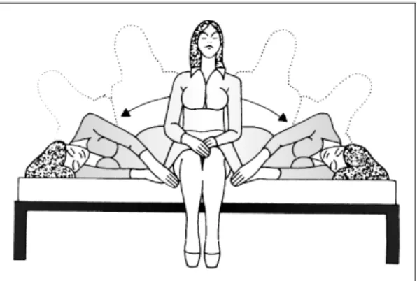
Brandt-Daroff 습관화 운동
앉은 자세에서 어지러움이 유발되는 쪽으로 갑작스럽게 쓰러지고 어지러움이 없어진 후 30초 정도 더 누워있다 가 앉는다. 약 30초 후 반대쪽으로의 운동을 반복하며 이 와 같은 운동을 하루 수 차례씩 반복 시행한다(Fig. 5).
반복된 운동으로 점차 유발되는 증세가 약화되어 연속되 는 이틀간 증세가 유발되지 않을 때까지 시행한다. 환자 의 증세가 호전되는 기전에 대한 설명은 명확하지는 않지 만 후반고리관의 이석 조각이 떨어져 나오고 작은 조각으 로 부스러져 증세를 유발시키지 않게 된다는 것과 반복되 는 자극에 의하여 중추에서 적응이 일어나기 때문인 것 으로 설명된다. 이러한 운동법은 환자가 시행하기 번거로 움 단점은 있지만,
12)15)팽대부릉정이석으로 인해 이석치 환술의 효과가 크지 않거나, 이석치환술로 안진은 없어졌 지만 주관적인 어지러움을 계속 느끼고 있는 경우 습관 화 운동을 시행하면 환자의 증상을 호전시킬 수 있다.
Fig. 5. Brandt-Daroff exercises. Start sitting upright. Then move into the side-lying position, with the head angled upward about half-way. Stay in the side-lying position for 30 seconds, or until the dizziness subsides, then go back to the sitting position. Stay there for 30 seconds and then go to the opposite side and follow the same routine.
이석유리술(Semont liberatory maneuver)
환자는 앉은 자세에서 환측의 반대쪽으로 얼굴을 돌리 고 갑작스럽게 환측으로 고개를 들고 눕는다. 5분이 지난 후 반대쪽으로 얼굴이 아래를 향하여 귀가 닿도록 빠르게 눕는다. 환자는 이 자세를 5~10분간 유지한 후 앉은 자세 로 서서히 돌아온다(Fig. 6).
Semont는 이 방법을 통하여 단 한 번의 운동으로 84%
의 치료효과를 보았고, 1주 후 한 번 더 시행함으로 93%
까지 치료효과를 높일 수 있었다고 보고하였다.
13)이후 다른 발표들에 의하면 52~90%의 치료 결과를 보이며 재 발율은 29% 정도로 보고 되었다.
8)16-18)Herdman 등
18)과 Cohen 등
19)에 의하면 뒤에 언급되는 이석치환술과 비 교하여 치료성적이 유사하지만 나이 많고 비만인 사람한 테 적용하기 어려운 점이 있고 이석치환술에 비하여 더 좋은 결과를 보이지는 않는다고 하였다.
이석치환술(Modified Epley maneuver)
1992년 Epley는 5가지 일련의 동작으로 이관 내의 이 석을 중력을 이용하여 난형낭 내로 재위치시키는 방법을
고안하여 높은 치료 성공률을 보였다.
14)그는 운동 시행 전에 환자에게 안정제를 투여하였고 진동기(mechanical skull vibrator)를 유양돌기부에 작동하여 이석의 부양을 유도하였으며 증세가 없어질 때까지 반복 시행하였다. Par - nes 등
20)21)은 이를 변형하여 안정제와 진동기를 사용하지 않고 3가지 자세 변화를 기본으로 한 번만 시행하는 변 형 Epley 이석치환술(modified Epley maneuver)을 제 안하였고, 현재 대부분의 의사들은 이 방법을 사용한다. 그 는 진동기의 사용이 오히려 이석의 탈출을 조장할 수 있 을 것이라 생각하였는데 이는 증명되지 않았고 최근의 Hain 등
22)은 진동기의 사용 여부와 치료 성공률은 관련이 없다 고 하였다.
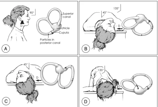
방법은 그림(Fig. 7)과 같이 앉은 자세에서 환측으로 Dix-Hallpike 자세를 취한 후 안진을 관찰한다(B). 다 음 단계로 천천히 머리를 반대쪽으로 90도 돌리며 이 때 고개는 최대한 신전된 상태를 유지한다(C). 이어서 몸전 체와 머리를 90도 더 반대쪽으로 돌리고(D), 이 자세에 서 안진(이차 안진)을 짧은 시간 안에 다시 확인한다. 첫 번째 보였던 안진과 동일한 모양으로 나타나면 이관 내 이
Utricle
Cupula Particles
in posterior canal
A
B C
A
B C
Fig. 6. Semont liberatory maneuver (right ear). A:The patient is sitting with the head turned horizontally 45°to the healthy (left) ear. B:Mo- ving to right side-lying position. C:
Moving to left side-lying position.
석이 공통각(common crus)을 통하여 난형낭 내로 제대 로 들어가고 있는 것이다. 마지막 단계로 환자를 앉도록 한다. 각 단계별로 자세를 1~2분 정도 유지하도록 되어 있으나, 안진의 지속시간과 이석이 움직이는 시간을 고려 할 때 안진이 사라지는 시점까지 자세를 유지시키면 충분 하리라 생각한다(약 30초).
이 방법은 시행하는데 5분 정도면 충분하고, 이석이 제 자리를 잡고 재발하지 않도록 시행 후 24~48시간 정도 눕지 말 것을 권고한다. 최근의 연구에서는 이석치환술을 시행한 후 엄격하게 앉아있는 체위를 일정시간 유지하는 것과 유지하지 않는 것 사이에 치유율에는 차이가 없다는
보고
17)23)도 있으므로 환자의 상태에 따라 유동적으로 적
용하여도 무방할 것으로 보인다.
환자 1회 방문 시 이석치환술 시행 횟수에는 이견이 있 다. 정해진 횟수만큼 시행하는 경우도 있고,
22)한 번만 시행하는 경우도 있다. 수 차례 이석치환술을 반복시키는 것은 피로현상을 초래하여 결과 판정이 어렵기 때문에 1 회만 시행하고 이석치환술 시행 중 이차 안진이 나타나 지 않는 경우와 마지막 단계인 앉은 자리로 돌아올 때 역
안진(reverse nystagmus)이나 어지럼증이 나타나는 환 자에서 선별적으로 재시행하는 방법
3)등이 있다. 치료 성공률은 저자마다 추적관찰기간과 시행 횟수, 안정제와 진동기 사용 등의 차이가 있어 정확하게 평가하기는 어 려우나 약 30~100%으로 보고되고 있다.
8)저자들의 경험에 의하면 후반고리관 BPPV에서 이석치환술을 진 단 당시 1회 시행하고, 1주일 후에 검사를 시행하여 치 료 판정을 시행하였을 때 82.2%의 치료 성공률을 보 였다.
24)수술적인 치료법
BPPV는 양성질환으로 수술적인 치료는 비수술적인 치 료법이 효과가 없거나 자주 재발하는 경우에 제한적으로 사용된다. 그리고 수술을 시행하기 전에 BPPV와 유사한 증상을 보이는 중추신경계의 질환이 없는지를 방사선학적 인 검사를 통하여 확인하여야 한다.
단신경 절제술(Singular neurectomy)
후반고리관으로부터 중추신경으로 자극을 전달하는 유
90° Superior
canal
Utricle Cupula Particles in posterior canal D
A
45°
D
C
135°
45°
D
B
135°
D
D
Fig. 7. Modified Epley maneuver (right ear). A:The patient is seated on a table as viewed from the right side. B:
Patient in normal Dix-Hallpike head-hanging position. The patient’s head is then rotated toward the opposite side with the neck in full extension through position (C) and into position (D) in a steady motion by rolling the patient onto the opposite lateral side and then the patient sits back up to position (A).
일한 후팽대부 신경(posterior ampullary nerve)을 자르 는 기법으로 1970년대 Gacek
25)에 의하여 처음 보고되 었으나 감각신경성난청이 발생할 수 있는 부작용이 있고 기술적으로도 어려워서 좀 더 단순한 후반고리관 폐쇄법 으로 대체되고 있다.
후반고리관 폐쇄법(Posterior semicircular canal occlusion) Parnes 등
26-28)에 의하여 1990년 제안된 방법으로 반 고리관을 눌러 막으면 내림프의 순환이 차단되고 팽대부 릉정이 효과적으로 고정되어 정상적인 각가속력에 의한 내림프의 움직임뿐만 아니라 이관 내의 유리된 이석이나 팽대부의 고정된 이석에 의해서도 팽대부릉정이 움직이지 않게 된다. 또한 예전의 침습적인 내이 수술에 비하여 후 반고리관 폐쇄법은 청력저하를 거의 일으키지 않는 안전 한 방법이다.
수술은 전신마취 하 2~3시간 정도 걸린다. 5~6 cm 정 도의 후이개 절개를 가하고 단순 유양돌기 절제술을 시행 하여 후반고리관에 접근한다. 드릴을 이용하여 골성 후반 고리관에 1×3 mm의 창을 내고 bone dust와 fibrinogen glue로 만든 plug를 밀어 넣어서 후반고리관을 막는다. 수 술이 끝난 후 2~3일이면 환자는 퇴원할 수 있다. 수술 후에 환자는 어지럼과 평형장애를 호소하나 중추성 보상 기전에 의하여 수술 후 수일에서 수주가 지나면서 회복 된다. 이 때 전정운동을 시행하여 회복을 도울 수 있다.
Agrawal 등
29)은 44예의 후반고리관 폐쇄 수술에서 모 두 성공하였고 재발은 1예에서 발생하였으며 다른 한 예 에서 수술 후 3개월에 심도의 돌발성 난청이 생겼다고 보 고하였다. 그 외 다수의 연구에 의해 후반고리관 폐쇄법 은 그 안정성과 효과가 입증되었다.
30-34)중심 단어 :양성돌발성 두위현훈·진단·치료.
REFERENCES
1) Brandt T, Strupp M. General vestibular testing. Clin Neuro- physiol 2005;116:406-26.
2) Froehling DA, Silverstein MD, Mohr DN, Beatty CW, Of- ford KP, Ballard DJ. Benign positional vertigo: incidence and prognosis in a population-based study in Olmsted County, Minnesota. Mayo Clin Proc 1991;66:596-601.
3) Parnes LS, Agrawal SK, Atlas J. Diagnosis and management of benign paroxysmal positional vertigo (BPPV). Cmaj 2003;169:681-93.
4) Dix MR, Hallpike CS. The pathology, symptomatology and diagnosis of certain common disorders of the vestibular sys- tem. Ann Otol Rhinol Laryngol 1952;61:987-1016.
5) Cohen HS. Side-lying as an alternative to the Dix-Hallpike test of the posterior canal. Otol Neurotol 2004;25:130-4.
6) Bronstein AM. Vestibular reflexes and positional manoeuvres.
J Neurol Neurosurg Psychiatry 2003;74:289-93.
7) Straumann D, Suzuki M, Henn V, Hess BJ, Haslwanter T.
Visual suppression of torsional vestibular nystagmus in rhe- sus monkeys. Vision Res 1992;32:1067-74.
8) Haynes DS, Resser JR, Labadie RF, Girasole CR, Kovach BT, Scheker LE, et al. Treatment of benign positional vertigo using the semont maneuver: efficacy in patients presenting without nystagmus. Laryngoscope 2002;112:796-801.
9) Tirelli G, D’Orlando E, Giacomarra V, Russolo M. Benign positional vertigo without detectable nystagmus. Laryngo- scope 2001;111:1053-6.
10) Weider DJ, Ryder CJ, Stram JR. Benign paroxysmal posi- tional vertigo: analysis of 44 cases treated by the canalith repositioning procedure of Epley. Am J Otol 1994;15:321-6.
11) McClure JA, Willett JM. Lorazepam and diazepam in the treatment of benign paroxysmal vertigo. J Otolaryngol 1980;
9:472-7.
12) Brandt T, Daroff RB. Physical therapy for benign paroxysmal positional vertigo. Arch Otolaryngol 1980;106:484-5.
13) Semont A, Freyss G, Vitte E. Curing the BPPV with a libera- tory maneuver. Adv Otorhinolaryngol 1988;42:290-3.
14) Epley JM. The canalith repositioning procedure: for treat- ment of benign paroxysmal positional vertigo. Otolaryngol Head Neck Surg 1992;107:399-404.
15) Banfield GK, Wood C, Knight J. Does vestibular habitua- tion still have a place in the treatment of benign paroxysmal positional vertigo? J Laryngol Otol 2000;114:501-5.
16) Norre ME, Beckers A. Comparative study of two types of exercise treatment for paroxysmal positioning vertigo. Adv Otorhinolaryngol 1988;42:287-9.
17) Nuti D, Nati C, Passali D. Treatment of benign paroxysmal positional vertigo: no need for postmaneuver restrictions.
Otolaryngol Head Neck Surg 2000;122:440-4.
18) Herdman SJ, Tusa RJ, Zee DS, Proctor LR, Mattox DE.
Single treatment approaches to benign paroxysmal positio- nal vertigo. Arch Otolaryngol Head Neck Surg 1993;119:
450-4.
19) Cohen HS, Jerabek J. Efficacy of treatments for posterior canal benign paroxysmal positional vertigo. Laryngoscope 1999;109:584-90.
20) Parnes LS, Price-Jones RG. Particle repositioning maneu- ver for benign paroxysmal positional vertigo. Ann Otol Rhi- nol Laryngol 1993;102:325-31.
21) Parnes LS, Robichaud J. Further observations during the particle repositioning maneuver for benign paroxysmal po- sitional vertigo. Otolaryngol Head Neck Surg 1997;116:
238-43.
22) Hain TC, Helminski JO, Reis IL, Uddin MK. Vibration does not improve results of the canalith repositioning procedure.
Arch Otolaryngol Head Neck Surg 2000;126:617-22.
23) Marciano E, Marcelli V. Postural restrictions in labyrintho- lithiasis. Eur Arch Otorhinolaryngol 2002;259:262-5.
24) Chung YJ, Choe JY, Chung WH, Hong SH. Outcome of canalith repositioning maneuver in benign paroxysmal ver- tigo. J Korean Balance Society 2002;1:118-23.
25) Gacek RR. Further observations on posterior ampullary nerve transection for positional vertigo. Ann Otol Rhinol Laryngol 1978;87:300-5.
26) Parnes LS, McClure JA. Posterior semicircular canal occlu- sion for intractable benign paroxysmal positional vertigo.
Ann Otol Rhinol Laryngol 1990;99:330-4.
27) Parnes LS, McClure JA. Posterior semicircular canal oc- clusion in the normal hearing ear. Otolaryngol Head Neck Surg 1991;104:52-7.
28) Parnes LS. Update on posterior canal occlusion for benign paroxysmal positional vertigo. Otolaryngol Clin North Am 1996;29:333-42.
29) Agrawal SK, Parnes LS. Human experience with canal plug- ging. Ann N Y Acad Sci 2001;942:300-5.
30) Walsh RM, Bath AP, Cullen JR, Rutka JA. Long-term results
of posterior semicircular canal occlusion for intractable benign paroxysmal positional vertigo. Clin Otolaryngol Al- lied Sci 1999;24:316-23.
31) Pace-Balzan A, Rutka JA. Non-ampullary plugging of the posterior semicircular canal for benign paroxysmal positio- nal vertigo. J Laryngol Otol 1991;105:901-6.
32) Hawthorne M, el-Naggar M. Fenestration and occlusion of posterior semicircular canal for patients with intractable be- nign paroxysmal positional vertigo. J Laryngol Otol 1994;
108:935-9.
33) Dingle AF, Hawthorne MR, Kumar BU. Fenestration and occlusion of the posterior semicircular canal for benign positional vertigo. Clin Otolaryngol Allied Sci 1992;17:
300-2.
34) Anthony PF. Partitioning the labyrinth for benign paroxy- smal positional vertigo: clinical and histologic findings. Am J Otol 1993;14:334-42.