레진 계열 근관봉함재 Adseal의 세포독성에 관한 연구
김희정∙백승호∙이우철∙박한수∙배광식*
서울대학교 치과대학 치과보존학교실
CYTOTOXICITY OF RESIN-BASED ROOT CANAL SEALER, ADSEAL
Hee-Jung Kim, Seung-Ho Baek, Woo-Cheol Lee, Han-Soo Park, Kwang-Shik Bae*
Department of Conservative Dentistry, College of Dentistry, Seoul National University
The properties of ideal root canal sealers include the ability of sealing the total root canal system and no toxic effects to periradicular tissues. Cytotoxicity test using cell culture is a common screening method for evaluation of the biocompatibility of root canal sealers. The purpose of this study was to investigate the cytotoxic effect of newly developed resin-based sealer (Adseal 1, 2, and 3) comparing with those commercial resin-based sealers (AH26 and AH Plus), ZOE-based sealers (Tubliseal EWT, Pulp Canal Sealer EWT) and calcium hydroxide based sealer (Sealapex). An indirect contact test of cytotoxicity by agar diffusion was performed according to the international standard ISO 10993-5. L929 fibroblast cells were incubated at 37℃ in humidified 5% CO2-containing air atmosphere. The freshly mixed test materials were inserted into glass rings of internal diameter 5 ㎜ and height 5 ㎜ placed on the agar. After the 24 hrs incubation period, the decolorization zones around the test materials were assessed using an inverted microscope with a calibrated screen. A Decolorization Index was determined for each specimen. Adseal 1, 2, and 3 did not exert any cytotoxic effects, whereas AH26, AH Plus, Tubliseal EWT, Pulp Canal Sealer EWT, and Sealapex produced mild cytotoxicity. [J Kor Acad Cons Dent 29(6):498-503, 2004]
Key words : Cytotoxicity, Agar diffusion test, Root canal sealer, Decolorization index, Adseal
Ⅰ. 서 론
근관 치료의 마지막 단계는 전체 근관계와 그와 관련된 복 잡한 해부학적 구조를 자극이 없는 무균의 충전물질로 충전 하는 것이다. 근관충전을 위한 다양한 방법이 제안되고 있 는데 가장 흔한 방법이 반고형 물질인 거터퍼쳐와 근관 봉
함재를 함께 혼합하여 사용하는 것이다. 이상적인 근관충전 물질은 상아질과의 접착, 근관계의 밀폐, 부피안정성이 있 어야 하며, 수분에 영향을 받지 말아야 하고, 독성이 없어야 한다1). 근관계를 침범하는 미생물들은 숙주조직과 작용하여 치수-치근 주위 병변을 일으킬 수 있다. 따라서 감염의 조절 이 치료의 핵심이 되는데, 오랜 기간의 성공을 기대하려면, 형성된 근관에 미생물이 침범하지 못하도록 근관 충전 물질 이 살균성을 가지고 있어야 한다. 하지만 이 충전 물질들이 근관 주위 조직과 오랫동안 직접 접촉하게 되므로 동시에 생체 적합성도 중요시 되어야 하며, 결국 근관치료의 결과 에도 영향을 준다2). Wayman 등3)은 근관치료의 성공이 근 관형성 뿐만 아니라 적절한 근관봉함재의 선택에도 있다고 기술하였다.
ABSTRACT
* Corresponding author: Kwang-Shik Bae Department of Conservative Dentistry, College of Dentistry, Seoul National University 28 Yoengun-dong, Chongro-gu, Seoul, Korea, 110-749 Tel : 82-2-2072-2650 Fax : 82-2-2072-3859 E-mail : [email protected]
※ 본 연구는 보건복지부 보건의료기술연구개발사업의 지원에 의하여 이루어진 것임. (01-PJ5-PG1-01CH12-0002)
레진계 근관봉함재는 다른 근관봉함재에 비해 근관 내 구 조에 잘 접착하고, 작업시간이 길며, 좋은 폐쇄효과를 가지 고, 또한 경화시 수축이 없어 최근 근관치료에 많이 도입되 고 있다. 가장 많이 사용되고 있는 에폭시 레진계 봉함재로 AH26(Dentsply DeTrey GmbH, Konstanz, Germany) 이 있으며 미국에서는 1957년 처음 소개되었다4). 그러나 AH26은 경화시 formaldehyde가 유리되어 강한 세포독성 을 나타낸다5). 이 후 AH26에 비해 세포독성 지속시간이 짧 아지고 물리적 성질이 개선된 AH Plus (Dentsply DeTrey GmbH, Konstanz, Germany)가 개발되었다. 그 러나 AH Plus의 경우 AH26에 비해서는 세포독성이 줄어 들었으나 몇몇 연구에서는 AH Plus 또한 강한 세포독성을 나타낸다고 보고하였다6).
생체적합성을 결정하는 단계 중 첫번째는 세포독성검사를 통해 주로 이루어 진다6). 일반적으로 세포독성검사는 세포 배양을 통해 실험실에서 시행되며, 동물실험과 비교하여 생 체 내 실험조건을 통제하기 쉽고, 실험실내 방법이 단순하 며 비용이 저렴하다는 장점이 있다. 그리하여 생체재료의 기본적인 생물학적 성상을 결정하는데 적합하다7).
본 연구의 목적은 최근 개발된 레진계 근관봉함재인 Adseal과 또 다른 레진계 근관봉함재인 AH26과 AH Plus 그리고 Zinc oxide eugenol 계의 Tubliseal EWT과 Pulp Canal Sealer EWT (Kerr U.S.A., Romulus, MI, USA), 수산화칼슘계의 Sealapex (Kerr U.S.A., Romulus, MI, USA)의 독성을 L-929 세포를 이용한 agar-diffusion test로 비교 평가하기 위한 것이다.
Ⅱ. 실험재료 및 방법 1. 실험재료
새로 개발된 레진계 근관봉함재 3종류 (Adseal 1, 2, 3:
Meta Biomed, Chungju, Korea)와 또 다른 레진계 봉함재 인 AH26과 AH Plus, ZOE계 봉함재인 Tubliseal EWT와 Pulp Canal Sealer EWT, 수산화칼슘계 봉함재인 Sealapex 가 본 실험에 사용되었다. 각각의 봉함재당 10개의 sample 을 사용하였다. Adseal의 성분은 Table 1에 나열하였다.
2. 세포 배양
Fibroblast (NCTC clone 929)을 10% fetal bovine serum (FBS)(GIBCO, Grand Island, NY, USA)와 antibiotics가 함유된 minimum essential medium (MEM)(GIBCO, Grand Island, NY, USA)으로 5%
CO2, 97% 의 습도의 조건을 갖춘 37℃의 배양기에서 배양 하였다.
3. 세포독성 실험
한천평판을 이용한 세포독성실험은 International Organization for Standardization에서 기술한 실험방법 을 기초로 하였다9). 1 ㎖ 당 2.5 × 10⁵ 개의 세포농도의 세포 배양액 10 ㎖을 지름 약 10 ㎝ 의 배양접시에 넣은 다 음 단층 배양되도록 5% CO2, 97%의 습도의 조건을 갖춘 37℃의 배양기에서 배양하였다. 배양 후 배양접시의 액체 성분을 흡입제거 후 Eagle 배양액에 한천을 1.5% 넣은 것 을 약 10 ㎖씩 배양접시에 중층한다. 이 한천평판 위에 0.02% 뉴트랄레드 인산완충 생리식염수액 10 ㎖를 넣은 다음 37℃에서 2-3 시간 배양하였다. 배양한 다음 과량의 염색액을 제거하고 다시 5% CO2, 97%의 습도의 조건을 갖춘 37℃의 배양기에서 20분간 배양하였다. 배양 후 세포 가 충분히 색소를 흡수한 평판에 지름 5 ㎜, 높이 5 ㎜의 유 리관을 한천평판 위에 놓고 준비한 sealer를 제조회사의 지 시대로 혼합한 직후 유리관내를 채웠다. Sample이 놓여진 평판을 5% CO2, 97%의 습도의 조건을 갖춘 37℃의 배양 기에서 다시 24시간 동안 배양한 후 결과를 관찰하였다.
4. 결과분석
배양세포의 반응은 배양접시를 백색의 배경에서 관찰할 때 적색으로 염색된 세포가 검체의 밑부분 및 주변에서 어 느 정도 염색성이 저하되어 있는가에 의하여 판정하였다.
염색성이 저하된 세포의 범위를 측정하고 Table 2의 기준 에 따라 지대지수를 기록하였다9). 측정된 결과를 Kruskal- Wallis test를 사용하여 분석을 시행하였다.
Ⅲ. 실험결과
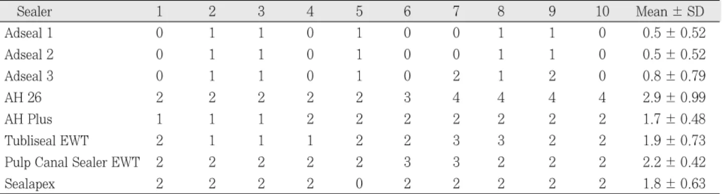
실험결과는 Table 3에 정리되어 있다. Table 3은 측정결 과에 따른 지대지수를 표시한 것이다.
Adseal 1, 2, 3은 나머지 다른 sealer에 비해 통계학적으 로 유의할 만한 낮은 세포독성을 보였으며 (p < 0.05), Adseal 1, 2, 3 상호간에는 통계학적으로 유의성 있는 차 이를 보이지 않았다. 레진계 근관봉함재의 경우, AH 26이 역시 가장 높은 결과를 나타내었다. AH Plus의 경우 AH26보다는 낮은 독성을 나타내었으나 Adseal과 비교시 높은 세포독성을 나타내었다. ZOE계 근관봉함재인 Tubliseal EWT와 Pulp Canal Sealer EWT, 수산화칼슘 계 근관봉함재인 Sealapex 역시 Adseal에 비해 높은 세포 독성을 보였다.
Figure 1은 각 근관봉함재의 decolorization zone을 현 미경을 통해 관찰한 것이다. Adseal 1, 2, 3이 다른 근관봉 함재에 비해 훨씬 정상적인 세포의 모습을 하고 있었다.
Table 1.Compositions of Adseal 1, 2, and 3
Name Type Compositions
Epoxy Oligomer
Ethylene glycol mono salicylate
Paste A Calcium Phosphate
Zirconium oxide
Adseal 1* Bismuth subcarbonate
4-aminobenzoate Calcium Phosphate
Paste B Zirconium oxide
Bismuth subcarbonate Calcium Oxide Epoxy Oligomer
Ethylene glycol mono salicylate
Paste A Calcium Phosphate
Zirconium oxide Bismuth subcarbonate
Adseal 2 4-aminobenzoate
Triethanol amine
Paste B Calcium Phosphate
Zirconium oxide Bismuth subcarbonate Calcium Oxide Epoxy Oligomer
Ethylene glycol mono salicylate
Paste A Calcium Phosphate
Zirconium oxide
Adseal 3* Bismuth subcarbonate
4-aminobenzoate
Paste B Calcium Phosphate
Zirconium oxide Bismuth subcarbonate
* Adseal 1 and Adseal 3 have same ingredients but they are different in the ratio of ingredients.
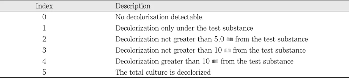
Table 2.Criteria for scoring of decolorization index
Index Description
0 No decolorization detectable
1 Decolorization only under the test substance
2 Decolorization not greater than 5.0 ㎜from the test substance 3 Decolorization not greater than 10 ㎜from the test substance 4 Decolorization greater than 10 ㎜from the test substance 5 The total culture is decolorized
Figure 1. Decolorization zone and cell appearance by agar diffusion test (inverted microscope, left: × 200, right: × 400).
Each sealer has two pictures. The left sides show the decolorization zone except Adseal 1, 2, and 3. Adseal 1, 2, and 3 show normal cell morphology and cell density.
Table 3.Decolorization index of experimental sealers (agar diffusion test)
Sealer 1 2 3 4 5 6 7 8 9 10 Mean ± SD
Adseal 1 0 1 1 0 1 0 0 1 1 0 0.5 ± 0.52
Adseal 2 0 1 1 0 1 0 0 1 1 0 0.5 ± 0.52
Adseal 3 0 1 1 0 1 0 2 1 2 0 0.8 ± 0.79
AH 26 2 2 2 2 2 3 4 4 4 4 2.9 ± 0.99
AH Plus 1 1 1 2 2 2 2 2 2 2 1.7 ± 0.48
Tubliseal EWT 2 1 1 1 2 2 3 3 2 2 1.9 ± 0.73
Pulp Canal Sealer EWT 2 2 2 2 2 3 3 2 2 2 2.2 ± 0.42
Sealapex 2 2 2 2 0 2 2 2 2 2 1.8 ± 0.63
A: Adseal 1 B: Adseal 2 C: Adseal 3
D: AH 26 E: AH Plus F: Pulp Canal Sealer EWT
G: Tubliseal EWT H: Sealapex I : Normal cell
A B C
D E F
G H I
Adseal을 제외한 나머지 실험군의 많은 sample에서 비교 적 넓은 decolorization zone이 관찰되었다.
Ⅳ. 총괄 및 고안
근관봉함재의 생체적합성을 평가하는 방법으로 실험실내 세포독성검사를 초기 검사방법으로 적용하여, 동물을 이용 한 생체 내 실험의 필요성을 감소시킬 수 있는 장점이 있다.
실험실내 세포독성검사 중 한천평판 검사는 실험재료가 세 포와 직접 접촉하지 않고 한천층을 통해 배양세포로 독성물 질이 확산되어 영향을 미치게 하는 시험조건으로 근관충전 물질이 용해되어 근관에서 치근단 주위조직으로 확산되는 상황과 유사하여 근관봉함재의 독성 평가에 적합하다고 할 수 있다10). 또한 ISO 7405에 의한 가이드에 따르면 두 종 류 이상의 세포독성검사를 권유하고 있는데 이 중 하나가 한천평판검사이다.
본 연구에서 산화아연유지놀계의 근관봉함재인 Tubliseal EWT와 Pulp Canal Sealer EWT는 높은 세포독성을 나 타내었는데, 이미 많은 연구로 알려진 바와 같이 산화아연 과 유지놀이 혼합시 유리되는 유지놀 때문에 독성을 나타낸 다고 볼 수 있다11,12). 또한 다른 연구가들은 ZOE계 봉함재 가 독성을 나타내는 이유가 아연이온과 관련될 수 있다고 주장하였다13,14).
독성이 미약하다고 알려진 수산화칼슘계 근관봉함재인 Sealapex 역시 Tubliseal EWT와 AH Plus와 유사한 세 포독성을 나타내었는데 Gordon 등15)은 이 봉함재의 세포독 성은 높은 알칼리성 때문에 기인한다고 보고하였다. 또한 Huang 등과 Leonardo 등은 Sealapex 가 경화 후 붕괴된 봉함재에서 적지않은 독성물질이 빠져나와 독성이 증가됨 을 보고하였다16,17).
AH26의 독성은 다른 연구에서와 유사한 결과로 본 연구 에서도 심한 독성을 나타내었다18,19). Leyhausen 등20)은 AH Plus에서는 AH26과는 달리 유전독성이 없다고 발표 하였고, Koulazodou 등21)은 AH26과 비교하여 더 낮은 독 성을 AH Plus가 나타내었다고 보고하였다. 반면 Cohen 등6)은 AH Plus도 AH26 과 마찬가지로 심한 독성을 가지 고 있으며, Huang TH 등22)또한 AH26 과 AH Plus 모두 쥐의 간세포에 적용 시 독성을 나타내었음을 보고하였다.
본 실험에서도 AH26에 비해 AH Plus가 독성이 낮게 나타 났으나, Adseal군과 비교시 비교적 높은 세포독성을 나타 내었다. AH Plus의 경우, 부산물로 formaldehyde가 유리 되는 양이 적음에도 불구하고 세포독성이 높게 나타나는 이 유는 주성분의 하나인 Epoxy resin이 독성인자로 확인되었 기 때문이다. 이것으로 보아 AH Plus가 AH 26에 비해 임 상적으로 더 나은 봉함재로 결론하기는 어렵다고 사료 된다.
Adseal 1, 2와 3은 AH 26과 AH Plus와 같이 resin계 봉함재이나 본 연구에서 가장 낮은 세포독성을 나타내었으 며, Adseal 1, 2와 3 사이에는 통계학적으로 유의할 만한 차이는 보이지 않았다(p>0.05). 본 교실에서 김 등23)은 Adseal을 쥐의 피하조직에 매식하여 염증반응을 관찰한 결 과 초기 1, 2주에 가장 약한 염증반응을 보였음을 보고하였 고, 박 등24)또한 세포의 viable ratio를 계산하고, Giemsa stain으로 염색하여 세포의 양상을 관찰한 결과 기존의 상 품화된 수종의 봉함재보다 Adseal이 가장 낮은 세포독성을 보였다. 이것으로 보아 Adseal이 기존의 resin계 봉함재보 다 생체친화성이 개선되었으며, 이는 calcium phosphate 를 첨가함으로써 얻을 수 있었다고 사료된다. Calcium phosphate는 높은 골 유도 효과와 생체적합성이 뛰어난 물 질이므로 치근단 조직에 접촉되어도 유해성이 적다25,26).
본 연구로 최근 국내에서 개발된 Adseal이 다른 resin계 봉함재보다도 생체적합성이 우수함을 확인하였다. 그러나 이상적인 근관봉함재가 되기 위해서는 생체적합성뿐만 아 니라 다른 많은 조건에도 충족되어야 하므로 지속적인 연구 와 개발이 이루어져야 할 것으로 사료된다.
Ⅴ. 결 론
본 연구에서 새로 개발된 레진계 근관충전용 봉함재인 Adseal과 상품화된 수종의 봉함재의 독성을 한천평판을 이 용하여 세포독성검사를 시행한 결과 다음과 같은 결론을 얻 었다.
1. Adseal 1, 2와 3은 실험에 사용된 다른 근관봉함재보다 낮은 세포독성을 나타내었다 (p < 0.05). Adseal 1, 2 와 3 사이에서는 통계학적으로 유의할 만한 차이가 없 었다.
2. 레진계 근관봉함재인 AH26의 독성이 가장 높게 나타났 으며, AH Plus의 경우 AH26보다는 낮은 독성을 나타 내었다. 그러나 통계학적으로 유의한 차이가 없었다 (p
> 0.05).
3. ZOE계 근관봉함재인 Tubliseal EWT와 Pulp Canal Sealer EWT, 수산화칼슘계 근관봉함재인 Sealapex 역 시 Adseal에 비해 높은 세포독성을 보였으나 통계학적 으로 유의한 차이가 없었다 (p > 0.05).
참고문헌
1. Grossman LI, Olivet S, Del-Rio CE. Endodontic Practice. 11thed. Philadelphia : Lea & Fabiger. 1998.
2. Geurtsen W, Leyhausen G . Biological aspects of root canal filling materials - histocompatibility, cytotoxicity, and mutgenicity. Clin Oral Invest 1:5-11, 1997.
3. Wayman BE, Murata SM, Almeida RJ, Fowler CB. A bacterial and histological evaluation of 58 periapical
lesions. J Endod 18:152-158, 1992.
4. Cohen BI, Pagnillo MK, Musikant BL, Deutsch AS.
Evaluation of the release of formaldehyde for three endodontic filing materials. Oral Health 88:37-39, 1998.
5. Spangberg L, Barbosa SV and Lavigne GD. AH26 releases formaldehyde. J Endod 19:596-598, 1993.
6. Cohen BI, Pagnillo MK, Musikant BL, and Deutsch AS. An in vitro study of the cytotoxicity of two root canal sealers.J Endod 26;228-229, 2000.
7. Schmatz G .Concepts in biocompatibility testing of dental restorative materials. Clin Oral Invest 1;154- 162, 1997.
8. Beltes P, Koulaouzidou E, Kotoula V, and Kortsaris AH. In vitro evaluation of the cytotoxicity of calcium hydroxide-based root canal sealers. Endod Dent Traumatol 11:245-249, 1995.
9. The Internatnal Organization for Standardization.
Biological evaluation of medical devices - Part 5. Tests for cytotoxicity: in vitro method. ISO 10993-5.
10. Mohammad AR, Mincer HH, Younis O, Dillingham E, Siskin M. Cytotoxicity evaluation of root canal sealers by the tissue culture--agar overlay technique. Oral Surg Oral Med Oral Pathol 45:768-773, 1978.
11. Briseno BM, Willershausen B. Root canal sealer cytotoxicity on human gingival fibroblasts. 1. Zinc oxide-eugenol-based sealers. J Endod 16:383-386, 1990.
12. Lindqvist L, Otteskog P. Eugenol: liberation from den- tal materials and effect on human diploid fibroblast cells. Scand J Dent Res 88:552-556, 1980.
13. Meryon SD, Johnson SG, Smith AJ. Eugenol release and the cytotoxicity of different zinc oxide-eugenol combination.J Dent 16: 66-70, 1988.
14. Meryon SD, Jakeman KJ. The effects in vitro of zinc released from dental restorative materials.Int Endod J 18:191-198, 1985.
15. Gordon TM, Ranly DM, Boyan BD. The effects of calci- um hydroxide on bovine pulp tissue: variations in pH
and calcium concentration. J Endod 11:156-160, 1985.
16. Huang FM, Tai KW, Chou MY, Chang YC. Cytotoxicity of resin-, zinc oxide-eugenol-, and calcium hydroxide- based root canal sealers on human periodontal liga- ment cells and permanent V79 cells. Int Endod J 35:153-158, 2002.
17. Leonardo RT, Consolaro A, Carlos IZ, Leonardo MR.
Evaluation of cell culture cytotoxicity of five root canal sealers.J Endod 26:328-330, 2000.
18. Briseno BM, Willershausen B. Root canal sealer cytotoxicity on human gingival fibroblasts. Part II.
Silicone- and resin-based sealers. J Endod 17:537- 540, 1991.
19. Cohen BI, Pagnillo MK, Musikant BL, Deutsch AS.
The evaluation of apical leakage for three endodontic fill systems. Gen Dent 46:618-623, 1998.
20. Leyhausen G, Heil J, Reifferscheid G, Waldmann P, Geurtsen W. Genotoxicity and cytotoxicity of the epoxy resin-based root canal sealer AH Plus. J Endod 25:
109-113, 1999.
21. Koulaouzidou EA, Papazisis KT, Beltes P, Geromichalos GD, Kortsaris AH. Cytotoxicity of three resin-based root canal sealers: an in vitro evaluation.
Endod Dent Traumatol 12:182-185, 1998.
22. Huang TH, Lii CK, Chou MY, Kao CT. Lactate dehy- drogenase leakage of hepatocytes with AH26 and AH Plus sealer treatments.J Endod 26:509-511, 2000.
23. 김용범, 백승호, 배광식. 신개발 레진 계열 봉함재의 생체친화 성에 관한 연구. 대한치과보존학회지 27:122-134, 2002.
24. 박소영, 이우철, 임성삼. 새로운 레진 계통의 근관봉함재의 독
성과 항균작용에 관한 연구. 대한치과보존학회지 28:162-
168, 2003.
25. Chohayeb AA, Chow LC, Tsaknis PJ. Evaluation of calcium phosphate as a root canal sealer-filler materi- al. J Endod 13:384-387,1987.
26. Sugawara A, Nishiyama M, Kusama K, Moro I, Nishimura S, Kudo I, Chow LC, Takagi S. Hist- opathological reactions of calcium phosphate cement.
Dent Mater J 11:11-16, 1992.