Ǖ⦝ӹྕ ᯯ ⇵⇽ྜྷ᮹ ᭥ᦵᖙ⡍ᨱ ݡ⦽ ᖙ⡍ᔍ໙ ᮁࠥ ⬉ŝ
ᯕ⩶ᖁ*
ᵲᬱݡ⦺Ʊ ᯥᔢᄲญ⦺ŝ
Received: February 25, 2021 / Revised: March 30, 2021 / Accepted: April 1, 2021
ᕽ ು
⩥ݡ ᮹⦺᮹ ၽݍŝ ᔾ⪽⪹Ğ᮹ }ᖁᮝಽ ᱥၹᱢᯙ ᔗ᮹ ḩᮡ ׳ᦥᲭḡอ
,ᩍᱥ⯩ ᦵᮡ ᱥ ᖙĥᱢᮝಽ ᯙe᮹ ᔾᮥ
᭥⩲⦹۵ ḩ⪹ᯕ݅
[1].ᱥ ᖙĥᱢᨱᕽ ⠱ᦵ ݅ᮭᮝಽ ฯᯕ ၽ
ᔾ⦹໑ ✚⯩ ᬑญӹᨱᕽࠥ ᱥℕ ᦵ ᵲᨱᕽ ⧎ᔢ ᔢ᭥ෝ ₉ ḡ⦹Ł ᯩ۵ ᦵᯕ ᭥ᦵᯕ݅
.⩥ᰍ ᭥ᦵᮥ ⊹ഭ⦹ʑ ᭥⦽ ႊ ჶᮝಽ۵ ᙹᚁᮥ ☖⦽ ᱩᱽ
,᧞ྜྷᮥ ᯕᬊ⦽ ⪵⦺ჶ
,ႊᔍ ᖁ ჶ ॒ᮥ ݅᧲⦹í ᄲ⧪⦹Ł ᯩ݅
.⦹ḡอ ᭥ᦵᮡ ᳑ʑ ၽčᯕ ᨕಖŁ ᙹᚁ ⬥ᨱࠥ ᰍၽ᮹ a܆ᖒᯕ ׳ᮝ໑
,⧎ᦵᱽ ᨱ ݡ⦽ ᇡ᯲ᬊᮝಽ ᪥⊹a ᨕಅᬕ ᦵᵲ᮹ ⦹ӹᯕ݅
[2].ᯕ
్⦽ ᨕಅᬡᮥ ɚᅖ⦹ʑ ᭥⦽ ᯝ⪹ᮝಽ ᇡ᯲ᬊᯕ ᱢᮝ໑ ᱶ ᔢᖙ⡍ᨱ۵ ᩢ⨆ᮥ ၙ⊹ḡ ᦫ۵ ⧎ᦵᱽ᮹ }ၽᯕ ᱩᝅ⦹໑
,ᦵ᮹ ၽᔾᮥ ᩩႊ⧁ ᙹ ᯩ۵ ᩑྜྷ ᮁ௹ ᮹᧞⣩᮹ }ၽᨱ ݡ⦽ ᩑǍa ⪽ၽ⦹í ᯕᨕḡŁ ᯩ݅
[3].ᖙ⡍ᔍ໙ᮡ ݅᧲
⦽ ✚ᖒᨱ Ⓧí
apoptosis᪡
necrosisಽ ӹڹᨕ ḥ݅
.v
⦽ ࠦᖒ ੱ۵ ྜྷญᱢ ᔢ⧕ᨱ ᮹⧕ ӹ┡ӹ۵
necrosis᪡۵ ݍ
ญ
apoptosis۵ ܆࠺ᱢᯙ ᖙ⡍ᔍ໙ ŝᱶ᮹ ⦹ӹಽ ᖙ⡍ ԕᇡ
᮹ ᝁ⪙ᱥݍᯕ ᱶƱ⦹í ᯕᨕḡ໕ᕽ ᔍ໙⦽݅
[4, 5].Apoptosis
۵ ᖙ⡍᮹ እᵲ qᗭ
,ᖙ⡍ส ❭ƕ
,ᖙ⡍ḩ ၰ ᩝ
ᔪḩ ᮲⇶
, DNA݉⠙⪵ ॒᮹ ⩥ᔢᯕ ᯝᨕӹ໑ đŝᱢᮝಽ
ᖙ⡍ෝ ᯱᔕಽ ᮁࠥ⦽݅
.⧎ᦵ⬉ŝෝ aḡ۵ ᧞ྜྷॅᯕ ᦵᖙ
⡍ᨱ ݡ⦹ᩍ
apoptosisᮁࠥᨱ ษӹ ⬉ŝᱢᯙḡෝ ⪶ᯙ⦹
۵ äᯕ ᦵ ⊹ഭ᮹ ᔩಽᬕ ᧞ྜྷ}ၽ᮹ đŝྜྷಽ v᳑ࡹŁ ᯩ
݅
[6]. Apoptosis۵ šಉࡽ ԕᯙᖒ Ğಽᯙ ၙ☁⎹ऽญᦥ ᮹
᳕ᖒ Ğಽ᪡ šಉ⦹ᩍ
Bcl-2 family᮹ ᔢ⪙ၽ⩥ŝ ᖙ⡍ᵝʑ
᳑ᱩᨱ ݡ⦽ ʑ܆ᱢ ᳑ᱩᮥ ⪶ᯙ⦹۵ äᯕ ๅᬑ ᵲ⦹݅
[7].Ǖ⦝ӹྕ
(Platycarya strobilacea)۵ a௹ӹྕŝᨱ ᗮ⦹۵ ᙹ
༊ᮝಽ
,ᩩಽᇡ░ ᩕๅ᪡ ᐭญ۵ ᧞ᬊᮝಽ ᯕᬊࡹᨕ ᪵ᮝ ໑
,ᐭญ᪡ ᙹ⦝۵ ᗭᩝ᯲ᬊᯕ ᯩ۵ äᮝಽ ಅᲙ ᪵݅
.Ǖ
⦝ӹྕ۵ ⧎ᖙɁ
,⧎ḥɁ
,⧎ᩝ᷾
,⧎ᔑ⪵ ၰ ⧎י⪵ ⪽ᖒ॒
ᯕ ᯩ۵ äᮝಽ ᩑǍa ᅕŁࡽ ၵ ᯩ݅
[8−
11].Ǖ⦝ӹྕ ᙹ
⦝ᨱᕽ ⇵⇽⦽ ᖒᇥᮝಽ۵
quercetin, myricetin᮹ ⥭ᅕי ᯕऽ᪡
afzelin, quercitrin, myricitrin᮹ ⥭ಽᅕיᯕऽ ɡญ
ऽෝ ⡍⧉⦽ ⥭ᅕיᯕऽඹ ၰ ə ႑ݚℕॅᯕ ⧉ᮁࡹᨕ
ᯩ݅Ł ᩑǍࡹᨩ݅
[12, 13].✚⯩
, quercetin, quercitrinᮡ ݅
Induction of Apoptosis in AGS Human Gastric Cancer Cells by Platycarya strobilacea Leaf ExtractHyeong-Seon Lee*
Department of Biomedical Laboratory Science, Jungwon University, Goesan 28024, Republic of Korea
This study investigated the anticancer activity of methanol extract from Platycarya strobilacea leaf in AGS human gastric cancer cells. We determined the cell viability effect of P. strobilacea using MTS assay. Apop- tosis induction and cell cycle arrest were confirmed by fluorescein isothiocyanate and propidium iodide staining using cellometer K2. The mRNA expression levels of the Bcl-2 family were confirmed by reverse transcription-polymerase chain reaction. The cell viability was decreased in a dose-dependent manner treated with different concentrations of P. strobilacea. Total, early, and late apoptotic cells were dramati- cally increased, and the cell cycle was arrested at the sub-G1 phase. The mRNA expressions of Bcl-2 and Bcl-xL were reduced, whereas pro-apoptotic factors, Bax and Bak, were increased in a dose-dependent manner. These results suggested that P. strobilacea leaf extract induced significant apoptotic activity through an intrinsic mitochondria pathway.
Keywords: Platycrya strobilacea, AGS human gastric cancer cells, Bcl-2 family, apoptosis
*Corresponding author
Tel.: +82-43-830-8861, Fax: +82-43-830-8679 E-mail: [email protected]
᧲⦽ ᦵᖙ⡍ᨱᕽ ᖙ⡍ᔍ໙ᮥ ᮁࠥ⦽ ⧎ᦵ ⬉ŝa ಅᲙ ᯩ
݅
[14, 15].ᯕ᪡ zᯕ Ǖ⦝ӹྕ ⇵⇽ྜྷᨱ ݡ⦽ ⪽ၽ⦽ ᩑǍ
ᨱࠥ ᇩǍ⦹Ł ᦵᖙ⡍ᨱ ݡ⦽ ⧎ᦵ ⪽ᖒᯕ ᯩ۵ḡᨱ ݡ⦽ ᩑ Ǎ۵ ᱥྕ⦽ ᝅᱶᯕ݅
.ᕽ ᅙ ᩑǍᨱᕽ۵ Ǖ⦝ӹྕ ᯯ
⇵⇽ྜྷᮥ ᯕᬊ⦹ᩍ ᦵᖙ⡍᮹ ᖙ⡍ᔍ໙ᨱ ݡ⦽ ⪽ᖒᯕ ᯩ۵ ḡෝ ⪶ᯙ⦹Ł
,ᖙ⡍ᔍ໙ŝ šಉࡽ
apoptosis⪽ᖒᨱ šᩍ⦹
۵ḡෝ ᖙ⡍᮹ ԕᯙᖒ Ğಽෝ ☖⧕ ᦥᅕŁ ⧎ᦵᱽಽᕽ a
܆ᖒᯕ ᯩ۵ḡෝ ┡ḥ⦹Łᯱ ⦽݅
.ᰍഭ ၰ ᝅ⨹
ᖙ⡍႑᧲
ᯙe ᭥ᦵ ᖙ⡍ᵝᯙ
AGSᖙ⡍۵ ⦽ǎ ᖙ⡍ᵝ ᮡ⧪
(KCLB, Korea)
ᨱᕽ Ǎ᯦⦹ᩍ ᔍᬊ⦹ᩡ݅
. AGSᖙ⡍۵
10%fetal bovin serum (FBS, Korea)
᪡
1%⧎ᔾᱽ
(penicillin/streptomycin)
ᯕ ℉aࡽ
RPMI 1640႑ḡෝ ᔍᬊ⦹ᩍ
37Ⳅ
, 5% CO2႑᧲ʑᨱᕽ
2−
3ᯝ eĊᮝಽ ĥݡ ႑᧲⦹ᩡ݅
.Ǖ⦝ӹྕ ᯯ ⇵⇽ྜྷ ᱽ᳑
ᅙ ⇵⇽ྜྷᮡ ⦽ǎྜྷ⇵⇽ྜྷᮡ⧪
(Korea Plant ExtractBank, Korea)
ᮝಽᇡ░ Ǎ᯦⦹ᩍ ᔍᬊ⦹ᩡ݅
.Ǖ⦝ӹྕ ᯯᮡ
2004
֥ ᱥԉࠥ ᇡᦩǑᨱᕽ ₥≉⦹ᩍ Õ᳑ࡽ ᯯ
(54 g)ᨱ
1 L᮹ ີ┥
(99.9%)ᮥ քŁ ᝅ᪉ᨱᕽ
30⫭ ⇵⇽⦹ᩡ݅
(1⫭ݚ
40 KHz, 1,500 W, 15ᇥ
, Ultrasonication).⦥░
(Filter paper NO. 2, Whatman, UK)⦽ ᩍᧂᮥ qᦶ ⇶⦹ᩍ ↽᳦
4.33 g᮹ ⇵⇽ྜྷᮥ ᨩ݅
.ᖙ⡍ᝅ⨹ᨱ ᔍᬊ⦹ʑᱥ
dimethyl sulfoxide (DMSO, USA)ᨱ
200 mg/mlಽ ᱽ᳑⦹ᩍ Ԫ࠺ ᅕ š⦹ᩡ݅
.ᖙ⡍ᔾ᳕ᮉ ⊂ᱶ
AGS
ᖙ⡍ᵝᨱ ݡ⦽ Ǖ⦝ӹྕ ᯯ ⇵⇽ྜྷ᮹ ᖙ⡍ࠦᖒᮥ ⪶ ᯙ⦹ʑ ᭥⧕
MTS᧞
(Promega, USA)ᮥ ᯕᬊ⦹ᩍ ᖙ⡍ ᔾ
᳕ᮉ ⊂ᱶᮥ ᝅ⦹ᩡ݅
. 96-well plateᨱ
2.5 × 10⁵/mlಽ ᖙ
⡍ෝ ⦹ ᇡ₊┉ ⬥
SFM (serum free media Daihan Science, Korea)ಽ Ʊℕ⦹ᩍ
24e ᦩᱶ⪵⎑݅
.ᖙ⡍ᨱ Ǖ⦝ӹྕ ᯯ ⇵⇽ྜྷᮥ ࠥᄥ
(0, 25, 50, 100, 150, 200 μg/ml)
ಽ
24e ญ⦹ᩡ݅
.ၹ᮲ᯕ Ҿӽ ᖙ⡍᮹ ႑ḡ
100 μlݚ
MTS᧞ᮥ
10 μlᦊ ⇵a⦹ᩍ
3e ⬥
490 nmᨱᕽ ⯂ Ųࠥෝ ⊂ᱶ⦹ᩡ݅
(FilterMax F5 Multi-Mode microplate reader, USA).ᖙ⡍ᔍ໙܆ ⊂ᱶ
Apoptosis
ᨱ ᮹⦽ ᖙ⡍ᔍ໙܆ ⊂ᱶᮡ
annexin V/FITC-PI staining kit (Biovision, USA)ෝ ᯕᬊ⦹ᩍ ⊂ᱶ⦹ᩡ݅
. 6- well plateᨱ
2.5 × 10⁵/mlಽ ᖙ⡍ෝ ⦹ ᇡ₊┉ ⬥
SFM(serum free media)
ಽ Ʊℕ⦹ᩍ
24e ᦩᱶ⪵⎑݅
.Ǖ⦝
ӹྕ ᯯ ⇵⇽ྜྷᮥ ࠥᄥ
(0, 50, 150, 200 μg/ml)ಽ քŁ
15e ၹ᮲⎑݅
.ᖙ⡍ෝ ⫭ᙹ⦹ᩍ
1 × PBSಽ ᦜᨕԙ ⬥
70%ᨱ┥ಽ
30ᇥe Łᱶ⦹ᩡ݅
. 1 × binding bufferᨱ ᇡ
ᮁࡽ ᖙ⡍ᨱ
annexin V/FITC-PI᧞ᮥ քᨕ
30ᇥe ᨕࢱ ᬕ Ŕᨱᕽ ၹ᮲┅Ł ᖙ⡍ᧂ
20 μlෝ ᯝ⫭ᬊ ᜍᯕऽ ₵ ქᨱ ᱢ⦹⦹ᩍ
Cellometer K2ᰆእ
(Nexelom bioscience, USA)ಽ ⊂ᱶ⦹ᩡ݅
.ᖙ⡍ᵝʑ ⊂ᱶ
Ǖ⦝ӹྕ ᯯ ⇵⇽ྜྷᨱ ᮹⦽ ᖙ⡍ᵝʑᨱ ၙ⊹۵ ᩢ⨆ᮡ
cell cycle kit (Biovision)ෝ ᯕᬊ⦹ᩍ ⊂ᱶ⦹ᩡ݅
. 6-well plateᨱ
2.5 × 10⁵/mlಽ ᖙ⡍ෝ ⦹ ᇡ₊┉ ⬥
SFM (serum freemedia)
ಽ Ʊℕ⦹ᩍ
24e ᦩᱶ⪵⎑݅
.Ǖ⦝ӹྕ ᯯ ⇵⇽
ྜྷᮥ ࠥᄥ
(0, 50, 150, 200 μg/ml)ಽ քŁ
15e ၹ᮲⎑
݅
.ᖙ⡍ෝ
1 × PBSಽ
2ჩ ᦜᨕԕŁ
trypsin/EDTAෝ ᯕᬊ
⦹ᩍ ⫭ᙹ⦹ᩡ݅
.ᖙ⡍۵
1 × cell cycle assay (CCA) bufferಽ ݅ ᦜᨕԕŁ
70%ᨱ┥ಽ
30ᇥe Łᱶ⎑݅
.Łᱶࡽ
ᖙ⡍ᨱ
PI᧞ᮥ ญ⦹ᩍ
DNAෝ
30ᇥe ᩝᔪ⦹ᩍ ᖙ⡍
ᇡᮁᧂ
20 μlෝ ᜍᯕऽ ₵ქᨱ ᱢ⦹⦹ᩍ
Cellometer K2ᰆ እ
(Nexelom bioscience)ಽ ⊂ᱶ⦹ᩡ݅
.Reverse transcription-polymerase chain reaction (RT- PCR)
⊂ᱶ
ᖙ⡍ᔍ໙᮹ ԕᯙᖒ Ğಽ᪡ šಉࡽ ┡Ā
mRNA᮹ ᙹᵡᮡ
RT-PCR
ᮥ ᯕᬊ⦹ᩍ ⊂ᱶ⦹ᩡ݅
. 6-well plateᨱ
2.5 × 10⁵/ mlಽ ᖙ⡍ෝ ⦹ ᇡ₊┉ ⬥
SFM (serum free media)ಽ Ʊℕ⦹ᩍ
24e ᦩᱶ⪵⎑݅
.Ǖ⦝ӹྕ ᯯ ⇵⇽ྜྷᮥ ࠥ
ᄥ
(0, 50, 150, 200 μg/ml)ಽ քŁ
24e ၹ᮲⍽ ᖙ⡍ෝ
⫭ᙹ⦹ᩡ݅
.ᖙ⡍ᨱ
1 ml᮹
Trizol᧞
(Ambion, USA)ෝ ℉ a⦹ᩍ
10ᇥe ၹ᮲┅Ł
200 μl᮹ ⓕಽಽ⡍ᮥ ℉a⦹ᩍ
5ᇥ eĊᮝಽ
3ჩ v⦹í ⮵ॅᨕᵡ ⬥
14,000 ×gಽ
15ᇥe ᬱᝍᇥญ ⦹ᩡ݅
.ᔢ⊖᮹ าᮡ ᧂᮥ ᔩಽᬕ ⨹šᮝಽ ʑ
Ł
500 μl᮹ ᯕᗭ⥥ಽ❱ᮥ ᕿᨕ
10ᇥe ݅ ၹ᮲ ⬥ ᬱ
ᝍᇥญ⦹ᩍ ᦥ௹ᨱ
pelletᮥ ᨩ݅
. 40 μl᮹
DEPC-DWಽ
pelletᮥ ךᩍ ⅾ
RNA 1 μlݚ ࠥsᮥ ⊂ᱶ⦹ᩍ
AccuPower RT Premix kit (Bioneer, Korea)ෝ ᯕᬊ⦹ᩍ
cDNAෝ ⧊ᖒ
⦹Ł
AccuPower Taq PCR Premix kit (Bioneer)᪡ ᵡእࡽ
┡Ā ⥥ᯕນ
(Bax, Bak, Bcl-2, Bcl-xL)ᮥ ℉a⦹ᩍ
PCRᮥ ᙹ⧪⦹ᩡ݅
. PCRᯕ Ҿӽ अᨱ۵
EtBrᯕ ℉aࡽ
1.5%ᦥa
ಽᷩ õᨱ ⮹ಅ ᯕၙḡ ᇥᕾ ᰆ⊹
(Davinch-K ImagingSystem, Korea)
ಽ đŝෝ ᇥᕾ⦹ᩡ݅
.☖ĥญ
ᅙ ᩑǍ᮹ ༉ु ᝅ⨹ᮡ
3⫭ ᯕᔢ ၹᅖ⦹ᩍ ⠪Ɂ ± ⢽ᵡ⠙
₉ෝ Ǎ⦹ᩡ݅
.ᝅ⨹ ࠥe᮹ ☖ĥᱢ ᮁ᮹ᖒᮡ
Student’s t-testෝ ᯕᬊ⦹ᩍ
p-valueෝ Ǎ⦹ᩍ á᷾⦹ᩡŁ
, p < 0.05, p < 0.01, p < 0.001ᯕ⦹ᯙ Ğᬑ
*, **, ***ಽ ⢽ʑ⦹ᩡ݅
.đŝ ၰ Łₑ
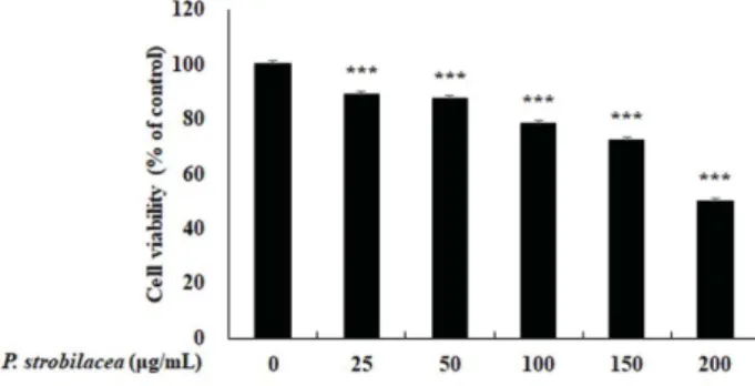
Ǖ⦝ӹྕ ᯯ ⇵⇽ྜྷᯕ ᭥ᦵᖙ⡍ AGS᮹ ᷾ᨱ ၙ⊹۵ ᩢ⨆
Ǖ⦝ӹྕ ᯯ ⇵⇽ྜྷᯕ ᭥ᦵᖙ⡍
AGSᔾ᳕ᨱ ၙ⊹۵ ᩢ⨆
ᮥ ⪶ᯙ⦹ʑ ᭥⧕ ࠥᄥ
(0, 25, 50, 100, 150, 200 μg/ml)ಽ
ญ⦹ᩍ
MTS assayෝ ᙹ⧪⦹ᩡ݅
.Ǖ⦝ӹྕ ᯯ ⇵⇽ྜྷᮥ
24
e ⊹⦹ᩡᮥ ভ ᦵᖙ⡍᮹ ᔾ᳕ᮉᮡ ݡ᳑Ǒᨱ እ⧕
89.3, 87.3, 78.4, 72.2, 50.0%
ಽ ࠥ ᮹᳕ᱢᮝಽ ᮁ᮹ᱢᯙ
qᗭෝ ᅕᩡ݅
(Fig. 1).ᯕ్⦽ ᖙ⡍ ᔾ᳕ᮉ᮹ qᗭa
apoptosis
ᨱ ᮹⦽ ᖙ⡍ᯱ໙ᔍ ᯙḡෝ ⪶ᯙ⦹ʑ ᭥⧕
annexinV/FITC-PI
ᩝᔪ ႊჶᮥ ᯕᬊ⦹ᩍ ə ᧲ᔢᮥ šₑ⦹ᩡ݅
.Ǖ⦝ӹྕ ᯯ ⇵⇽ྜྷᯕ ᭥ᦵᖙ⡍ AGSᨱᕽ᮹ apoptosis
ᮁࠥ
Ǖ⦝ӹྕ ᯯ ⇵⇽ྜྷᮥ ᭥ᦵᖙ⡍
AGSᨱ ญ⦹ᩡᮥ ভ ᖙ
⡍᮹ ᷾ᯕ ⩥Ċ⦹í qᗭࡽ äᮥ ⪶ᯙ⦹Ł ᯕ్⦽ đŝa
ᖙ⡍᮹
apoptosisᮁၽಽ ᯕᨕḡŁ ᯩ۵ḡ ᩍᇡෝ ⪶ᯙ⦹ʑ
᭥⧕ ᖙ⡍ෝ ⩶Ų ᩝᔪ⦹ᩍ ᇥᕾᮥ ᝅ⦹ᩡ݅
. Apoptosisᝁ⪙ෝ ၼᮡ ᦵᖙ⡍۵ ᖙ⡍ ⢽໕ᮝಽ
phosphatidylserine(PS)
ᯕ י⇽ࡹŁ
annexin V/FITC⩶Ų ᧞ŝ ✚ᯕᱢᮝಽ v⦹í đ⧊⦹í ࡽ݅
.ੱ⦽
,ᖙ⡍᮹ ᔍ໙ᯕ ᯝᨕӹ໕ ᖙ⡍
สᮥ ☖ŝ⦽
PI᧞ᯕ
DNA᪡ đ⧊⦹ᩍ Ⅹʑ
(early)᪡ ⬥ʑ
(late) apoptosis᮹ ᧲ᔢᮥ ⪶ᯙ⧁ ᙹ ᯩ݅
[16, 17].Ǖ⦝ӹྕ
ᯯ ⇵⇽ྜྷᮥ ࠥᄥ
(0, 50, 150, 200 μg/ml)ಽ ญ⦹ᩍ ᖙ⡍
ෝ
Cellometer K2ಽ ⩶Ų᮹ ᧲ᔢᮥ ⪶ᯙ⦽ đŝ
Fig. 2᪡ z
ᮡ đŝෝ ⪶ᯙ⧁ ᙹ ᯩᨩ݅
.Ⅹʑ
apoptosisᖙ⡍᮹ ᧲ᔢᯕ
Fig. 1. Cell viability of AGS cells by MTS assay. The cells weretreated with PSE for 24 h and added with MTS solution for 3 h.
Significant evaluation was indicated from control by t-test (*p < 0.05, **p < 0.01, ***p < 0.001).
Fig. 2. Apoptotic activity of AGS cells by annexin V/FITC-PI stain. The cells were treated with PSE (0, 50, 150, and 200 μg/ml) for 15 h, and stained with annexin V/FITC-PI solution for 0.5 h. (A) The cells were determined by cellometer K2. (B) and (C) Percent of early, late and total apoptosis was indicated using bar charts. Significant evaluation was indicated from control by t-test (*p < 0.05,
**p < 0.01, ***p < 0.001, n = 6).
ݡ᳑Ǒᨱ እ⧕
4.53, 7.53, 15.74%ಽ ࠥ ᮹᳕ᱢᮝಽ ᮁ᮹
ၙ⦽ ᷾aෝ ᅕᯕŁ ᯩ݅
.ੱ⦽
,⬥ʑ
apoptosisᖙ⡍᮹ ᧲ᔢ
ࠥ ݡ᳑Ǒᨱ እ⧕ ᷾a⦹Ł ᯩᮝӹ ☖ĥᱢᯙ ᮁ᮹ᖒᮥ ૽ಘ
⦹í ᅕᯕḡ۵ ᦫᦹ݅
.đŝᱢᮝಽ Ǖ⦝ӹྕ ᯯ ⇵⇽ྜྷ᮹ ญa ᱥℕ ᱢᮝಽ ᭥ᦵᖙ⡍ෝ
apoptosisᖙ⡍ಽ ᮁࠥ⦹ᩍ ᔾ
᳕ᮉᯕ qᗭ⦹ᩡᮭᮥ ⪶ᯙ⧁ ᙹ ᯩᨩ݅
.Ǖ⦝ӹྕ ᯯ ⇵⇽ྜྷᯕ ᭥ᦵᖙ⡍ AGSᨱᕽ᮹ ᖙ⡍ᵝʑᨱ
ၙ⊹۵ ᩢ⨆
ᖙ⡍ᵝʑ۵ ⦹ӹ᮹ ᖙ⡍a ࢱ }᮹ ঙ ᖙ⡍ಽ ᱢᱩ⦽ ᇥ
ᩕᯕ ᯝᨕӹ۵ ᳑Ḣ⪵ࡽ ŝᱶᯕ݅
.ᖙ⡍ ᖒᰆᨱ ⦥ᙹᱢᯙ
G1 (⧊ᖒᵡእʑ
), S (DNA⧊ᖒʑ
), G2 (ᇥᩕᵡእʑ
), M (ᇥᩕʑ
)ʑ᮹ ᙽ₉ᱢᯙ
4݉ĥ ᖙ⡍ᵝʑෝ Ñ⊽݅
.b ݉ĥᄥ ᵝʑᨱ ۵
cyclin-dependent kinases (CDKs)⬉ᗭa ᯲ᬊ⦹ᩍ ᖙ⡍
⪽ᖒᮥ ᳑ᱩ⦽݅
.እᱶᔢᱢᯙ ᦵᖙ⡍۵ b ݉ĥᄥ ᵝʑᨱ š ಉࡽ ⬉ᗭa ᱶᔢᱢᮝಽ ᳑ᱩࡹḡ ༜⦹Ł ྕ⦽ ᷾ᯕ Ⅹ௹
ࡽ݅
[18−
20].݅ᙹ᮹ ⧎ᦵ ᩑǍᨱᕽ᪡ zᯕ ᖙ⡍ ᷾ ᨖᱽ
۵ ᖙ⡍ᵝʑ᳑ᱩŝ šಉᖒᯕ ᯩᮝ໑
apoptosisa ᮁࠥࡹ໕ ᖙ⡍ᵝʑ᮹ ݅᧲⦽ ݉ĥᨱᕽ ᷾ᯕ ຩ⇵۵ äᮥ ᙹ ᯩ
݅
[21].ᯕᨱ ᅙ ᩑǍᨱᕽ۵ Ǖ⦝ӹྕ ᯯ ⇵⇽ྜྷ᮹
ญᨱ ᮹⦽ ᖙ⡍᷾ ᨖᱽ đŝa ᖙ⡍ᵝʑ ᱶḡᨱ ᮹⦽ ä
ᯕʑ ᦥᅕʑ ᭥⧕ Ǖ⦝ӹྕ ᯯ ⇵⇽ྜྷᮥ ࠥᄥ
(0, 50, 150,200 μg/ml)
ಽ
15e ญ⦹ᩍ
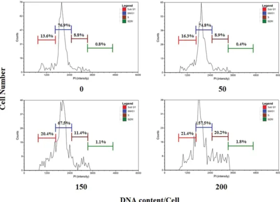
PIᩝᔪᮥ ☖⧕ ᖙ⡍ᵝʑෝ
⊂ᱶ⦹ᩡ݅
.ə đŝ
,ᦥྕäࠥ ญ⦹ḡ ᦫᮡ ݡ᳑Ǒᨱ እ
⧕
16.3, 20.4, 21.4%ಽ
sub-G1ʑᨱ ᖙ⡍a ນྜྷ్ Ǖ⦝ӹྕ
ᯯ ⇵⇽ྜྷᨱ ࠥ ᮹᳕ᱢᮝಽ ᷾a⦹۵ ᙹ⊹ෝ ⪶ᯙ⧁ ᙹ ᯩ ᨩ݅
(Fig. 3). Sub-G1ʑ۵
DNA᮹ ᧲ᯕ ᱶᔢᖙ⡍ᅕ݅ ᱢᮡ
ᔢ┽᮹ ᖙ⡍ॅᯕ ⊂ᱶࡹ۵ ݉ĥಽ Ǖ⦝ӹྕ ᯯ ⇵⇽ྜྷᨱ ᮹
⧕
apoptosisŝᱶᯕ ᯝᨕӹŁ ᯩ۵ ᖙ⡍a ᯕᔢ ᇥᩕᯕ
ӹ ᖒᰆ⦹ḡ ༜⦹Ł ᇥ⧕ࡹ۵ ŝᱶᨱᕽ
sub-G1ʑ᮹ ᷾a ᧲
ᔢᯕ ૽ಘ⦹í šₑࡹŁ ᯩ݅
.đŝᱢᮝಽ Ǖ⦝ӹྕ ᯯ ⇵⇽
ྜྷ ญᨱ ᮹⧕ ᭥ᦵ ᖙ⡍۵
sub-G1ʑᨱ ນྜྷ్ ᖙ⡍ᔍ໙ᯕ
ᮁࠥࡹᨕ ᦵᖙ⡍᮹ ᷾ᯕ ᨖᱽ ࢁ äᮝಽ ❱݉ࡽ݅
.Ǖ⦝ӹྕ ᯯ ⇵⇽ྜྷᯕ Bcl-2 family ၽ⩥ᨱ ၙ⊹۵ ᩢ⨆
ᖙ⡍᮹ ᔍ໙ᮡ
death receptor (DR)ᨱ ᮹⦽ ᯙᖒ Ğಽ
ੱ۵ ԕᯙᖒ Ğಽ ࢱ aḡಽ ᯙ⧕ ᯝᨕӽ݅
.ə ᵲ ԕᯙᖒ Ğ ಽ۵ ၙ☁⎹ऽญᦥෝ Ğᮁ⦹໑
Bcl-2 family݉႒ḩ᮹ ᔢ⪙
᯲ᬊᯕ ᵲ⦹݅
[22, 23]. Bcl-2 family۵
anti-apoptosisᨱ ᗮ⦹۵
Bcl-2, Bcl-xL, Bcl-w᪡
pro-apoptosisᨱ ᗮ⦹۵
Bax, Bak, Bid, Bad, Noxa, PUMA॒ᮝಽ Ǎᖒࡹᨕ ᯩ݅
[24].ᖙ
⡍ᔍ໙ᯕ ᮁࠥࡹ໕
Bcl-2 familyᯙᯱ᮹ ᔢ⪙᯲ᬊᨱ ᮹⧕ ၙ
☁⎹ऽญᦥ ԕ᮹
cytochrome ca ႊ⇽ࡹᨕ
caspase-3᮹ ⪽
ᖒ⪵ෝ ☖⧕
DNA᮹ ᮲⇶ŝ ᇥ⧕ෝ ᮁၽ⦽݅
[25].Ǖ⦝ӹྕ
Fig. 3. Cell cycle analysis in AGS cells by PI stain. The cells were treated with PSE (0, 50, 150, and 200 μg/ml) for 15 h, fixed in 70%
ethanol, and stained with PI solution for 0.5 h.
ᯯ ⇵⇽ྜྷᮥ ญ⦹ᩡᮥ ভ
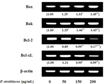
Bcl-2 family᮹ ၽ⩥ ᄡ⪵ෝ š
ₑ⦹ʑ ᭥⧕
RT-PCRᮥ ☖⧕ đŝෝ ⪶ᯙ⦹ᩡ݅
.ᝅ⨹ đŝ
Bax
۵ ࠥ᮹᳕ᱢᮝಽ ᷾a⦹ᩡŁ ੱ⦽
, Bakࠥ ᦥྕäࠥ ญ⦹ḡ ᦫᮡ ݡ᳑Ǒᨱ እ⧕ ᷾a⦹۵ Ğ⨆ᮥ ᅕᯕ۵ ၹ໕ᨱ
Bcl-2, Bcl-xL
ᮡ ࠥ᮹᳕ᱢᮝಽ qᗭ⦹۵ ᧲ᔢᮥ ૽ಘ⦹í
šₑ⧁ ᙹ ᯩᨩ݅
(Fig. 4).ᯕॅ đŝෝ ᳦⧊⧕ᅕ໕
pro-apoptosis
ᯙᯱ᮹ ᷾a᪡
anti-apoptosisᯙᯱ᮹ qᗭa ᔢ⪙
ᱢᮝಽ ᯕᨕḡ໑ ၙ☁⎹ऽญᦥ᮹ ส ⚍ŝᖒᯕ ʑࡹᨕ ԕᯙᖒ ᖙ⡍ᔍ໙᮹ ᝁ⪙ᱥݍ Ğಽa ḥ⧪ࢉᮥ ᙹ ᯩ݅
.Ǖ⦝ӹྕ ᯯ ⇵⇽ྜྷ᮹ ญ۵ ԕᯙᖒ Ğಽෝ ☖⦽ ᖙ⡍ᔍ໙
⪽ᖒᮥ ᵝࠥ⦹ᩍ ᭥ᦵᨱ ݡ⦽ ⧎ᦵ ⪽ᖒᮥ ᅕᯕ۵ äᮝಽ ᔍ ഭࡽ݅
. ᧞
ᅙ ᩑǍᨱᕽ۵ Ǖ⦝ӹྕ ᯯ ⇵⇽ྜྷಽᇡ░ ᔍ௭ ᭥ᦵᖙ⡍
ᵝᯙ
AGSᖙ⡍ᨱ ݡ⦽
apoptosisa ᮁࠥࡹᨕ ⧎ᦵ ⪽ᖒᮥ
aḡ۵ḡෝ ⪶ᯙ⦹Łᯱ ⦽݅
. AGSᖙ⡍ᨱ Ǖ⦝ӹྕ ᯯ ⇵
⇽ྜྷᮥ ࠥᄥ
(0, 50, 150, 200 μg/ml)ಽ ญ⦹ᩍ ᖙ⡍ᨱᕽ ӹ┡ӹ۵ ᧲ᔢॅᮥ ⪶ᯙ⦹ᩡ݅
. MTS⊂ᱶᮝಽ ᦵᖙ⡍ ᔾ᳕
ᮉᮥ ⪶ᯙ⦽ đŝ Ǖ⦝ӹྕ ᯯ ⇵⇽ྜྷᨱ ࠥ᮹᳕ᱢᮝ
ಽ ᖙ⡍ᨱ ࠦᖒᮥ ᅕᩡᮝ໑
,ᯕ్⦽ ᖙ⡍ ᵞᮭᯕ
apoptosisᮁࠥᨱ ᮹⦽ äᯙḡෝ
annexin V/FITC-PIᩝᔪᮥ ☖⧕ ⪶ ᯙ⦹ᩡ݅
.ə đŝ
, early apoptosisෝ ᅕᯕ۵ ᖙ⡍᮹ ᧲ᔢᯕ
ᮁ᮹ᖒ ᯩí ᷾a⦹ᩡ݅
.Ǖ⦝ӹྕ ᯯ ⇵⇽ྜྷ᮹ ญ۵ ᖙ⡍
ᵝʑᨱᕽ
sub-G1ʑ᮹ ᷾aಽ ᦵᖙ⡍a ᯕᔢ ᇥᩕ⦹ḡ ༜
⦹Ł ᷾ᯕ ᨖᱽࢉᮥ ᅕᩍᵡ݅
.ԕᯙᖒ Ğಽᨱ šᩍ⦹۵
Bax, Bak, Bcl-2, Bcl-xL᮹ ᷾qᮥ
mRNAᙹᵡᨱᕽ
RT- PCRಽ ⪶ᯙ⦽ đŝ
, Bax, Baka ᷾a⦹Ł
, Bcl-2, Bcl-xLᯕ qᗭෝ ࠺ၹ⦹໑ ၙ☁⎹ऽญᦥෝ ☖⦽ ᖙ⡍ᔍ໙᮹ ᝁ⪙ᱥݍ Ğಽa ḥ⧪ࡹŁ ᯩᮭᮥ ⪶ᯙ⧁ ᙹ ᯩᨩ݅
.ᯕ్⦽ đŝෝ
᳦⧊⦹໕ Ǖ⦝ӹྕ ᯯ ⇵⇽ྜྷᮡ ᔍ௭ ᭥ᦵᖙ⡍ᨱ ݡ⦽ ⧎ᦵ
⪽ᖒᮥ aḡ۵ ⬉܆ᱢ a⊹a ᯩᮭᨱ a܆ᖒᮥ ᱽ⧁ ᙹ ᯩ
ᮥ äᮝಽ ᔾbࡹ໑
,⇵aᱢᯙ ࠺ྜྷᝅ⨹ᮥ ☖⦽ ᯙℕᱢᬊ
⨹᮹ ⬉܆ á᷾ᮥ ☖⧕ ᮹᧞⣩ }ၽ a܆ᖒᮥ ⪶ᯙ⧕ ⦽݅
.Acknowledgments
This work was supported by the Jungwon University Research Grant (No. : 2020-003).
Conflict of Interest
The authors have no financial conflicts of interest to declare.
References
1. Song M, Shin GY. 1987. Efficaey of progressive muscle relaxation training in reducing nausea and vomiting, anxiety and symptom distress of cancer chemotherapy. J. Nurs. Acad. Soc. 17: 195-203.
2. Shin SA, Lee HN, Choo GS, Kim SJ, Kim HJ, Park YS, et al. 2016.
Induction of apoptosis and inhibition of growth in human gastric cancer by piperine. J. Korean Soc. Food Sci. Nutr. 45: 1589-1594.
3. Kim EJ, Park H, Lim SS, Kim JS, Shin HK, Yoon JH. 2008. Effect of the hexane extract of Saussurea lappa on the growth of HT-29 human colon cancer cells. Korean J. Food Sci. Technol. 40: 207- 214.
4. Hsu HF, Houng JY, Kuo CF, Tsao N, Wu YC. 2008. Glossogin, a novel phenylpropanoid from Glossogyne tenuifolia, induced apoptosis in A549 lung cancer cells. Food Chem. Toxicol. 46:
3785-3791.
5. Kim GM, Lee AR, Lee KW, Park JY, Chun J, Cha J, et al. 2009. Char- acterization of a 27 kDa fibrinolytic enzyme from Bacillus amylo- liquefaciens CH51 isolated from chenggukjang. J. Microbiol.
Biotechnol. 19: 997-1004.
6. Kim SR, Lee HH, Kim MJ, Seo MJ, Hong SH, Choi YH, et al. 2010.
Induction of apoptosis by ethanol extracts of fermented aga- beans in AGS human gastric carcinoma cells. J. Life Sci. 20: 1872- 1881.
7. Oda E, Ohki R, Murasawa H, Nemoto J, Shibue T, Yamashita T, et al. 2000. Noxa, a BH3-only member of the Bcl-2 family and can- didate mediator of p53-induced apoptosis. Science 288: 1053- 1058.
8. Yang HJ, Kim EH, Kang ST, Park SN. 2009. Antibacterial activity of Platycarya strobilacea extract and stability of the extract-con- taining cream. Korean J. Microbiol. Biotechnol. 37: 170-175.
Fig. 4. mRNA level analysis in AGS cells treated with P.
strobilacea extract (0, 50, 150, and 200 μg/ml). And, density of mRNA band was expressed as a value compared to the con- trol. Significant evaluation was indicated from control by t-test (*p <G0.05, **p < 0.01, ***p < 0.001, n = 3).
9. Choi YH, Chae SG, Kim JH, Gang SJ, Baeg NI, Han JT. 2003. Isola- tion of an antifungal compound from aerial parts of Platycarya strobilacea. J. Korea Soc. Agric. Chem. Biotechnol. 46: 268-270.
10. Babu D, Lee JS, Park SY, Thapa D, Choi MK, Kim AR, et al. 2008.
Involvement of NF-kappaB in the inhibitory actions of Platy- carya strobilacea on the TNF-alpha-induced monocyte adhesion to colon epithelial cells and chemokine expression. Arch. Pharm.
Res. 31: 727-735.
11. Yang HJ, Park SN. 2008. Antioxidative and antiaging effects of Platycarya strobilacea extract and clinical trial. J. Soc. Cosmet. Sci.
Korea 34: 275-286.
12. Lee HJ, Lee SK, Choi YJ, Jo HJ, Kang HY, Choi DH. 2007. Extractives from the bark of Platycarya strobilacea. J. Korean Soc. Forest Sci.
96: 408-413.
13. Lee JH, Kwon YS, Kim CM. 1998. Flavonoids from the stem bark of Platycarya strobilacea. Korean J. Pharmacogn. 29: 353-356.
14. Hashemzaei M, Far AD, Yari A, Heravi RE, Tabrizian K, Taghdisi SM, et al. 2017. Anticancer and apoptosis-inducing effects of quercetin in vitro and in vivo. Oncol. Rep. 38: 819-828.
15. Cincin ZB, Unlu M, Kiran B, Bireller ES, Baran Y, Cakmakoglu B.
2014. Molecular mechanisms of quercitrin-induced apoptosis in non-small cell lung cancer. Arch. Med. Res. 45: 445-454.
16. Ryu DS, Kim SH, Kwon JH, Lee DS. 2014. Orostachys japonicus induces apoptosis and cell cycle arrest through the mitochondria- dependent apoptotic pathway in AGS human gastric cancer cells. Int. J. Oncol. 45: 459-469.
17. Kaufmann SH, Henqartner MO. 2001. Programmed cell death:
alive and well in the new millennium. Trends Cell Biol. 11: 526-534.
18. Li B, Geo Y, Rankin GO, Rojanasakul Y, Cutler SJ, Tu Y, et al. 2015.
Chaetoglobosin K induces apoptosis and G2 cell cycle arrest through p53-dependent pathway in cisplatin-resistant ovarian cancer cells. Cancer Lett. 356: 418-433.
19. Buolamvini JK. 2000. Cell cycle molecular targets in novel anti- cancer drug discovery. Curr. Pharm. Des. 6: 379-392.
20. Diaz-Moralli S, Tarrado-Casterllamau M, Miranda A, Cascante M.
2013. Targeting cell cycle regulation in cancer therapy. Pharmacol.
Ther. 138: 255-271.
21. Bae JT, Chang JS, Lee KR. 2002. Effect of Sarcodon aspratus extract on expression of cell cycle174 associated proteins in HepG2 cells. J. Korean Soc. Food Sci. Nutr. 31: 329-332.
22. Xie QC, Yang YP. 2014. Anti-proliferative of physcion 8-O-β- glucopyranoside isolated from japonicus Houtt. on A549 cell lines via inducing apoptosis and cell cycle arrest. BMC Complement.
Altern. Med. 14: 377.
23. Fulda S, Debatin KM. 2006. Extrinsic versus intrinsic apoptosis pathways in anticancer chemotherapy. Oncogene 25: 4798- 4811.
24. Lavrik IN. 2010. Systems biology of apoptosis signaling net- works. Curr. Opin. Biotechnol. 21: 551-555.
25. Moon DO, Kim MO, Choi YH, Kim ND, Chang JH, Kim GY. 2008.
Bcl-2 overexpression attenuates SP600125-induced apoptosis in human leukemia U937 cells. Cancer Lett. 264: 316-325.
26. Park C, Hyun SK, Shin WJ, Chung KT, Choi BT, Kwon HJ, et al.
2009. Esculetin induces apoptosis through caspase-3 activation in human leukemia U937 cells. J. Life Sci. 19: 249-255.