Induction of Apoptotic Cell Death by Cordycepin, an Active Component of the Fungus Cordyceps militaris, in AGS Human Gastric Cancer Cells
Hye Hyeon Lee
1, Jin-Woo Jeong
1and Yung Hyun Choi
1,2*
1Anti-Aging Research Center, Dongeui University, Busan 614-714, Korea
2Department of Biochemistry, Dongeui University College of Korean Medicine, Busan 614-714, Korea
Received March 1, 2016 /Revised May 26, 2016 /Accepted June 2, 2016
Cordycepin, a derivative of the nucleoside adenosine, is one of the active components extracted from fungi of genus Cordyceps, and has been shown to have many pharmacological activities. In this study, we investigated the effects of cordycepin on proliferation and apoptosis of human gastric cancer AGS cells, and its possible mechanism of action. Treatment of cordycepin resulted in significant decrease in cell viability of AGS cells in a concentration-dependent manner. A concentration-dependent apop- totic cell death was also measured by agarose gel electrophoresis and flow cytometery analysis.
Molecular mechanistic studies of apoptosis unraveled cordycepin treatment resulted in an enhanced expression of tumor necrosis factor-related apoptosis-inducing ligand, death receptor 5 and Fas ligand.
Furthermore, up-regulation of pro-apoptotic Bax, and down-regulation of anti-apoptotic Bcl-2 and Bcl-xL expression were also observed in cordycepin-treated AGS cells. These were followed by activa- tion of caspases (caspase-9, -8 and -3), subsequently leading to poly (ADP-ribose) polymerase cleavage.
Taken together, these findings indicate that cordycepin induces apoptosis in AGS cells through regu- lation of multiple apoptotic pathways, including death receptor and mitochondria. Although further mechanical studies are needed, our results revealed that cordycepin can be regarded as a new effec- tive and chemopreventive compound for human gastric cancer treatment.
Key words :
AGS cells, apoptosis, caspase, cordycepin
*Corresponding author
*Tel : +82-51-850-7413, Fax : +82-51-853-4036
*E-mail : [email protected]
This is an Open-Access article distributed under the terms of the Creative Commons Attribution Non-Commercial License (http://creativecommons.org/licenses/by-nc/3.0) which permits unrestricted non-commercial use, distribution, and reproduction in any medium, provided the original work is properly cited.
Journal of Life Science 2016 Vol. 26. No. 7. 847~854 DOI : http://dx.doi.org/10.5352/JLS.2016.26.7.847
서 론
Cordycepin (3’-deoxyadenosine)은 전통 동양의학에서 임 상적으로 허약 증상, 만성기관지염, 거담과 천식, 폐결핵, 빈혈 및 병후 허약 등에 처방되어 온 동충하초에 속하는 Cordyceps 속에서 처음 분리되었으며, 구조적으로 deoxyadenosine과 매 우 유사한 adenosine nucleoside의 ribose 3번 탄소에 산소가 없는 adenosine analogue이다[6, 27]. Cordycepin의 효능은 1970년대 polyadenylation 억제 효과가 알려지면서 다양한 연 구가 활발히 진행되어 왔다[28]. 다양한 생리활성 중 특히 cor- dycepin의 강력한 항암활성은 이미 많은 보고가 있어 왔으며, 이는 암세포의 능동적 죽음에 속하는 apoptosis 유도와 매우 밀접한 관계를 가진다[2, 31, 32]. 일반적으로 apoptosis가 유도 되는 동안 나타나는 가장 특징적인 현상이 cysteine-depend- ent aspartate-directed protease 군에 속하는 caspase의 활성 증가이다[10, 12]. 일반적으로 apoptosis는 extrinsic 및 in-
trinsic pathway로 크게 대별되는데, 두 경로에서 caspase는 initiator caspases (caspase-8, -9 및 -10)와 effector caspases (caspase-3, -6 및 -7)로 구분된다. 즉 initiator caspases의 활성 화에 의해 effector caspases가 활성형으로 전환되는 caspase cascade 경로를 거치게 된다. 특히 initiator caspases 중에서 caspase-8과 -9는 apoptosis의 주요한 두 가지 경로인 extrinsic 및 intrinsic pathway의 개시를 알리는데 관여하는 대표적인 caspase이다[11, 19]. 활성화된 effector caspases는 DNA re- pair에 관여하는 poly (ADP-ribose)-polymerase (PARP)를 포 함한 다양한 할성형 caspase의 기질 단백질 절단을 초래하며 궁극적으로 apoptosis를 종결짓는다[1, 9].
Caspase의 활성 유도와 연관된 암세포에서의 cordycepin 에 의한 apoptosis 유도는 이미 몇몇 암세포에서 확인된 바 있다. 이러한 caspase 활성에는 calcium-calpain system 경로 가 관여하기도 하며[3], caspase의 활성이 활성산소종(reactive oxygen species) 의존적으로 일어나기도 한다[16, 20]. Cordy- cepin에 의한 apoptosis 유도 과정에는 세포주기의 교란과 동 반되기도 하는데, 암세포의 종류에 따라 S기 arrest [33] 또는 G1기 arrest [21]를 유발하기도 하여 암세포에 따른 차이를 보 여주고 있다[24, 25]. 또한 유방암세포에서는 estrogen re- ceptor 비의존적으로 apoptosis를 유도하였으며[21], apopto- sis 유도에는 autophagy가 관여하기도 하지만 이러한 autoph- agy의 동반이 암세포의 죽음에 관여할 것인지 생존에 관여할
- Note -
것인지에 대한 결론은 아직 내리기 힘든 상황이다[5, 23, 26].
아울러 cordycepin은 항암제 저항성의 극복 가능성[4, 22]과, phosphoinositide 3-kinase (PI3K)/Akt 신호계 의존적으로 te- lomerase 활성 억제[13] 및 암세포의 전이 차단 효과[14, 15]
등도 보고된 바 있다.
비록 상기에서 언급되지 않은 다양한 cordycepin의 항암활 성이 추가적으로 보고되고 있으나, 암세포 증식억제에 따른 apoptosis 유도 기전에 대한 연구 결과는 여전히 미비한 상태 이다. 본 연구에서는 cordycepin의 항암활성과 apoptosis 신호 계의 역할에 대한 추가적인 자료를 얻기 위하여 인체 위암세 포를 대상으로 cordycepin에 의한 세포증식 억제에 따른 apoptosis 유도 관련 다양한 유전자들의 발현 변화를 조사하 였다.
재료 및 방법
세포 배양 및 생존율 측정
본 연구에 사용된 cordycepin (MW, 251.2; product no.
C3394)은 Sigma-Aldrich Chemical Co. (St. Louis, MO, USA) 에서 구입하였으며 dimethyl sulfoxide (DMSO, Sigma-Aldrich Chemical Co.)에 녹인 후 적정 농도로 배지에 희석하여 처리 하였다. AGS 인체 위암세포는 American Type Culture Collection (Manassas, VA, USA)에서 구입하였으며, 10% fe- tal bovine serum, penicillin 및 streptomycin 등이 혼합된 RPMI 1640 배지(Gibco-BRL, Grand Island, NY, USA)를 이용 하여 37
oC, 5% CO
2조건하에서 배양하였다. Cordycepin 처리 에 따른 AGS 세포의 생존에 미치는 영향을 비교하기 위하여 세포 배양용 6 well plate에 3X10
5개/ml로 세포를 분주하고 적정 농도의 cordycepin을 배지에 처리하였다. 24시간 후 배지 를 제거하고 tetrazolium bromide salt (MTT, Ameresco, Solon, Ohio, USA)를 0.5 mg/ml 농도로 희석하여 37
oC에서 3시간 동안 처리하였다. 반응이 끝난 뒤 MTT 시약을 제거하고 DMSO로 생성된 formazan을 모두 녹인 후 ELISA reader (Molecular Devices, Sunnyvale, CA, USA)로 540 nm에서 흡 광도를 측정하였다. Cordycepin 처리에 의한 AGS 세포의 형 태 변화 정도를 살펴보기 위하여 cordycepin을 적정 농도로 희석 처리하여 배양 24시간 경과 후 도립 현미경(inverted mi- croscope, Carl Zeiss, Germany)을 이용하여 200배의 배율로 관찰하였다.
Agarose gel 전기영동에 의한 DNA 단편화 분석 Apoptosis 유발의 핵심적인 증거인 DNA 단편화 현상의 분석을 위하여 정상 및 cordycepin이 24시간 처리된 세포들을 모은 다음 lysis buffer [5 mM Tris-HCl (pH 7.5), 5 mM ethyl- enediaminetetraacetic acid (EDTA), 0.5% Triton X-100]를 첨 가하여 상온에서 30분간 lysis 시킨 다음 14,000 rpm, 4
oC에서
20분간 원심분리하여 찌꺼기가 제거된 상층액 750 μl를 회수 하였다. 회수된 상층액에 proteinase K solution (Sigma-Aldrich Chemical Co.)을 0.5 mg/ml의 농도로 처리하여 50
oC에서 3시 간 동안 반응시킨 후 750 μl의 phenol : chloroform : isoamyl alcohol 혼합 용액(25 : 24 : 1, Sigma-Aldrich Chemical Co.)을 첨가하고 30분간 회전 교반시킨 다음 14,000 rpm에서 10분간 원심 분리하였다. 여기서 얻어진 상층액 600 μl에 300 μl의 iso- propanol (Sigma-Aldrich Chemical Co.)과 100 μl의 5 M NaCl 를 첨가하여 4
oC에서 24시간 동안 반응시킨 후, 14,000 rpm, 4℃에서 30분간 원심분리시켜 상층액을 제거하고 DNA pellet 을 추출하였다. DNA pellet에 RNase A가 적당량 들어있는 buffer를 이용하여 녹인 후, 6X gel loading dye (Bioneer, Daejeon, Korea)를 혼합하였다. DNA pellet은 1.5% agarose gel을 이용하여 1시간 동안 50 V로 전기영동 시킨 후 ethidium bromide (EtBr, Sigma-Aldrich Chemical Co.)로 염색하여 DNA 단편화 유도 여부를 확인하였다.
Flow cytometry 분석에 의한 apoptosis 유발의 정량화 AGS 세포에서 cordycepin이 유발하는 apoptosis 정도를 정 량적으로 분석하기 위하여 정상 및 cordycepin이 함유된 배지 에서 24시간 동안 배양된 세포들을 모은 다음 2,000 rpm으로 5분간 원심분리하여 상층액을 제거한 후 phosphate-buffered saline (PBS)를 이용하여 2~3회 정도 세척하였다. 준비된 세포 는 CycleTEST PLUS DNA REAGENT Kit (Becton Dickinson, San Jose, CA, USA)를 이용하여 고정 및 염색하여 4
oC, 암실에 서 30분 동안 반응시켰다. 반응시킨 세포를 35-mm mesh를 이용하여 단일세포로 분리한 후 FACSCalibur (Becton Dickin- son)를 적용시켜 형광반응에 따른 Cellular DNA content 및 histogram을 CellQuest software 및 ModiFit LT (Becton Dickinson) 프로그램을 이용하여 분석하였다.
Reverse transcription-polymerase chain reaction (RT-PCR)에 의한 mRNA 발현 분석
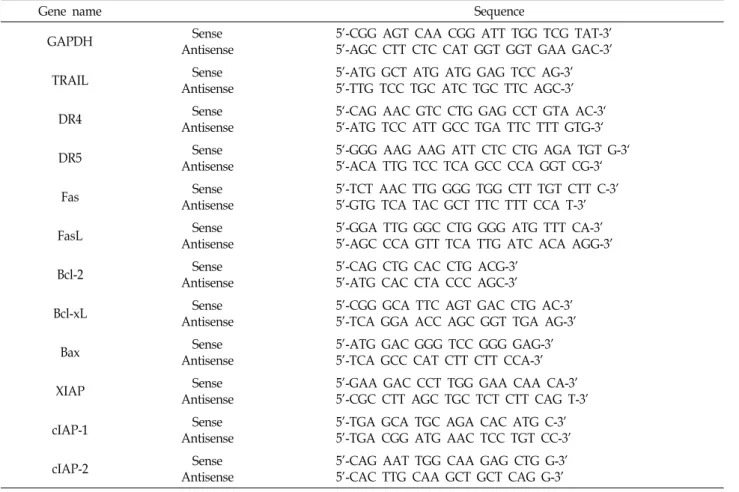
동일한 조건에서 배양된 AGS 세포들에 TRIzol reagent (Invitrogen Co., Carlsbad, CA, USA)를 이용하여 RNA를 분 리하였다. 분리된 RNA를 정량한 후, 각각의 primer (Table 1), DEPC water 그리고 ONE-STEP RT-PCR PreMix Kit (Intron, Seoul, Korea)를 넣고 Mastercycler gradient (Eppen- dorf, Hamburg, Germany)를 이용하여 증폭하였다. 각 PCR 산물들의 양적 차이를 확인하기 위하여 1X TAE buffer로 1.5%
agarose gel을 만들고 well 당 각각의 primer에 해당하는 PCR
산물에 DNA gel loading solution을 섞어서 loading 한 후
50 V에서 전기영동을 행하였다. 전기영동으로 DNA 분리가
끝난 gel을 EtBr로 염색한 후 UV 하에서 관찰하였으며, glyc-
eraldehyde-3-phosphate dehydrogenase (GAPDH)를 in-
ternal control로 사용하였다.
Table 1. Sequences of the primer pairs employed in the RT-PCR reactions
Gene name Sequence
GAPDH Sense
Antisense
5'-CGG AGT CAA CGG ATT TGG TCG TAT-3' 5'-AGC CTT CTC CAT GGT GGT GAA GAC-3'
TRAIL Sense
Antisense
5'-ATG GCT ATG ATG GAG TCC AG-3' 5'-TTG TCC TGC ATC TGC TTC AGC-3'
DR4 Sense
Antisense
5‘-CAG AAC GTC CTG GAG CCT GTA AC-3‘
5‘-ATG TCC ATT GCC TGA TTC TTT GTG-3‘
DR5 Sense
Antisense
5‘-GGG AAG AAG ATT CTC CTG AGA TGT G-3‘
5‘-ACA TTG TCC TCA GCC CCA GGT CG-3‘
Fas Sense
Antisense
5'-TCT AAC TTG GGG TGG CTT TGT CTT C-3' 5'-GTG TCA TAC GCT TTC TTT CCA T-3'
FasL Sense
Antisense
5'-GGA TTG GGC CTG GGG ATG TTT CA-3' 5'-AGC CCA GTT TCA TTG ATC ACA AGG-3'
Bcl-2 Sense
Antisense
5'-CAG CTG CAC CTG ACG-3' 5'-ATG CAC CTA CCC AGC-3'
Bcl-xL Sense
Antisense
5'-CGG GCA TTC AGT GAC CTG AC-3' 5'-TCA GGA ACC AGC GGT TGA AG-3'
Bax Sense
Antisense
5'-ATG GAC GGG TCC GGG GAG-3' 5'-TCA GCC CAT CTT CTT CCA-3'
XIAP Sense
Antisense
5'-GAA GAC CCT TGG GAA CAA CA-3' 5'-CGC CTT AGC TGC TCT CTT CAG T-3'
cIAP-1 Sense
Antisense
5'-TGA GCA TGC AGA CAC ATG C-3' 5'-TGA CGG ATG AAC TCC TGT CC-3'
cIAP-2 Sense
Antisense
5'-CAG AAT TGG CAA GAG CTG G-3' 5'-CAC TTG CAA GCT GCT CAG G-3'
Western blot analysis에 의한 단백질 발현의 분석 준비된 세포에 적당량의 lysis buffer [25 mM Tris-Cl (pH 7.5), 250 mM NaCl, 5 mM EDTA, 1% NP-40, 1 mM phenyme- thylsulfonyl fluoride (PMSF), 5 mM dithiothreitol (DTT)]를 첨가하여 4
oC에서 1시간 동안 반응시킨 후, 총 단백질을 분리 하였다. 단백질 농도는 Bio-Rad 단백질 정량 시약(Bio-Rad, Hercules, CA, USA) 사용 방법에 준하여 정량 후 다음 동량의 Laemmli sample buffer (Bio-Rad)를 섞어서 sample을 만들었 다. 동량의 sample을 sodium dodecyl sulphate (SDS)-poly- acrylamide gel을 이용하여 전기영동으로 분리한 후, PVDF membrane (Schleicher and Schuell, Keene, NH, USA)으로 electroblotting에 의해 전이시켰다. 분리된 단백질이 전이된 membrane을 5% skim milk를 처리하여 비특이적인 단백질들 에 대한 blocking을 실시하고 1차 항체를 처리하여 상온에서 2시간 이상 또는 4
oC에서 over night시킨 다음 PBS-T로 세척 하고 처리된 1차 항체에 맞는 2차 항체를 사용하여 상온에서 1시간 정도 반응시켰다. 반응이 끝난 후 암실에서 enhanced chemiluminoesence (ECL) solution (Amersham Life Science Corp., Arlington Heights, IL, USA)을 적용시킨 다음 X-ray film에 감광시켜 특정단백질의 발현 양을 분석하였다. 본 실험 에 사용된 1차 항체들은 Santa Cruz Biotechnology Inc. (Santa
Cruz, CA, USA) 및 Calbiochem (Cambridge, MA, USA)에서 구입하였으며, 2차 항체들은 Amersham Corp.에서 구입하였 다.
In vitro caspase activity 측정
Apoptosis 유발 연계 caspase 활성 정도의 정량적인 비교를
위하여 준비된 세포를 모은 뒤 상기와 동일한 방법으로 단백
질을 추출하고 정량하였다. Caspase activity assay kit는 R&D
Systems (Minneapolis, MN, USA)에서 구입하였으며, 150 μg
의 단백질이 함유된 50 μl의 sample에 기질 100 μM이 함유된
reaction buffer [40 mM HEPES (pH 7.4), 20% glycerol (v/v),
1 mM EDTA, 0.2% NP-40 and 10 mM DL-DTT] 50 μl를 혼합
하여 각 sample 당 총 양이 100 μl가 되게 하였다. 여기에 각
caspase 기질 5 μl를 첨가하여 37℃, 암실에서 3시간 동안 반응
시킨 후 ELISA reader를 이용하여 405 nm의 흡광도를 이용하
여 반응의 정도를 측정하였다. 본 실험에 사용된 기질은 cas-
pase-3의 경우에는 Asp-Glu-Val-Asp (DEVD)-p-nitroaniline
(pNA)이었고, caspase-8의 경우에는 Ile-Glu-Thr-Asp (IETD)-
pNA이었으며, caspase-9은 Leu-Glu-His-Asp (LEHD)-pNA
였다.
A
B
C
Time (hr)
Fig. 1. Inhibition of cell proliferation by cordycepin in AGS cells.
Cells were seeded in 6 well plate at 3×10⁵ cells/ml and treated with the indicated concentrations of cordycepin for 24 hr (A) or 30 μg/ml cordycepin for the indicated times (B). Cell viability was measured by the metabol- ic-dye-based MTT assay. Results are expressed as per- centage of the un-treated control ± SD of three separate experiments (*p<0.05 vs. untreated control). (C) The cells were photographed with an inverted microscope (Mag- nification, ×200).
A
B
C
Time (hr)
Fig. 2. Induction of apoptosis by cordycepin in AGS cells. (A) Cells were seeded in 6 well plate at 3×10⁵ cells/ml and treated with the indicated concentrations of cordycepin for 24 hr. DNA fragmentation was analyzed by extract- ing the fragmented DNA and separating it by electro- phoresis in a 1.5% agarose gel containing EtBr. (B and C) Cells were seeded in 6 well plate at 3×10⁵ cells/ml and treated with the indicated concentrations of cordyce- pin for 24 hr (B) or 30 μg/ml cordycepin for the in- dicated times (C). To quantify the degree of apoptosis induced by cordycepin, the cells were evaluated for sub-G1 DNA content, which represents the fractions un- dergoing apoptotic DNA degradation, using a DNA flow cytometer. Results are expressed as the means ± SD of three independent experiments. The statistical sig- nificance of the results was analyzed by Student’s t-test (*p<0.05 vs. untreated control).
통계 처리
모든 실험결과는 평균 ± 표준편차로 표시하였고 SigmaPlot (Systat Software Inc., San Jose, CA, USA)을 이용하여 Student
t-test를 이용하여 통계적 유의성을 얻었다.결과 및 고찰
AGS 세포에서 cordycepin에 의한 apoptosis 유도 비록 다양한 인체 암세포들을 이용한 다수의 선행 연구들에 서 cordycepin에 의한 증식 억제가 apoptosis 유도와 연관성이 있음이 이미 잘 알려져 있으나[2, 3, 5, 16, 20-26, 31], 인체 위암 세포에 대한 보고는 전무한 실정이다. 본 연구에서는 cordyce- pin 또한 위암세포에서도 apoptosis를 유도할 것으로 예측하 여 AGS 세포를 대상으로 세포의 증식에 미치는 영향을 먼저 조사하였다. 이를 위하여 cordycepin이 처리된 AGS 세포를 대상으로 MTT assay를 실시하였으며, cordycepin 처리 농도
및 처리 시간이 증가할수록 세포의 증식이 유의적으로 억제되
었음을 확인하였으며, 그에 따른 세포의 밀도 및 염색질 응축
과 같은 형태적 변화 동반을 관찰하였다(Fig. 1). 따라서 cordy-
cepin 처리에 따른 AGS 세포의 생존율 억제가 apoptosis 유발
과 직접적인 연관이 있는지를 확인하기 위하여 전형적인
apoptosis가 일어난 세포에서 볼 수 있는 가장 일반적인 변화
인 DNA 단편화의 유도 여부를 조사하였다. Fig. 2A의 결과에
A B
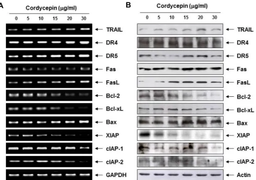
Fig. 3. Effects of cordycepin on the expression of apoptosis-related genes in AGS cells. (A) After treatment with the indicated concen- trations of cordycepin for 24 hr, total RNA was isolated, and RT-PCR was performed using the indicated primers. (B) The cells grown under the same conditions as (A) were lysed and then equal amounts of cell lysates were separated on SDS-poly- acrylamide gels and transferred to PVDF membranes. The membranes were probed with the indicated antibodies and the proteins were visualized using an ECL detection system. Actin was used as an internal control. GAPDH and actin were used as internal controls for the RT-PCR and western blot assays, respectively.
서 알 수 있듯이, cordycepin의 처리 농도 증가에 따라 DNA 분절에 의한 단편화의 형성이 현저히 증가되었다. 아울러 cor- dycepin 처리에 따른 apoptosis 유발 정도를 flow cytometry 분석을 통하여 확인한 결과, cordycepin 처리 농도 및 시간의 증가에 따라 apoptosis가 유발된 세포의 빈도에 해당되는 sub-G1기에 속하는 세포의 빈도가 특히 15 μg/ml 처리군 이 상에서 매우 증가되었다(Fig. 2B 및 C). 이는 cordycepin 처리 에 의한 AGS 세포의 생존율 억제가 apoptosis 유도와 직접 연계되었음을 보여주는 것이다. 아울러 본 연구의 결과에서 나타난 AGS 위암세포에서의 apoptosis 유도 활성의 정도는 LnCap 전립선 암세포[23, 34]나 HCT116 대장암세포[14] 등에 비해서는 저농도에서 apoptosis 유도능을 보였으나, 백혈병세 포들과 비교했을 경우 유사한 정도의 감수성을 나타내었다 [16].
AGS 세포에서 apoptosis 조절 인자들의 발현에 미치는 cordycepin의 영향
상기에서 관찰된 AGS 세포에서 cordycepin에 의한 apop- tosis 유발에 관여하는 기전 연구를 수행하기 위하여 apopto- sis 조절과 연관된 다양한 유전자들의 발현을 총체적으로 조사 하였다. 이를 위하여 apoptosis 유발 과정에서 extrinsic path- way를 경유하는 과정에서 중요한 역할을 하는 death receptor (DR) 및 ligand에 속하는 유전자들의 발현에 cordycepin이 어
떠한 영향을 미치는지를 조사하였다. Fig. 3의 결과에서 알 수 있듯이 DR 그룹에 속하는 대표적인 유전자인 DR4 및 Fas의 mRNA 및 단백질의 발현에는 cordycepin이 큰 영향을 미치지 못하였다. 그러나 DR5와 tumor necrosis factor (TNF)-related apoptosis-inducing ligand (TRAIL) 및 Fas ligand (FasL)의 발현은 cordycepin 처리 농도 의존적으로 전사 및 번역 수준에 서 다소 증가하였다. 일반적으로 extrinsic pathway의 개시가 세포막에 존재하는 DR에 TRAIL 및 FasL 등과 같은 apoptotic ligand가 결합함으로서 시작된다는 점을 고려할 경우[11, 17], 본 연구의 결과는 cordycepin의 처리에 의한 AGS 세포에서의 apoptosis 유발에 extrinsic pathway의 활성화가 관여하였음 을 보여주는 것이다.
다음은 intrinsic pathway 조절에 중요한 역할을 하는 Bcl-2
family 유전자의 발현에 미치는 cordycepin의 영향을 조사하
였다. Bcl-2 family에 속하는 단백질은 mitochondria 외막에서
mitochondria 보존과 mitochondria에 의해 유도되는 apopto-
sis를 조절하는 intrinsic pathway의 중요한 조절자로서, Bcl-2
및 Bcl-xL 등과 같은 apoptosis를 억제하는 anti-apoptotic 단백
질과 Bax 및 Bad 등과 같이 apoptosis를 유발하는 pro-apop-
totic 단백질로 구성되어 있다[17, 29]. 하지만 anti-apoptotic
및 pro-apoptotic 단백질들 사이의 균형이 깨어지게 되면 mi-
tochondria의 기능 이상을 유발하게 되고 mitochondria로 내
막에 존재하고 있는 apoptosis 유발 촉진 인자들이 세포질로
A
B
Fig. 4. Activation of caspases and the degradation of the PARP by cordycepin treatment in AGS cells. (A) Cells were in- cubated with various concentrations of cordycepin for 24 hr, lysed and cellular proteins were separated by SDS-polyacrylamide gels and transferred onto PVDF membranes. The membranes were probed with indicated antibodies. Proteins were visualized using an ECL de- tection system. Actin was used as a loading control. (B) After 24 hr incubation with PVDF, aliquots (150 μg pro- tein) were incubated with substrates, DEVD-pNA, IETD- pNA and LEHD-pNA, for in vitro caspase-3, -8 and -9 activity, respectively, at 37℃ for 3 hr. The released fluo- rescent products were measured. Results are expressed as the means ± SD of three independent experiments.
The statistical significance of the results was analyzed by Student’s t-test (*p<0.05 vs. untreated control).
방출됨으로서 apoptosis 유발 신호를 증폭시키는 것으로 알려 져 있다[17, 29]. Fig. 3의 결과에서 알 수 있듯이, cordycepin의 처리 농도 증가에 따라 anti-apoptotic 유전자인 Bcl-2 및 Bcl-xL의 mRNA 및 단백질의 발현이 점차 감소하였으며, 대표 적인 pro-apoptotic 유전자인 Bax의 발현은 전사 및 번역 수준 에서 점차 증가되었다. 따라서 cordycepin에 의한 AGS 세포 의 apoptosis 유발에는 mitochondria 기능 이상에 따른 in- trinsic pathway도 관여하고 있음을 알 수 있었다.
한편 inhibitors of apoptosis protein (IAP) family에 속하는 단백질들은 apoptosis 유도에 핵심적인 역할을 하는 caspase 의 활성을 차단함으로서 apoptosis를 억제하는 것으로 알려져 있다[7, 8]. 특히 IAP family 중 활성화된 caspase와 높은 친화 력을 가지는 것으로 알려진 XIAP는 caspase-9 및 -3과 결합하 여 apoptosis를 직접 억제하는 것으로 알려져 있으며, cIAP-1 및 cIAP-2도 각각 다양한 caspase와의 결합을 통하여 apopto- sis를 억제하는 것으로 알려져 있다[7, 8]. 따라서 cordycepin이 IAP family 인자들의 발현에 어떠한 영향을 미치는지를 확인 한 결과, 본 연구에서 조사된 3가지 IAP family 유전자들의 발현이 다소 차이는 있었으나 전사 및 번역 수준에서 모두 감소되어 caspase 활성화에 기여하였을 것으로 추정된다.
AGS 세포에서 cordycepin에 의한 caspase의 활성화 Caspase는 세포가 정상적으로 성장 및 생존할 경우 핵과 mitochondria의 외막에 불활성 상태로 존재하고 있지만 세포 내외부의 다양한 자극에 의하여 활성화되어 기질 단백질들의 분해를 유발함으로서 apoptosis를 유발한다[18, 30]. 그 중 ex- trinsic 및 intrinsic pathway의 initiator caspase는 caspase-8과 -9로 구분되며, effector caspase에는 caspase-3 및 -7 등이 포함 되어 있다. 즉 initiator caspase가 활성화 되면 하위 단계에 있는 effector caspase를 활성화시킴으로서 세포의 성장 및 생 존에 중요한 역할을 하는 여러 종류의 기질 단백질들은 분해 함으로서 apoptosis를 유발한다[10, 12]. 특히 PARP는 활성화 된 effector caspase에 의하여 분해되는 대표적인 기질 단백질 로서 정상적인 DNA 수복이나 유전자 안정성 유지에 핵심인 자로 작용한다. 116 kDa의 분자량을 가진 PARP 단백질이 85 및 24 kDa의 단편으로 잘리게 되면 PARP의 효소적 기능의 상실로 인하여 정상적인 DNA repair 과정이 억제된다. 따라 서 PARP의 단편화는 caspase 활성 의존적 apoptosis 유발의 생화학적 표식자로 사용되고 있다[1, 9]. 따라서 본 연구에서는 대표적인 apoptosis 유발 두 경로의 initiator caspase인 cas- pase-8과 -9 및 effector caspase인 caspase-3의 발현에 cordy- cepin의 영향을 조사하였으며, Fig. 4A의 immunoblotting 결 과에서 알 수 있듯이, cordycepin 처리에 의하여 불활성형인 pro-caspase-3, -8 및 -9 단백질의 발현은 cordycepin 처리 농도 의존적으로 감소되었으나 뚜렷한 활성형의 단백질 발현 증가 를 정확히 관찰하기는 어려웠다. 따라서 in vitro caspases ac-
tivity assay를 통하여 이들 caspase의 활성 정도를 직접 분석 한 결과, Fig. 4B에서 나타난 바와 같이 cordycepin 처리 농도 의 증가에 따라 caspase-3, -8 및 -9의 활성이 유의적으로 상승 하는 것으로 나타났다. 또한 활성화된 caspase-3의 표적 기질 단백질 중의 하나인 PARP 단백질의 단편화도 매우 증가되어 (Fig. 4A), cordycepin에 의한 apoptosis 유발에는 caspase 활 성의 증가가 핵심적인 역할을 함을 알 수 있었다. 아울러 이러 한 현상은 mitochondria에서 세포질로의 cytochrome c의 유 리와 연관성이 있었다(Fig. 5).
이상의 결과를 종합해 보면 인체 위암 AGS 세포에 cordy-
cepin을 처리하였을 경우에 유발되는 apoptosis는 extrinsic
pathway의 개시를 통한 caspase-8의 활성화와, Bcl-2 family
인자들의 발현 변화와 연관된 mitochondria 기능 손상에 의한
Fig. 5. Release of cytochrome c from cytosol to nucleus by cor- dycepin treatment in AGS cells. Cells were incubated with various concentrations of cordycepin for 24 hr. The cytosolic and mitochondrial proteins were extracted and Western blot analyses were performed using the in- dicated antibodies and an ECL detection system. Actin and cytochrome c oxidase subunit 4 (COX IV) were used as internal controls for the cytosolic and mitochondrial fractions, respectively.
caspase-9의 활성 증가를 통한 세포질로의 cytochrome c의 유 리 및 caspase-3의 활성에 따른 기질 단백질들의 분해가 중요 한 역할을 하는 것으로 생각되어지며, IAP family의 발현 감소 로 인하여 caspase의 활성이 억제되지 못하는 것도 apoptosis 유도에 어느 정도 관여했을 것으로 생각되어진다. 비록 이에 대한 추가적인 연구가 필요하겠지만, cordycepin 처리에 의하 여 유발되는 apoptosis는 extrinsic 및 intrinsic pathway를 모 두 경유하는 multiple apoptotic pathway에 의하여 조절될 것 으로 생각된다.
감사의 글
이 논문은 2015년도 정부(미래창조과학부)의 재원으로 한 국연구재단의 지원을 받아 수행된 기초연구사업임(No. 2015 R1A2A2A01004633).
References
1. Agarwal, A., Mahfouz, R. Z., Sharma, R. K., Sarkar, O., Mangrola, D. and Mathur, P. P. 2009. Potential biological role of poly (ADP-ribose) polymerase (PARP) in male gametes. Reprod. Biol. Endocrinol. 7, 143-163.
2. Chen, L. S., Stellrecht, C. M. and Gandhi, V. 2008. RNA-di- rected agent, cordycepin, induces cell death in multiple myeloma cells. Br. J. Haematol. 140, 682-391.
3. Chen, Y., Chen, Y. C., Lin, Y. T., Huang, S. H. and Wang, S. M. 2010. Cordycepin induces apoptosis of CGTH W-2 thyroid carcinoma cells through the calcium-calpain-caspase 7-PARP pathway. J. Agric. Food Chem. 58, 11645-11652.
4. Chen, Y. H., Hao, L. J., Hung, C. P., Chen, J. W., Leu, S.
F. and Huang, B. M. 2014. Apoptotic effect of cisplatin and cordycepin on OC3 human oral cancer cells. Chin. J. Integr.
Med. 20, 624-632.
5. Choi, S., Lim, M. H., Kim, K. M., Jeon, B. H., Song, W. O.
and Kim, T. W. 2011. Cordycepin-induced apoptosis and autophagy in breast cancer cells are independent of the es- trogen receptor. Toxicol. Appl. Pharmacol. 257, 165-173.
6. Cunningham, K. G., Manson, W., Spring, F. S. and Hutchin- son, S. A. 1950. Cordycepin, a metabolic product isolated from cultures of Cordyceps militaris (Linn.) Link. Nature 166, 949.
7. Danson, S., Dean, E., Dive, C. and Ranson, M. 2007. IAPs as a target for anticancer therapy. Curr. Cancer Drug Targets 7, 785-794.
8. de Graaf, A. O., de Witte, T. and Jansen, J. H. 2004. Inhibitor of apoptosis proteins: new therapeutic targets in hemato- logical cancer? Leukemia 18, 1751-1759.
9. Duriez, P. J. and Shah, G. M. 1997. Cleavage of poly (ADP-ribose) polymerase: a sensitive parameter to study cell death. Biochem. Cell. Biol. 75, 337-349.
10. Fiandalo, M. V. and Kyprianou, N. 2012. Caspase control:
protagonists of cancer cell apoptosis. Exp. Oncol. 34, 165-175.
11. Fulda, S. and Debatin, K. M. 2006. Extrinsic versus intrinsic apoptosis pathways in anticancer chemotherapy. Oncogene 25, 4798-4811.
12. Hensley, P., Mishra, M. and Kyprianou, N. 2013. Targeting caspases in cancer therapeutics. Biol. Chem. 394, 831-843.
13. Jang, K. J., Kwon, G. S., Jeong, J. W., Kim, C. H., Yoon, H. M., Kim, G. Y., Shim, J. H., Moon, S. K., Kim, W. J. and Choi, Y. H. 2015. Cordyceptin induces apoptosis through repressing hTERT expression and inducing extranuclear ex- port of hTERT. J. Biosci. Bioeng. 119, 351-357.
14. Jeong, J. W. and Choi, Y. H. 2014. Cordycepin inhibits the migration and invasion of HCT116 human colorectal carci- noma cells through the tightening of tight junctions and in- hibition of matrix metalloproteinase activity. J. Kor. Soc. Food Sci. Nutr. 43, 86-92.
15. Jeong, J. W., Jin, C. Y., Park, C., Han, M. H., Kim, G. Y., Moon, S. K., Kim, C. G., Jeong, Y. K., Kim, W. J., Lee, J.
D. and Choi, Y. H. 2012. Inhibition of migration and in- vasion of LNCaP human prostate carcinoma cells by cordy- cepin through inactivation of Akt. Int. J. Oncol. 40, 1697- 1704.
16. Jeong, J. W., Jin, C. Y., Park, C., Hong, S. H., Kim, G. Y., Jeong, Y. K., Lee, J. D., Yoo, Y. H. and Choi, Y. H. 2011.
Induction of apoptosis by cordycepin via reactive oxygen species generation in human leukemia cells. Toxicol. In Vitro 25, 817-824.
17. Kimberley, F. C. and Screaton, G. R. 2004. Following a TRAIL: update on a ligand and its five receptors. Cell Res.
14, 359-372.
18. Lavrik, I. N., Golks, A. and Krammer, P. H. 2005. Caspases:
pharmacological manipulation of cell death. J. Clin. Invest.
115, 2665-2672.
19. Lavrik, I. N. 2010. Systems biology of apoptosis signaling networks. Curr. Opin. Biotechnol. 21, 551-555.
20. Lee, H. H., Park, C., Jeong, J. W., Kim, M. J., Seo, M. J., Kang, B. W., Park, J. U., Kim, G. Y., Choi, B. T., Choi, Y.
H. and Jeong, Y. K. 2013. Apoptosis induction of human
초록:동충하초 유래 cordycepin에 의한 AGS 인체 위암세포의 apoptosis 유발
이혜현
1․정진우
1․최영현
1,2*
(1동의대학교 항노화연구소, 2동의대학교 한의과대학 생화학교실)
Nucleoside adenosine 유도체의 하나인 cordycepin (3'-deoxyadenosine)은 Cordyceps 속에서 유래된 활성 물질 중의 하나로서 항염증, 항산화 및 항암활성을 포함한 다양한 약리학적 효능이 있는 것으로 잘 알려져 있다. 본 연구에서는 AGS 인체 위암세포의 증식에 미치는 cordycepin의 영향과 관련 기전 연구를 시도하였다. Cordycepin의 처리에 따라 AGS 세포의 생존율이 처리 농도 의존적으로 감소되었으며, DNA 단편화 및 flow cytometery 분석에 따른 apoptosis 유발 또한 유의적으로 증가하였음을 확인하였다. 이러한 cordycepin 처리에 따른 AGS 세포의 apoptosis 유도에는 TRAIL, DR5 및 FasL의 mRNA 및 단백질의 발현 증가가 연관되어 있었다. 아울러 cordycepin은 Bcl-2 family 중 pro-apoptotic 인자인 Bax의 발현은 증가시켰으며, anti-apoptotic 인자인 Bcl-2 및 Bcl-xL의 발현은 전사 및 번역 수준에 서 억제시켰다. 이러한 현상들은 extrinsic 및 intrinsic apoptosis의 initiator caspase (caspase-8 및 -9) 뿐만 아니라 effec- tor caspase인 caspase-3의 활성과 PARP 단백질의 절단 증가와 연관성이 있었다. 따라서 AGS 세포에서 cordycepin에 의한 apoptosis의 유발은 death receptor 활성과 mitochondria 기능 손상을 포함한 multiple apoptotic pathway가 관여 할 것으로 생각된다. 비록 좀 더 세심한 기전 연구의 결과가 뒤따라야 되겠지만, 본 연구의 결과는 cordycepin의 항암작 용을 이해하는데 중요한 자료가 될 것이며 향후 수행될 추가 실험을 위한 기초 자료로서 그 가치가 매우 높을 것으로 생각된다.
prostate carcinoma cells by cordycepin through reactive oxygen species mediated mitochondrial death pathway. Int.
J. Oncol. 42, 1036-1044.
21. Lee, S. Y., Debnath, T., Kim, S. K. and Lim, B. O. 2013.
Anti-cancer effect and apoptosis induction of cordycepin through DR3 pathway in the human colonic cancer cell HT-29. Food Chem. Toxicol. 60, 439-447.
22. Lee, H. H., Jeong, J. W., Lee, J. H., Kim, G. Y., Cheong, J., Jeong, Y. K., Yoo, Y. H. and Choi, Y. H. 2013. Cordycepin increases sensitivity of Hep3B human hepatocellular carci- noma cells to TRAIL-mediated apoptosis by inactivating the JNK signaling pathway. Oncol. Rep. 30, 1257-1264.
23. Lee, H. H., Kim, S. O., Kim, G. Y., Moon, S. K., Kim, W.
J., Jeong, Y. K., Yoo, Y. H. and Choi, Y. H. 2014. Involvement of autophagy in cordycepin-induced apoptosis in human prostate carcinoma LNCaP cells. Environ. Toxicol. Pharmacol.
38, 239-250.
24. Seong da, B., Hong, S., Muthusami, S., Kim, W. D., Yu, J.
R. and Park, W. Y. 2016. Cordycepin increases radio- sensitivity in cervical cancer cells by overriding or prolong- ing radiation-induced G2/M arrest. Eur. J. Pharmacol. 771, 77-83.
25. Lee, H. H., Hwang, W. D., Jeong, J. W., Park, C., Han, M.
H., Hong, S. H., Jeong, Y. K. and Choi, Y. H. 2014. Induction of apoptosis and G2/M cell cycle arrest by cordycepin in human prostate carcinoma LNCap cells. J. Life Sci. 24, 92-97.
26. Li, Y., Li, R., Zhu, S., Zhou, R., Wang L, Du, J, Wang, Y., Zhou, B. and Mai, L. 2015. Cordycepin induces apoptosis and autophagy in human neuroblastoma SK-N-SH and BE(2)-M17 cells. Oncol. Lett. 9, 2541-2547.
27. Liang, Z. C., Liang, C. H. and Wu, C. Y. 2014. Various grain substrates for the production of fruiting bodies and bio- active compounds of the medicinal caterpillar mushroom, Cordyceps militaris (Ascomycetes). Int. J. Med. Mushrooms 16, 569-578.
28. Nair, C. N. and Owens, M. J. 1974. Preliminary observations pertaining to polyadenylation of rhinovirus RNA. J. Virol.
13, 535-537.
29. Ola, M. S., Nawaz, M. and Ahsan, H. 2011. Role of Bcl-2 family proteins and caspases in the regulation of apoptosis.
Mol. Cell. Biochem. 351, 41-58.
30. Stennicke, H. R. and Salvesen, G. S. 1999. Catalytic proper- ties of the caspases. Cell Death Differ. 6, 1054-1059.
31. Thomadaki, H., Scorilas, A., Tsiapalis, C. M. and Havredaki, M. 2008. The role of cordycepin in cancer treatment via in- duction or inhibition of apoptosis: implication of poly- adenylation in a cell type specific manner. Cancer Chemother.
Pharmacol. 61, 251-265.
32. Thomadaki, H., Tsiapalis, C. M. and Scorilas, A. 2005.
Polyadenylate polymerase modulations in human epi- thelioid cervix and breast cancer cell lines, treated with eto- poside or cordycepin, follow cell cycle rather than apoptosis induction. Biol. Chem. 386, 471-480.
33. Wang, X. A., Xiang, S. S., Li, H. F., Wu, X. S., Li, M. L., Shu, Y. J., Zhang, F., Cao, Y., Ye, Y. Y., Bao, R. F., Weng, H., Wu, W. G., Mu, J. S., Hu, Y. P., Jiang, L., Tan, Z. J., Lu, W., Wang, P. and Liu, Y. B. 2014. Cordycepin induces S phase arrest and apoptosis in human gallbladder cancer cells. Molecules 19, 11350-11365.