Available at http://www.formulastudy.com
HFS
Original Article / 원저
18α-Glycyrrhetinic acid의 위암 세포 사멸 효과에 관한 연구
김정남, 김병주
*부산대학교 한의학전문대학원 양생기능의학부
18α-Glycyrrhetinic acid induces apoptosis of AGS human gastric cancer cells
Jeong Nam Kim, Byung Joo Kim
*Division of Longevity and Biofunctional Medicine School of Korean Medicine, Pusan National University
ABSTRACT
Objectives : The purpose of this study was to investigate the anti-cancer effects of 18α-Glycyrrhetinic acid (18α -GA), a hydrolyzed metabolite of glycyrrhizin, in AGS human gastric adenocarcinoma cells.
Methods : We used human gastric adenocarcinoma cell line, AGS cells. We examined cell death by MTT assay and caspase 3 and 9 assay with 18α-GA. To examine the inhibitory effects of 18α-GA, sub-G1 analysis was done the AGS cells after 24 hours with 18α-GA. Also, to investigate the inhibitory mechanisms of 18α-GA, mitogen-activated protein kinase pathways and reactive oxygen species (ROS) generation were examined.
Results :
1. 18α-GA inhibited the growth of AGS cells in a dose-dependent fashion.
2. Sub-G1 fractions were significantly and dose-dependently increased by 18α-GA.
3. 18α-GA increased the caspase 3 and 9 activities in AGS cells.
4. 18α-GA inhibited proliferation of AGS cells via the modulation of c‑Jun N‑terminal kinase (JNK) signaling pathways, which results in the induction of apoptosis.
5. 18α-GA enhanced ROS accumulation in AGS cells.
Conclusions : Our findings provide insight into unraveling the effects of 18α-GA in human gastric adenocarcinoma cells and developing therapeutic agents against gastric cancer.
ⓒ 2020 The Korean Medicine Society For The Herbal Formula Study
This paper is available at http://www.formulastudy.com which permits unrestricted non-commercial use, distribution, and reproduction in any medium, provided the original work is properly cited.
Key words : 18α-Glycyrrhetinic acid, Human gastric adenocarcinoma cell, AGS, Anti-cancer.
Ⅰ. 서론
a)암은 전 세계에서 가장 생명을 위협하는 질병 중 하나이며 그 중에서 위암은 세계에서 두번째로 암과 관련된 사망 원인이다1,2). 위암은 매년 거의 100만 명 의 새로운 환자에게서 진단되고 있으며, 동아시아, 동유럽, 중남미 일부 지역에서 가장 높은 발병률을 보이고 있다3). 위암 치료법은 수술만이 유일한 치료 법이라고 알려져 있지만 최근에는 위암 수술 후 각종 화학요법으로 위암 치료 결과를 좋게 하고 있다4). 따 라서, 새롭고 더 효과적인 항암제를 연구하고 개발해 야 한다.
감초 (Glycyrrhiza uralensis)의 성분인 Glycyrrhetic acid (GA)은 간보호 같은 여러 가지 질병의 치료에 자주 처방되어 온 잘 알려진 한약제제이다5,6). GA의 항암 활동 메커니즘은 세포 증식 감소, 세포 전이 억 제, 세포 주기 변화 등을 통해 나타나는 것으로 알려 져 왔다7,8). 18α-Glycyrrhetinic acid (18α-GA;
Glycyrrhizin의 가수 분해 대사물)는 항염증, 항바이 러스, 항바이러스, 항암 효능 등을 보이는 것으로 나 타났다9,10). 최근에는 18α-GA가 gingival fibroblast 의 성장을 억제해서 세포사멸을 일으키고, 인간백혈병 세포주인 HL-60세포를 카스파제 (caspase)와 미토 콘드리아 (mitochondria) 의존적으로 세포사멸을 일 으키는 것으로 보고 되었다11,12). 하지만, 18α-GA가 인간 위암세포에 미치는 작용에 대한 연구는 많이 부 족한 현실이다. 본 연구에서는 18α-GA의 위암세포 에서의 효능 및 작용기전을 확인하는 기초적 연구를 수행하였다.
Ⅱ. 재료 및 방법
1. 재료 (1) 약재
실험에 사용한 18α-Glycyrrhetinic acid (18α
-GA)는 시그마 알드리치 (Sigma-Aldrich, St. Louis, MO, USA)에서 구입한 후 사용하였다.
(2) 대상 세포주
이 실험에 사용한 인체 위암세포는 서울대학교 암 세포주 은행에서 구입한 위암 세포주 AGS이며, adenocarcinoma의 histopathology 특징을 나타내고 있다.
2. 방법
(1) 암세포의 배양
암세포주는 10% fetal bovine serum (Gibco BRL, MD, USA), 1% broad-spectrum antibiotics (Gibco BRL, MD, USA)가 함유된 RPMI-1640 (Gibco BRL, MD, USA)배지를 이용하여 37℃, 5% CO2 incubator (Precision Scientific Inc. NY, USA)에서 배양하였 고, 세포는 0.1% Trypsin-EDTA (Sigma, MD, USA)를 이용하여 37℃에서 5분간 처리한 후 회수하 였다.
(2) MTT assay
세포 생존율 측정은 세포 배양판 (12-well)에 암세 포를 3✕104 cells/well 씩 분주하여 24시간 이상 배 양 후 18α-GA를 다양한 농도로 처리한 다음, 37℃, 5% CO2 하에서 48시간 또는 72시간 배양한 후, MTT용액 (5 mg/ml)을 배양액 최종 부피의 1/10되게 첨가 하였다. 3시간 후 배양액을 제거하고 형성된 formazan 침전물을 dimethyl sulfoxide (DMSO) 1 ml로 녹여서 540 nm에서 흡광도를 측정하였다.
(3) Caspase 3과 9 assay 측정
Caspase 3 과 9 assay kits를 BioMol (Plymouth, PA, USA)에서 구입하여 측정하였다. 405 nm의 흡 광도에서 여러 번 측정하였다.
*Corresponding author : Byung Joo Kim. Division of Longevity and Biofunctional Medicine School of Korean Medicine, Pusan National University, 49, Busandaehak-ro, Mulgeum-eup, Yangsan-si, Gyeongsangnam-do, 50612, Republic of Korea.
Tel : +82-51-510-8469, Fax : +82-51-510-8420, E-mail : [email protected]
∙Received : November 7, 2019 / Revised : November 18, 2019 / Accepted : February 25, 2020
(4) Flow cytometry를 이용한 세포주기 분석 18α-GA이 AGS세포의 증식을 억제하는데 있어서 세포주기의 어느 단계를 지연시키는지 flow cytometry 를 이용하여 분석하였다. 18α-GA을 첨가한 배지에 서 3일간 배양된 AGS세포를 PBS용액으로 세척한 후, 4℃에서 30분간 70% ethanol로 고정하였다. 고 정후 propidium iodide (Sigma-Aldrich, St. Louis, MO, USA)로 DNA를 염색하여 Becton Dickinson FACStar Flow Cytometry를 이용하여 세포주기를 비교 분석하였다.
(5) 활성산소 (Reactive Oxygen Species; ROS) 측정 AGS세 포의 ROS 생성은 DCF-DA (2’,7’- dichlorodihydrofluorescein diacetate; Molecular Probes, Eugene, OR, USA)를 사용하여 측정하였다.
이 세포들은 37°C에서 30분 동안 20 μl DCF-DA로 처리되었고 PBS로 세척하였다. 488/525 nm의 excitation/emission 파장에서 FACS (Becton- Dickinson, Mountain View, CA, USA)를 사용하여 측정하였다.
(6) 통계 분석
실험성적은 평균 ± 표준편차 (mean ± SD)로 나
타내었으며, 결과의 차이를 검정할 때에는 Student's t-test로 검정하여 p 값이 0.05 미만일 때 유의한 차이가 있는 것으로 판정하였다.
Ⅲ. 결과
1. 위암세포 사멸 효과
위암세포 사멸 효과를 측정하기 위하여 18α-GA 50, 100, 150, 200 μM 농도로 AGS세포에 시험한 결과 24시간 후인 (A)에서는 150 μM이상에서 뚜렷 한 위암세포 사멸 효과를 볼 수 있었으며, 48시간 후 인 (B)에서는 50 μM 이상에서 뚜렷한 위암세포 사 멸 효과를 볼 수 있었다 (Fig. 1). MTT assay로 24 시간 후인 (A) 경우에는 50 μM에서 76.5 ± 2.7%
(P<0.01), 100 μM 에서 81.4 ± 3.6% (P<0.01), 150 μM에서 56.6 ± 4.6% (P<0.01), 200 μM에서 26.8 ± 2.3% (P<0.01)의 세포 생존 결과를 보였다.
48시간 후인 (B) 경우에는 50 μM에서 67.3 ± 4.0% (P<0.01), 100 μM 에서 67.6 ± 3.2%
(P<0.01), 150 μM에서 35.7 ± 0.7% (P<0.01), 200 μM에서 2.6 ± 0.1% (P<0.01)의 세포 생존 결 과를 보였다.
Fig. 1. 18α-GA inhibited the cell viability in AGS cells. Cell viabilities were determined by 3-[4,5-dimethylthiazol-2-yl]-2,5-diphenyltetrazolium bromide (MTT) aassay. (A) 18α-GA reduced cell viabilities in a dose-dependent manner for 24 h. (B) 18α-GA reduced cell viabilities in a dose-dependent manner for 48 h. Bars represent mean values±S.D. **P <
0.01. CTRL: Control.
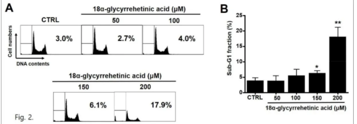
2. Flow cytometry를 이용한 세포주기 분석
18α-GA에 의한 세포사가 apoptosis에 의한 것인
지 확인하기 위하여 flow cytometry를 이용한 세포 주기 sub-G1분석을 실시하였다. 18α-GA에 의한
Sub-G1 분석 결과 18α-GA 농도에 비례해서 sub-G1 peak가 증가함을 알 수 있다. Sub-G1 비율 은 50 μM에서 3.9 ± 1.6%, 100 μM 에서 5.5 ±
2.1%, 150 μM에서 6.3 ± 0.8% (P<0.05), 200 μ M에서 18.1 ± 3.1% (P<0.01)로 나타났다 (Fig. 2).
Fig. 2. Effect of 18α-GA on AGS cell apoptosis. (A) Cells were treated with 18α-GA for 24 h. Cell cycle analysis was conducted by flow cytometry. (B) Sub-G1 fractions were significantly and dose-dependently increased by 18α-GA. Sub-G1 fractions are expressed as percentages. Bars represent the mean values ± S.D. **P < 0.01. CTRL: Control.
3. Caspase 3과 caspase 9를 통한 암세포 사멸 확인 18α-GA에 의한 세포사가 apoptosis에 의한 것인 지 확인하기 위하여 농도별로 세포사멸의 진행 정도 를 알아보았다. Caspase 3과 9의 활성도를 측정한
결과 18α-GA에 의해 caspase 3과 9의 활성도가 농 도에 의존적으로 증가함을 알 수 있고 이러한 활성도 는 pan-caspase inhibitor인 zVAD-fmk에 의해서 억제됨도 알 수 있었다 (Fig. 3).
Fig. 3. 18α-GA increased the caspase 3 and 9 activities in AGS cells. Caspase assays were performed following the addition of the indicated 18α-GA concentration for 24 h. 18α-GA and zVAD‑fmk as a pan-caspase inhibitor were simultaneously treated. Bars represent the mean values ± S.D. *P < 0.05. **P < 0.01. CTRL: Control.
4. 18α-GA에 의한 apoptosis에서 c‑Jun N‑terminal kinase (JNK)와 p42/44 mitogen-activated protein kinase (MAPK)의 관련성 확인
18α-GA에 의한 AGS세포 사멸에서 JNK와 p42/44 MAPK 기전 관련성을 알아보기 위해서 JNK 억제제 인 SP600125와 p42/44 MAPK 억제제인 PD98059을 이용하여 MTT 방법으로 세포 사멸 변화여부를 알아 보았다. 18α-GA와 SP600125를 함께 투여할 경우 18α-GA 단독투여시에 비해서 세포 사멸 효과가 감 소함을 보였지만, PD98059인 경우는 세포 사멸 변화
가 없었다. MTT assay로 SP600125 처치시 50 μM 에서 79.9 ± 4.9%, 100 μM에서 76.6 ± 0.2%, 150 μM에서 58.8 ± 4.3%, 200 μM에서 35.5 ± 2.1% (P<0.05)의 세포 생존 결과를 보였다 (Fig.
4A). 또한 PD98059 처치시에는 50 μM에서 76.5
± 2.1%, 100 μM에서 62.8 ± 1.6%, 150 μM에서 35.9 ± 3.9%, 200 μM에서 21.1 ± 4.7%의 세포 생존 결과를 보였다 (Fig. 4B). 이러한 결과는 통계 적으로 유의하게 JNK기전이 18α-GA에 의한 apoptosis 에 관여함을 알 수 있다.
Fig. 4. Effect of 18α-GA on the JNK and p42/44 MAPK pathway in AGS cells. MTT assays were used to determine cell viabilities in the presence of (A) SP600125 (JNK II inhibitor) or (B) PD98059 (a p42/44 MAPK inhibitor). Cells were treated with the indicated concentrations of 18α-GA with SP600125 or PD98059 for 24 h. Bars represent the mean values ± S.D. **P <
0.01. #p<0.05 compared with each other. CTRL: Control. 18α-GA: 18α-Glycyrrhetinic acid.
5. 18α-GA에 의한 apoptosis에서 세포내 활성산소 의 관련성 확인
18α-GA에 의한 AGS세포 사멸에서 활성산소 발 생 여부를 알아보기 위해서 형광 염색제 DCF-DA를
이용하였다. AGS세포에 18α-GA를 주고 flow cytometry 를 이용하여 활성산소를 측정해 보면 18α-GA 농도 의존적으로 활성산소발생이 증가하였다 (Fig. 5).
Fig. 5. 18α-GA enhanced ROS accumulation in AGS cells. (A) Cells were stained with DCF-DA and intracellular ROS levels were measured. (B) Intracellular ROS was detected in AGS cells treated with the indicated concentrations of 18α-GA for 24 h. Bars represent the mean values ± S.D. *P < 0.05. **P < 0.01. CTRL: Control. ROS: Reactive Oxygen Species.
Ⅳ. 고찰
전통 의학은 다양한 질병을 치료하기 위한 다양한 많은 약초들의 조합으로 구성되어 있다. 많은 질병들 중에서, 특히 암에 대한 유용한 보완적인 치료법으로 서 이러한 약초들과 구성성분들이 효과적이라고 인식 되고 있다13).
감초 추출물은 여러 가지 질병의 치료에 가장 자주 처방되어 온 잘 알려진 한약제제이다14).
감초는 부상이나 붓기 치료, 해독작용에 사용되고, 약제, 담배, 껌, 사탕, 음료등에서도 향신료와 감미료 로 사용된다15). 감초의 주요 활성 성분은 glycyrrhizin 이다16). Glycyrrhizin은 항염증, 면역조절, 항암작용 등 여러 가지 약리활동을 하고 있는 것으로 보고되고 있다17-19). Glycyrrhizin은 장에서 glycyrrhetinic acid로 바뀌고 더 좋은 활성을 나타낸다20). Glycyrrhetinic acid는 trans 형태의 18α-glycyrrhetinic acid (18 α-GA)와 cis 형태의 18β-glycyrrhetinic acid (18 β-GA)가 있다21-23). 18α-GA는 gingival fibroblast 의 성장을 억제해서 세포사멸을 일으키고, 인간백혈병 세포주인 HL-60세포에서도 caspase와 mitochondria 의 존적으로 세포사멸을 일으키는 것으로 보고 되었다
11,12). 또 염증관련 유전자를 조절해서 전립선 암세포
사멸 효능을 나타내고 있다24). 18β-GA는 COX-2 발현을 억제하고, miR-149-3p-Wnt-1 기전을 활성 화 시켜 위암세포의 성장을 억제한다는 보고가 있고, ROS/protein kinase C (PKC)-α/extracellular- signal-regulated kinase (ERK) 기전을 통해서 위 암세포의 이동과 전이를 억제한다고 알려지고 있다
25,26). 하지만 18α-GA에 의한 위암세포 사멸에 대한
연구는 거의 되어 있지 않다. 이번 연구에서 위암세 포주에 대한 18α-GA의 효능을 연구하여 caspase, JNK에 의존적이며 활성산소를 일으켜 세포 사멸을 일으키는 것을 확인하였다.
세포사멸은 발달과 노화 단계에서 정상적으로 일어 나는 항상성 유지 메커니즘의 하나로 면역반응과 같 은 방어기제로서도 발생할 수 있고, 세포가 질병이나 유해물질에 의해 손상되었을 때도 발생할 수 있다
27,28). 이러한 세포사멸의 이해는 항암효능을 나타내
는 다양한 물질의 반응과 작용기전을 이해할수 있으 며, 새로운 항암제 개발에 중요한 아이디어를 제공할 수 있다고 생각된다.
세포 내부 또는 세포 상호간 신호를 담당하는 막 단 백질중 하나인 이온 채널 또한 암세포 사멸에 중요한 역할을 한다29). 이온 채널 중 일시적 수용체 전압 멜 라스타틴 타입 7 (transient receptor potential melastatin 7; TRPM7) 채널은 위암세포의 생존에 필수적이며, 따라서 위암 치료 타겟 중 하나이다30,31). TRPM2 채널도 AKT 신호 경로를 통한 위암 이동 및 전이에 중요한 역할을 한다32). 또한 human ether à-go-go-related gene 1 (hERG1)은 인간의 위암에 서 비정상적으로 표현되며, 위암의 예측마커로 이용 될 수 있는 것으로 알려지고 있다33). 따라서 이러한 이온통로가 18α-GA에 의한 위암 성장 억제 효과에 관여하고 있는지 여부를 연구할 필요가 있다고 생각 된다.
본 연구에서는 18α-GA에 의한 위암 세포인 AGS 세포에서의 항암작용을 조사해 보았다. 24시간 후에 18α-GA 농도 의존적으로 AGS세포가 사멸되는 효 과를 보았고, sub-G1 analysis와 caspase 3 또는 9 를 통해서 세포 사멸에 apoptosis가 관여함을 알 수 있었다. 또 18α-GA에 의한 세포사멸에 JNK 기전이 관여함을 확인하였고, 활성산소가 발생되어 세포사멸 을 조절할 수 있음을 알 수 있었다. 따라서 18α-GA 에 의한 인체 위암세포에서의 효과를 확인할 수 있었 고, 이런 결과를 바탕으로 한의학에서 사용하는 감초 의 성분 중 하나인 18α-GA 효능에 대한 적응증을 확대시킬 수 있는 좋은 자료가 될 수 있을 것으로 생 각된다.
감사의 글
이 성과는 2017년도 정부(과학기술정보통신부)의 재원으로 한국연구재단의 지원을 받아 수행된 연구임 (No. 2017R1A2B2003764).
References
1. Luo Z, Zeng H, Ye Y, Liu L, Li S, Zhang J, et al. Safflower polysaccharide inhibits the proliferation and metastasis of MCF-7 breast cancer cell. Mol Med Rep. 2015;11:4611-4616.
2. Sitarz R, Skierucha M, Mielko J, Offerhaus GJA, Maciejewski R, Polkowski WP. Gastric
cancer: epidemiology, prevention, classification, and treatment. Cancer Manag Res. 2018;10: 239-248.
3. Siegel R, Ma J, Zou Z, Jemal A. Cancer statistics, 2014. CA Cancer J Clin. 2014;64:9–29.
4. Orditura M, Galizia G, Sforza V, Gambardella V, Fabozzi A, Laterza MM, et al. Treatment of gastric cancer. World J Gastroenterol. 2014;
20:1635-1649.
5. Sharma G, Kar S, Palit S, Das PK. 18beta- glycyrrhetinic acid induces apoptosis through modulation of Akt/FOXO3a/Bim pathway in human breast cancer MCF-7 cells. J Cell Physiol. 2012;227:1923–1931.
6. Xiao Y, Xu J, Mao C, Jin M, Wu Q, Zou J, et al. 18Beta-glycyrrhetinic acid ameliorates acute Propionibacterium acnes-induced liver injury through inhibition of macrophage inflammatory protein-1alpha. J Biol Chem.
2010;285:1128–1137.
7. Jayasooriya RG, Dilshara MG, Park SR, Choi YH, Hyun JW, Chang WY, et al. 18β -Glycyrrhetinic acid suppresses TNF-α induced matrix metalloproteinase-9 and vascular endothelial growth factor by suppressing the AKT-dependent NF-κB pathway. Toxicol In Vitro. 2014;28:751–758.
8. Lin D, Zhong W, Li J, Zhang B. Song G, Hu T.
Involvement of BID translocation in glycyrrhetinic acid and 11-deoxy glycyrrhetinic acid-induced attenuation of gastric cancer growth. Nutr Cancer. 2014;66:463–473.
9. Agarwal MK, Iqbal M, Athar M. Inhibitory effect of 18beta-glycyrrhetinic acid on 12-O-tetradecanoyl phorbol-13-acetate-induced cutaneous oxidative stress and tumor promotion in mice. Redox Rep. 2005;10:151–157.
10. Hoever G, Baltina L, Michaelis M, Kondratenko R, Baltina L, Tolstikov GA, et al. Antiviral activity of glycyrrhizic acid derivatives against SARS-coronavirus. J Med Chem. 2005; 48:
1256–1259.
11. Takeuchi R, Hiratsuka K, Arikawa K, Ono M,
Komiya M, Akimoto Y, et al. Possible pharmacotherapy for nifedipine-induced gingival overgrowth: 18alpha-glycyrrhetinic acid inhibits human gingival fibroblast growth. Br J Pharmacol. 2016;173:913–924.
12. Huang YC, Kuo CL, Lu KW, Lin JJ, Yang JL, Wu RS, et al. 18α-glycyrrhetinic acid induces apoptosis of HL-60 human leukemia cells through caspases- and mitochondria- dependent signaling Pathways. Molecules.
2016;21:872.
13. Yin SY, Wei WC, Jian FY, Yang NS.
Therapeutic applications of herbal medicines for cancer patients. Evid Based Complement Alternat Med. 2013;2013;302426.
14. Wang ZY, Nixon DW. Licorice and cancer.
Nutr Cancer. 2001;39:1-11.
15. Nomura T, Fukai T. Phenolic constituents of licorice (Glycyrrhiza species). Fortschr Chem Org Naturst. 1998;73:1-158.
16. Baltina LA. Chemical modification of glycyrrhizic acid as a route to new bioactive compounds for medicine. Curr Med Chem. 2003;10:155-171.
17. Rackova L, Jancinova V, Petrikova M, Drábiková K, Nosál R, Stefek M, et al.
Mechanism of anti-inflammatory action of liquorice extract and glycyrrhizin. Nat Prod Res. 2007;21:1234-1241.
18. Thirugnanam S, Xu L, Ramaswamy K, Gnanasekar M. Glycyrrhizin induces apoptosis in prostate cancer cell lines DU-145 and LNCaP.
Oncol Rep. 2008;20:1387-1392.
19. Niwa K, Lian Z, Onogi K, Yun W, Tang L, Mori H, et al. Preventive effects of glycyrrhizin on estrogen-related endometrial carcinogenesis in mice. Oncol Rep. 2007;
17:617-622.
20. Ikeda T, Yokomizo K, Okawa M, Tsuchihashi R, Kinjo J, Nohara T, et al. Anti-herpes virus type 1 activity of oleanane-type triterpenoids.
Biol Pharm Bull. 2005;28: 1779-1781.
21. Zeng CX, Yang Q, Hu Q. A comparison of the distribution of two glycyrrhizic acid epimers in rat tissues. Eur J Drug Metab Pharmacokinet.
2006;31:253-258.
22. Ha YM, Cheung AP, Lim P. Chiral separation of glycyrrhetinic acid by high-performance liquid chromatography. J Pharm Biomed Anal.
1991;9:805-809.
23. Rossi T, Castelli M, Zandomeneghi G, Ruberto A, Benassi L, Magnoni C, et al. Selectivity of action of glycyrrhizin derivatives on the growth of MCF-7 and HEP-2 cells. Anticancer Res.
2003;23:3813-3818.
24. Shetty AV, Thirugnanam S, Dakshinamoorthy G, Samykutty A, Zheng G, Chen A, et al. 18α -glycyrrhetinic acid targets prostate cancer cells by down-regulating inflammation- related genes. Int J Oncol. 2011;39:635-640.
25. Cai H, Chen X, Zhang J, Wang J. 18β -glycyrrhetinic acid inhibits migration and invasion of human gastric cancer cells via the ROS/PKC-α/ERK pathway. J Nat Med.
2018;72:252-259.
26. Cao D, Jia Z, You L, Wu Y, Hou Z, Suo Y, et al. 18β-glycyrrhetinic acid suppresses gastric cancer by activation of miR-149-3p- Wnt-1 signaling. Oncotarget. 2016;7:71960-71973.
27. Elmore S. Apoptosis: a review of programmed cell death. Toxicol Pathol. 2007;35:495-516.
28. Debnath J, Baehrecke EH, Kroemer G. Does autophagy contribute to cell death?. Autophagy.
2005;1:66–74.
29. Prevarskaya N, Skryma R, Shuba Y. Ion Channels in Cancer: Are Cancer Hallmarks Oncochannelopathies?. Physiol Rev. 2018;98:
559-621.
30. Kim BJ, Park EJ, Lee JH, Jeon JH, Kim SJ, So I. Suppression of transient receptor potential melastatin 7 channel induces cell death in gastric cancer. Cancer Sci. 2008;99:
2502-2509.
31. Kim MC, Lee HJ, Lim B, Ha KT, Kim SY, So I, et al. Quercetin induces apoptosis by inhibiting MAPKs and TRPM7 channels in AGS cells. Int J Mol Med. 2014;33:1657- 1663.
32. Almasi S, Sterea AM, Fernando W, Clements DR, Marcato P, Hoskin DW, et al. TRPM2 ion channel promotes gastric cancer migration, invasion and tumor growth through the AKT signaling pathway. Sci Rep. 2019;9:4182.
33. Crociani O, Lastraioli E, Boni L, Pillozzi S, Romoli MR, D'Amico M, et al. hERG1 channels regulate VEGF-A secretion in human gastric cancer: clinicopathological correlations and therapeutical implications. Clin Cancer Res.
2014;20:1502-1512.
![Fig. 1. 18α-GA inhibited the cell viability in AGS cells. Cell viabilities were determined by 3-[4,5-dimethylthiazol-2-yl]-2,5-diphenyltetrazolium bromide (MTT) aassay](https://thumb-ap.123doks.com/thumbv2/123dokinfo/5453053.435454/3.892.110.734.663.974/inhibited-viability-viabilities-determined-dimethylthiazol-diphenyltetrazolium-bromide-aassay.webp)