Increased Apoptotic Efficacy of Decitabine in Combination with an NF-kappaB Inhibitor in Human Gastric Cancer AGS Cells
Won Kyung Choe1* and Yung Hyun Choi2,3*
1Department of Sports Rehabilitation, Gimcheon University, Gimcheon 39528, Korea
2Department of Biochemistry, Dongeui University College of Korean Medicine, Busan 47227, Korea
3Anti-Aging Research Center, Dongeui University, Busan 47340, Korea
Received July 30, 2018 /Revised October 30, 2018 /Accepted October 31, 2018
The cytidine analog decitabine (DEC) acts as a nucleic acid synthesis inhibitor, whereas ammonium pyrrolidine dithiocarbamate (PDTC) is an inhibitor of nuclear factor-κB. The aim of this study was to investigate the possible synergistic inhibitory effect of these two inhibitors on proliferation of hu- man gastric cancer AGS cells. The inhibitory effect of PDTC on AGS cell proliferation was significantly increased by DEC in a concentration-dependent manner, and this inhibition was associated with cell cycle arrest at the G2/M phase and the induction of apoptosis. This induction of apoptosis by the co-treatment with PDTC and DEC was related to the induction of DNA damage, as assessed by H2AX phosphorylation. Further studies demonstrated that co-treatment with PDTC and DEC induced the disruption of mitochondrial membrane potential, increased the generation of intracellular reactive oxygen species (ROS) and the expression of pro-apoptotic Bax, and down-regulated the expression of anti-apop- totic Bcl-2, ultimately resulting in the release of cytochrome c from the mitochondria into the cytoplasm.
Co-treatment with PDTC and DEC also activated caspase-8 and caspase-9, which are representative caspases of the extrinsic and intrinsic apoptosis pathways. Co-treatment also activated caspase-3, which was accompanied by proteolytic degradation of poly (ADP-ribose) polymerase. Taken together, these data clearly indicated that co-treatment with PDTC and DEC suppressed the proliferation of AGS cells by increasing DNA damage and activating the ROS-mediated extrinsic and intrinsic apoptosis pathways.
Key words : Apoptosis, co-treatment, decitabine, DNA damage, PDTC
*Corresponding authors
*Tel : +82-54-420-4085, Fax : +82-54-420-4481
*E-mail : [email protected] (Won Kyung Choe)
*Tel : +82-51-850-7413, Fax : +82-51-853-4036
*E-mail : [email protected] (Yung Hyun Choi)
This is an Open-Access article distributed under the terms of the Creative Commons Attribution Non-Commercial License (http://creativecommons.org/licenses/by-nc/3.0) which permits unrestricted non-commercial use, distribution, and reproduction in any medium, provided the original work is properly cited.
Journal of Life Science 2018 Vol. 28. No. 11. 1268~1276 DOI : https://doi.org/10.5352/JLS.2018.28.11.1268
서 론
능동적 세포죽음의 대표적인 양식인 세포사멸(apoptosis) 은 개체의 항상성 유지와 발생 과정에서 중요한 역할을 할 뿐만 아니라 다양한 질환의 발병과 진행에 핵심적인 역할을 한다[11, 15]. 따라서 표적 질환 예방과 치료를 위한 apoptosis 조절 경로의 파악은 필수적인 연구 분야이다. 특히 대부분의 암세포에서는 apoptosis를 촉진시키는 유전자들의 발현이 낮 은 반면, 이를 억제하는데 관여하는 유전자들의 활성은 상대 적으로 높은 경향을 보인다[21, 26]. 이러한 apoptosis 조절 유 전자들은 그들의 전사활성을 조절하는 상위 신호계의 영향을 받으며, 이는 항암활성기전을 연구하는 표적이 될 수 있다[14, 37].
암세포의 증식 제어를 위한 수단으로 개발되는 약물은 크게 핵산 생성의 저해와 세포 내 신호전달 교란 유도제로 대별될 수 있다. 그 중 decitabine (5-aza-2'-deoxycytidine, DEC)은 cy- tidine 유도체로서 핵산 합성의 대표적인 억제제이다[1, 6].
DEC는 암 억제 유전자의 활성을 방해하는 demethylation을 저해하여 암 억제유전자가 정상적인 기능을 갖게 하는 hypo- methylating agent의 일종으로[1, 35], 특정 혈액 세포가 기능 을 못하는 골수 이형성 증후군(myelodysplastic syndromes) 의 치료를 위해 개발된 약물이다. 비록 DEC는 급성 골수성 백혈병(acute myeloid leukemia)의 치료제로 널리 사용되고 있으나, 혈구 수의 감소를 포함한 다양한 부작용을 유발한다 [6, 30, 39]. 한편 세포 내 다양한 신호전달계 중에서 nuclear factor-κB (NF-κB)는 세포 생존에 필수적인 유전자들의 발현 이나 apoptosis 억제 및 세포 증식 촉진 유전자들의 발현에 관여하는 전사 인자의 일종이다[18, 34]. Ammonium pyrroli- dine dithiocarbamate (PDTC)는 다양한 세포에서 transcrip- tion factor인 nuclear factor-κB (NF-κB)의 활성억제를 통해 apoptosis를 유도하며, 산화질소 생성에 관여하는 유전자의 전사활성을 억제하는 것으로도 알려져 있다[23, 36]. 많은 선행 연구에 의하면 PDTC에 의한 다양한 암세포의 증식억제가 apoptosis 유도와 직접 관련이 있음이 밝혀져 왔으며, 산화
환원 반응성의 증가가 연관되어 있는 것으로 보고되었다[5, 8, 38].
최근 효율적인 암세포의 증식 억제나 항암제의 저항성 극복 및 부작용의 최소화를 위하여 특정 항암제의 병용투여가 암환 자의 항암요법으로 시도되고 있다[16, 17, 28, 41]. 예를 들어 Gong 등[12]은 췌장암세포에서 항암제 gemcitabine에 대한 저항성에 NF-κB가 핵심적인 역할을 하며, PDTC에 의한 NF-κ B의 활성 차단은 췌장암의 치료에 잠재적 활용성이 매우 높음 을 보고하였다. 이는 췌장암세포에서뿐만 아니라 많은 암세포 에서 NF-κB의 활성이 일반적으로 높게 유지되기 때문으로 다 양한 암세포의 증식제어에 NF-κB의 표적 동시 적용이 가능함 을 의미하는 것이다[2, 20, 27]. 비록 암세포의 증식 차단과 apoptosis 유발 수단으로 NF-κB의 활성 저해제 및 핵산 합성 저해제의 개발이 광범위하게 이루어지고 있으나, 이러한 표적 차단제는 정상세포에서의 세포 독성과 암세포의 저항성을 유 발할 수 있어 암세포 증식 제어를 위한 약물로서 한계점을 가진다. 이러한 한계점을 극복하고 표적 치료제의 독성을 최 소하면서 암세포 증식 억제 효능을 증진시키기 위한 수단을 발굴하기 위하여 본 연구에서는 인체 위암세포를 대상으로 PDTC와 DEC의 병용 처리 가능성을 조사하였다.
재료 및 방법
세포배양 및 생존율 분석
본 실험에 사용한 인체 위암 AGS 세포는 American Type Culture Collection (Manassas, VA, USA)에서 분양 받았으며, 10% fetal bovine serum (FBS, WelGENE Inc., Daegu, Repu- blic of Korea) 와 L-glutamine (2 mM), streptomycin (100 mg/ml) 및 penicillin (100 U/ml)이 함유된 RPMI 1640 배지 (WelGENE Inc.)를 사용하여 37℃, 5% CO2 조건 하에서 배양 하였다. PDTC와 DEC는 Sigma-Aldrich Chemical Co. (St.
Louis, MO, USA)에서 구입하였으며 dimethyl sulfoxide (DMSO, Sigma-Aldrich Chemical Co.)에 녹여 stock 용액을 만들었다. PDTC와 DEC가 AGS 세포에 미치는 생존율을 조사 하기 위하여 세포 배양용 6 well plate에 AGS 세포를 5×10⁴ cells/ml로 분주하고 24시간 동안 안정화시킨 다음 500 nM의 PDTC와 DEC를 적정농도(0-50 μM)로 처리하였다. 48시간 후 0.5 mg/ml 농도의 3-(4,5-Dimethylthiazol-2-yl)-2,5-diphenyl- tetrazolium bromide (MTT, Sigma-Aldrich Chemical Co.)를 성장배지로 희석하여 2 ml씩 분주하고 3시간 동안 반응시킨 다음 DMSO로 well에 생성된 formazan을 녹인 후 ELISA reader (Molecular Devices, Sunnyvale, CA, USA)로 540 nm 에서 흡광도를 측정하였다. 실험에 사용된 초순수는 Milli-Q Water를 이용하였다.
DAPI staining에 의한 apoptosis 분석
PDTC와 DEC 처리에 의한 AGS 세포의 apoptosis 유발여 부를 확인하기 위한 핵의 형태적 변화를 관찰하기 위하여 준 비된 세포를 3.7% formaldehyde 용액을 처리하여 상온에서 10분 동안 고정하였다. 이들 세포를 phosphate buffered saline (PBS)로 수세 후 4',6-diamidino-2-phenylindole (DAPI, Sigma- Aldrich Chemical Co.) 용액(1 mg/ml)으로 염색하였다. 이들 세포를 PBS 및 증류수로 다시 세척한 다음 형광 현미경(Carl Zeiss, Oberkochen, Germany)을 이용하여 400배의 배율로 핵 의 형태 변화를 관찰하였다.
DNA flow cytometry 분석
정상 및 PDTC와 DEC가 함유된 배지에서 배양된 AGS 세 포를 모아서 PBS로 충분히 수세 후 1,000 rpm으로 10분간 원 심분리하여 모은 세포를 PBS로 잘 부유시키고, 차가운 etha- nol을 첨가하여 4℃에서 1시간 고정시켰다. 고정된 세포들을 핵산에 특이적으로 결합하는 형광물질인 propidium iodide (PI, concentration, 50 μg/ml, Sigma-Aldrich Chemical Co.)와 10 kunit의 RNase A (Sigma-Aldrich Chemical Co.)를 처리하 여 암실, 4℃에서 1시간 동안 염색하였다. PBS로 두 번 수세 후 DNA flow cytometry (Becton Dickinson, San Jose, CA, USA)에 적용시켜 형광반응에 따른 histogram을 ModiFit LT (Becton Dickinson) program을 사용하여 분석하였다.
Reactive oxygen species (ROS) 생성의 측정 PDTC와 DEC의 병용 처리에 의한 apoptosis 유도 과정에 ROS의 생성이 관여하는지를 조사하기 위하여 fluorescent probe 인 2‘,7’-di-chlorodihydrofluorescein diacetate (DCF- DA, Molecular Probes, Leiden, Netherlands) 염색을 이용한 flow cytometry (Becton Dickinson) 분석을 실시하였다. 이를 위하여 다양한 조건에서 배양된 세포들을 PBS로 수세 후 10 μM의 DCF-DA 용액으로 20분간 염색 후 flow cytometer를 적용시켜 ROS 값의 변화를 분석하였다.
Western blot 분석
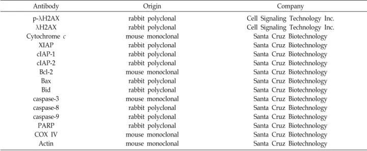
정상 및 PDTC와 DEC가 포함된 배지에서 배양된 세포들을 200 μl의 lysis buffer [25 mM Tris-Cl (pH 7.5), 250 mM NaCl, 5 mM ethylenediaminetetraacetic acid, 1% NP-40, 1 mM phe- nymethylsulfonyl fluoride, 5 mM dithiothreitol]로 용해한 후, 동량의 단백질을 sodium dodecyl sulphate (SDS)-polyacryl- amide gel을 이용하여 전기영동으로 분리하였다. 분리된 단백 질을 polyvinylidene fluoride (PVDF) membrane (Schleicher and Schuell, Keene, NH, USA)으로 전이시킨 후, 특정 단백질 에 대한 항체와 그에 대한 이차 항체 반응을 실시한 후 en- hanced chemiluminoesence (ECL) 용액(Amersham Bioscien- ces Co., Arlington Heights, IL, USA)을 적용시킨 다음 암실에
Table 1. Antibodies used in the present study
Antibody Origin Company
p-λH2AX λH2AX Cytochrome c
XIAP cIAP-1 cIAP-2 Bcl-2
Bax Bid caspase-3 caspase-8 caspase-9 PARP COX IV
Actin
rabbit polyclonal rabbit polyclonal mouse monoclonal
rabbit polyclonal rabbit polyclonal rabbit polyclonal mouse monoclonal
rabbit polyclonal rabbit polyclonal mouse monoclonal
rabbit polyclonal rabbit polyclonal rabbit polyclonal mouse monoclonal mouse monoclonal
Cell Signaling Technology Inc.
Cell Signaling Technology Inc.
Santa Cruz Biotechnology Santa Cruz Biotechnology Santa Cruz Biotechnology Santa Cruz Biotechnology Santa Cruz Biotechnology Santa Cruz Biotechnology Santa Cruz Biotechnology Santa Cruz Biotechnology Santa Cruz Biotechnology Santa Cruz Biotechnology Santa Cruz Biotechnology Santa Cruz Biotechnology Santa Cruz Biotechnology
서 X-ray film에 감광시켜 특정단백질의 양을 분석하였다. 본 실험에 사용된 항체들(Table 1)은 Santa Cruz Biotechnology Inc. (Santa Cruz, CA, USA) 및 Cell Signaling Technology Inc. (Beverly, MA, USA)에서 구입하였으며, 2차 항체로 사용 된 peroxidase-labeled donkey anti-rabbit 및 peroxidase-la- beled sheep anti-mouse immunoglobulin G는 Amersham Life Science에서 구입하였다. 동시에 미토콘드리아와 세포질 단백질을 분리하기 위해서는 Active Motif (Carlsbad, CA, USA)의 mitochondria isolation kit를 사용하였다.
미토콘드리아 막 전위(mitochondrial membrane poten- tial, MMP, Δψm) 분석
MMP를 측정하기 위해 dual-emission fluorescent dye인 5,5',6,6'-tetrachloro-1,1',3,3'-tetraethylbenzimidazolylcarbo- cyanine lodide (JC-1, Amersham Biosciences Co.)을 사용하였 다. 이를 위하여 PDTC와 DEC가 단독 또는 병합 처리된 AGS 세포를 모아 PBS로 수세 후, 10 μM의 JC-1 용액을 처리하여 20분 동안 상온에서 반응시켰다. 반응이 끝난 후 상층액을 제 거하고 PBS를 첨가하여 세포를 부유시킨 다음 flow cytometer 에 적용시켜 MMP의 변화를 측정하였다.
In vitro caspase 활성 측정
PDTC와 DEC 처리에 의한 caspase의 활성 변화 측정을 위 한 colorimetric assay kit는 R&D Systems (Minneapolis, MN, USA)에서 구입하였다. Caspase의 활성 측정을 위하여 정상 및 PDTC와 DEC가 처리된 배지에서 48시간 배양된 세포를 모은 뒤 단백질을 추출하고 정량하여 각각 150 μg의 단백질을 fluorogenic peptide 기질 100 μM이 함유된 extraction buffer 50 μl에 혼합하였으며, microtiter plate에 다시 extraction buf- fer에 희석하여 각 sample 당 총 volume이 100 μl가 되게 하였
다. 실험에 사용된 기질은 caspase-3의 경우에는 Asp-Glu- Val-Asp (DEVD)-p-nitroaniline (pNA)이었고 caspase-8의 경 우에는 Ile-Glu-Thr-Asp (IETD)-pNA이었으며, caspase-9은 Leu-Glu-His-Asp (LEHD)-pNA였다. 준비된 plate를 37℃에 서 2시간 동안 반응시킨 후 ELISA reader를 이용하여 405 nm 의 흡광도에서 활성의 정도를 측정하였다.
통계 분석
모든 실험결과는 평균±표준편차(standard deviation, SD) 로 표시하였고 SigmaPlot (Systat Software Inc., San Jose, CA, USA)을 이용하여 Student t-test를 이용하여 통계적 유의성을 얻었다.
결과 및 고찰
AGS 세포의 증식에 미치는 PDTC 및 DEC의 영향 AGS 세포의 증식에 미치는 PDTC 및 DEC의 병용 처리 조건 설정을 위하여 먼저 AGS 세포의 증식에 미치는 PDTC의 영향을 조사하여 80% 정도의 생존력을 보이는 농도인 500 nM 을 설정하였다(data not shown). 아울러 DEC가 AGS 세포의 증식에 미치는 영향을 조사한 결과, 설정된 농도의 범위 내 (5-50 μM)에서 유의적인 증식억제 효과를 관찰할 수 없었다 (Fig. 1A). 그러나 500 nM의 PDTC가 처리된 조건에서 DEC의 병용 처리 농도의 증가에 따라 AGS 세포의 증식이 DEC 처리 농도 의존적으로 감소되었으며(Fig. 1A), AGS 세포의 분지 형 성과 부착력 상실과 같은 형태적 변화가 유발되었다(Fig. 1B).
이러한 PDTC 및 DEC의 병용 처리에 따른 AGS 세포의 증식 억제가 apoptosis 유도와 연관성이 있는지를 조사한 결과, 전 형적인 apoptosis가 유도된 세포에서 관찰되는 염색질의 응축 (chromatin condensation)에 의한 apoptotic body의 형성과
A
B
C
Fig. 1. Inhibition of cell viability and induction of apoptosis by co-treatment with PDTC and DEC in AGS gastric cancer cells. (A) The cell viability was measured by an MTT assay. The data are expressed as the mean ± SD of three independent experiments (*p<0.05 vs. untreated control;
#p<0.05 vs. PDTC-treated group). (B) The morphological changes of AGS cells were observed under an inverted microscope. Representative photomicrographs of the morphological changes are presented (magnification,
×200). (C) The cells were stained with DAPI, and then the nuclei were photographed using a fluorescence mi- croscope using a blue filter (magnification, ×400).
Fig. 2. Effects of PDTC and DEC on the cell cycle progression in AGS gastric cancer cells. Cells were fixed and stained with PI for cell cycle distribution. The percentage of cells in each phase is presented. The data represent the aver- age of two independent experiments.
A
B
Fig. 3. Generation of ROS and induction of DNA damage by co-treatment with PDTC and DEC in AGS gastric cancer cells. (A) The production of ROS was measured using a flow cytometer. The data are the means of the two different experiments. (B) The membranes were probed with specific antibodies against p-γH2AX and γH2AX, and the proteins were visualized using an ECL detection system. Actin was used as an internal control.
같은 핵의 형태적 변형이 PDTC 및 DEC 단독 처리군에 비하 여 병용 처리군에서 높은 빈도로 나타났다(Fig. 1C). 아울러 세포주기 분포에서 apoptosis 유발을 의미하는 sub-G1기의 빈 도가 PDTC 및 DEC 단독 처리군(각각 8.14% 및 5.18%)에서는 대조군(2.67%)에 비하여 낮은 수준으로 증가되었으나, 병용 처리군(21.31%)에서는 대조군에 비하여 약 8배 이상 증가되었 다(Fig. 2).
이상에서 관찰된 PDTC 및 DEC의 병용 처리에 따른 AGS 세포 증식억제가 세포주기 진행에 어떤 영향을 주는지를 조사 한 결과, 500 nM의 PDTC가 단독 처리된 AGS 세포에서는 대조군에 비하여 S 및 G2/M기에 속하는 세포의 빈도가 다소 증가되었으며, 반면에 G1기의 빈도는 감소되었다(Fig. 2). 그 리고 50 μM의 DEC 단독 처리군에서도 G2/M기에 속하는 세 포의 빈도가 다소 증가되었으나, PDTC 단독 처리군에서 비하 여 변화의 정도가 상대적으로 낮게 나타났다. 아울러 PDTC 및 DEC의 병용 처리군에서는 PDTC 단독 처리군에서와 유사 한 경향성을 보여 주었다. 따라서 PDTC와 DEC의 병용 처리 가 단독 처리에 비하여 AGS 위암세포의 증식 억제에 상승 효과를 보여주었으며, 비록 세포주기 특정 시기의 arrest 현상 에 대한 증폭 효과는 미비하였으나, PDTC와 DEC의 병용 처
리에 의한 AGS 세포의 증식 억제는 apoptosis 유발과 연관이 있음을 알 수 있었다.
AGS 세포에서 ROS의 생성 및 DNA 손상에 미치는 PDTC 및 DEC의 영향
PDTC 및 DEC의 병용 처리에 따른 AGS 세포 증식억제와
A
B
Fig. 4. Effects of combined treatment with PDTC and DEC on the MMP values and cytochrome c expression in AGS gastric cancer cells. (A) The cells were stained with JC-1 dye and then analyzed to evaluate the changes in MMP.
An example of representative results according to each treatment concentration is presented. (B) The cytosolic and mitochondrial proteins were prepared, separated by SDS polyacrylamide gel electrophoresis and transferred to PVDF membranes. The membranes were probed with anti-cytochrome c antibody. Proteins were visualized us- ing an ECL detection system. Equal protein loading was confirmed by analysis of actin and cytochrome oxidase subunit VI (COX VI) in the each protein extract.
연관된 apoptosis 유도 과정에 ROS 생성이 관여하는지를 DCF-DA 염색을 통하여 조사하였다. Fig. 3A에 나타낸 바와 같이, PDTC와 DEC 단독 처리군에 비하여 병용 처리군에서 ROS의 축적이 매우 증가되어 산화적 스트레스가 강하게 유발 되었음을 알 수 있었다. 세포 내 산화적 스트레스는 미토콘드 리아 기능의 교란과 연계된 핵산의 손상을 동반할 가능성이 매우 높기 때문에[25, 40] DNA 손상이 병용 처리군에서 증가 되었는지를 조사하였다. 이를 위하여 DNA 이중 나선 손상 지표인 γH2AX 단백질의 인산화(serine 139) [32, 33]에 미치는 PDTC와 DEC의 단독 및 병용 처리에 따른 영향을 조사하였 다. Fig. 3B의 Western blot 분석 결과에 의하면 γH2AX 단백질 의 전체적인 발현에는 큰 변화 없이 PDTC와 DEC 단독 처리 군에서 γH2AX 단백질의 인산화 정도가 대조군에 비하여 다 소 증가되었으나, 병용 처리군에서는 더욱 증가되어 두 억제 제의 단독 처리에 비하여 병용 처리군에서는 DNA 손상 유발 이 증대되었음을 알 수 있었다. 따라서 PDTC와 DEC의 병용 처리에 따른 DNA 손상의 증가는 산화적 스트레스 및 apopto- sis 유발의 증가와 밀접한 연관성이 있음을 알 수 있었다.
AGS 세포의 미토콘드리아 기능 손상에 미치는 PDTC 및 DEC의 영향
다양한 자극에 의한 apoptosis 유도는 외인성(extrinsic) 및 내인성(intrinsic) 경로로 대별된다[9, 11]. 세포 내에서 생산되 는 ROS의 대부분은 미토콘드리아에서 만들어지며, 과도한 ROS의 생성은 DNA 손상과 연계된 미토콘드리아의 기능 손 상과 연관되며, 이는 intrinsic apoptosis 신호 경로의 직접적인 개시로 이어진다[3, 29]. 따라서 PDTC와 DEC의 병용 처리에 따른 apoptosis 및 DNA 손상의 증가가 미토콘드리아 기능 손상과 연계되어 있는지를 조사하기 위하여 MMP (Δψm)의 변화를 관찰하였으며, MMP의 소실은 미토콘드리아의 기능 이 손상되었음을 나타내는 지표이다[10, 22]. Fig. 4A에 나타낸 JC-1 염색에 의한 결과에 따르면, PDTC와 DEC의 단독 처리군 에서 MMP의 소실이 각각 12.7% 및 8.3%로 대조군(5.9%)에 비하여 각각 2.2 및 1.4배 정도 증가된 반면, 두 저해제의 병용 처리군에서는 54.8%로 나타나 대준군에 비하여 9.3배 이상 증 가되어 미토콘드리아 손상이 매우 증가되었음을 알 수 있었다.
한편, intrinsic apoptosis 경로의 활성은 미토콘드리아의 기 능 손상에 따른 미토콘드리아의 내막과 외막 사이에 존재하는 대표적인 apoptosis 유도 인자인 cytochrome c의 세포질 방출 이 필수적이다[11, 13]. 따라서 세포질과 미토콘드리아 분획 단백질을 이용한 immunoblotting을 수행하여 cytochrome c 의 분포에 미치는 PDTC와 DEC의 단독 및 병용 처리 효과를 비교하였다. Fig. 4B의 결과에 의하면, PDTC 및 DEC의 단독 처리군에서 세포질에서의 cytochrome c의 발현이 대조군에 비하여 유사하거나 다소 증가하였음에 비하여, 병용 처리군에 서는 매우 증가되었다. 아울러 미토콘드리아에서의 cyto-
chrome c 발현은 PDTC와 DEC의 병용 처리군에서 거의 검출 되지 않아, 병용 처리군에서 미토콘드리아에서 세포질로의 cytochrome c 유리 현상이 현저히 증가하였음을 알 수 있었다.
따라서 이러한 병용 처리군에서 단독 처리군에 비하여 cyto- chrome c의 세포질로의 유출이 증가한 것은 MMP의 소실 증 대와 직접적인 연관성이 있음을 의미한다.
AGS 세포의 caspase 활성에 미치는 PDTC 및 DEC의 영향
Apoptosis 유도의 두 대표적인 경로에는 caspase cascade가 중심적인 역할을 하는데, extrinsic 경로는 death receptor 분자 에 death ligand가 결합하여 caspase-8을 활성화시키고 이는 다시 caspase-3 및 caspase-7과 같은 effect caspase의 활성을 통하여 다양한 세포 내 poly (ADP-ribose) polymerase (PARP)을 포함한 기질 단백질의 분해를 유도한다[9, 13]. 반면
A B
Fig. 5. Activation of caspases and degradation of PARP by combined treatment with PDTC and DEC in AGS MCF-7 gastric cancer cells. (A) The membranes were probed with the indicated antibodies, and the proteins were visualized using an ECL detection system. Actin was used as an internal control. (B) The activities of caspases were evaluated using caspases colorimetric assay kits. The data are expressed as the mean ± SD of three independent experiments (*p<0.05 vs. untreated control; #p<0.05 vs. PDTC-treated group).
에 미토콘드리아가 관여하는 intrinsic 경로는 세포질로 유출 된 cytochrome c가 pro-caspase-9 및 apoptotic protease acti- vating factor-1 등과 apoptosome을 형성하여 caspase-9와 caspase-3의 활성을 촉진시킴으로써 기질 단백질의 절단을 촉 진하고 apoptosis를 종결하는 공동 경로로 유입된다[13, 31].
따라서 apoptosis 유도의 핵심 기전인 caspase cascade의 활성 이 PDTC와 DEC의 병용 처리 조건에서도 관여하는지의 여부 를 조사하였다. 이를 위하여 대표적인 extrinsic 및 intrinsic 경로의 개시 caspase에 해당되는 caspase-8 및 caspase-9를 포 함한, 두 caspase의 활성에 의하여 활성이 증가되는 effector caspase에 해당되는 caspase-3의 활성 여부를 조사한 결과, PDTC 및 DEC의 단독 처리군에서는 이들 caspase의 활성을 의미하는 active form의 발현이 미비하거나 검출되지 않았으 나, 병용 처리군에서는 active form 발현의 증가 또는 비활성 pro-form의 발현이 현저하게 감소되어 병용 처리군에서 단독 처리군에 비하여 3가지 caspase의 활성이 모두 증가되었음을 확인하였다(Fig. 5A). 이러한 각각의 caspase 활성의 정도를 정량적으로 평가하기 위하여 in vitro caspase 활성을 해당 cas- pase의 기질을 이용하여 조사한 결과에서도 단독 처리군에 비하여 병용 처리군에서 그들의 활성이 유의적으로 증가되었 음을 알 수 있었다(Fig. 5B). 아울러 활성화된 caspase-3의 기질 단백질로서 전형적인 apoptosis 유도 과정에서 분해가 관찰되 는 PARP의 발현[7, 11]을 조사해 본 결과, 단독 처리군에서는 관찰되지 않았던 PARP의 단편화가 병용 처리군에서 매우 증 가되어 PDTC와 DEC 병용 처리에 의한 AGS 세포의 apopto- sis 유도에는 caspase의 활성 증가가 관여하고 있음을 유추할 수 있다. 특히 PARP의 단편화는 산화적 스트레스에 의한 DNA damage 손상과도 밀접한 연관이 있는 것으로 알려져 [7], PDTC와 DEC 병용 처리에 의한 ROS의 발생 증가는 apop- tosis 유도 촉진 인자로 작용할 가능성을 보여 주었다.
AGS 세포에서 apoptosis 조절 주요 인자들의 발현에 미 치는 PDTC 및 DEC의 영향
한편 미토콘드리아 기능 손상과 연계된 intrinsic apoptosis 유도 조절에 가장 중요하게 작용하는 유전자군이 Bcl-2 family 이다[9, 14]. Bcl-2 family에 속하는 단백질은 중, Bax를 포함한 pro-apoptotic 단백질이 Bcl-2와 같은 anti-apoptotic 단백질과 비교하여 상대적으로 발현이 증가하면 Bax가 미토콘드리아로 이동하여 MMP의 소실과 세포질로의 cytochrome c 방출을 유도하여 intrinsic apoptosis 경로의 활성을 촉진한다[11, 14].
아울러 Bcl-2 family에 속하는 pro-apoptotic BH3-interacting domain death agonist인 Bid는 extrinsic과 intrinsic 경로의 신호 전달 증폭에 관여할 수 있는데, 활성화된 caspase-8에 의 하여 Bid가 활성형인 tBid (truncated form of Bid)로 전환하게 되면 미토콘드리아에서 세포질로의 cytochrome c 방출을 촉 진한다[9, 37]. 따라서 PDTC와 DEC 병용 처리에 의한 AGS 세포의 apoptosis 유도에서 이들 Bcl-2 family 단백질의 발현 변화를 조사한 결과, Bcl-2의 발현은 단독 처리군에 비하여 병 용 처리군에서 현저하게 감소된 반면, Bax의 발현은 매우 증가 되었다(Fig. 6). 또한 Bid의 발현은 PDTC와 DEC 병용 처리군 에서 단독 처리군에 비하여 현저한 감소를 보여 tBid로 전환되 었을 가능성을 보여 주었다. 한편 inhibitor of apoptosis pro- tein (IAP) family에 속하는 단백질들은 caspase와의 직접적인 결합을 통하여 그들의 활성을 억제하여 apoptotic 활성을 억제 할 수 있는 것으로 알려져 있다[4, 24]. 본 연구의 결과에 의하 면 PDTC와 DEC가 함께 처리된 AGS 세포에서 IAP family에 해당되는 단백질들(XIAP, cIAP-1 및 cIAP-2)의 발현이 매우 감소되었으며(Fig. 6), 이러한 감소는 caspase의 활성 증가에 기여하여 apoptosis 유도를 촉진하였을 것으로 생각된다.
이상의 결과를 종합하면, 세포독성을 나타내지 않는 범위의 농도에서 DEC는 PDTC에 의한 AGS 세포 증식 억제를 유의적
Fig. 6. Effects of combined treatment with PDTC and DEC on the expression of apoptosis regulatory proteins in AGS gastric cancer cells. The membranes were probed with the indicated antibodies. Proteins were visualized using an ECL detection system. Equal protein loading was confirmed by the analysis of actin in the protein extracts.
으로 촉진시켰으며, 이는 apoptosis 유도 효과의 증가와 연관 성이 있었다. 아울러 PDTC와 DEC의 병용 처리는 단독 처리 군에 비하여 산화적 스트레스를 향상시키면서 DNA 손상을 유도하였다. 또한 두 억제제의 병용 처리에 의한 산화적 스트 레스는 미토콘드리아 기능 손상을 촉진하였을 것으로 추정되 며 이로 인한 미토콘드리아에서 세포질로의 cytochrome c 유 리 증가는 intrinsic apoptosis 신호 개시에 기여하였을 것이다.
아울러 PDTC와 DEC의 병용 처리에 의한 apoptosis 유발에서 caspase-8의 활성 증가에 따른 extrinsic 경로의 활성을 촉진하 였으며, 이는 Bid 경로 활성을 통한 intrinsic 신호의 증폭에 기여하였을 것으로 추정된다. 비록 핵산 합성 및 NF-κB 신호 계와 연계된 지속적인 연구가 요구되지만, 본 연구의 결과는 두 억제제의 독성을 최소화하면서 암세포 증식 제어를 위한 가능성을 보여준 것으로 항암제 병용 처리에 대한 근거 자료 로서의 활용도가 매우 높을 것으로 생각된다.
감사의 글
이 논문은 2017년도 김천대학교 교내학술연구비지원에 의 한 것임.
References
1. Appleton, K., Mackay, H. J., Judson, I., Plumb, J. A., McCor- mick, C., Strathdee, G., Lee, C., Barrett, S., Reade, S., Jadayel, D., Tang, A., Bellenger, K., Mackay, L., Setanoians, A., Schatzlein, A., Twelves, C., Kaye, S. B. and Brown, R. 2007.
Phase I and pharmacodynamic trial of the DNA methyl- transferase inhibitor decitabine and carboplatin in solid tumors. J. Clin. Oncol. 25, 4603-4609.
2. Arlt, A., Gehrz, A., Müerköster, S., Vorndamm, J., Kruse,
M. L., Fölsch, U. R. and Schäfer, H. 2003. Role of NF-kappaB and Akt/PI3K in the resistance of pancreatic carcinoma cell lines against gemcitabine-induced cell death. Oncogene 22, 3243-3251.
3. Birch-Machin, M. A., Russell, E. V. and Latimer, J. A. 2013.
Mitochondrial DNA damage as a biomarker for ultraviolet radiation exposure and oxidative stress. Br. J. Dermatol. 169, 9-14.
4. Chaudhary, A. K., Yadav, N., Bhat, T. A., O'Malley, J., Kumar, S. and Chandra, D. 2016. A potential role of X-linked inhibitor of apoptosis protein in mitochondrial membrane permeabilization and its implication in cancer therapy. Drug Discov. Today 21, 38-47.
5. Chen, D., Peng, F., Cui, Q. C., Daniel, K. G., Orlu, S., Liu, J. and Dou, Q. P. 2005. Inhibition of prostate cancer cellular proteasome activity by a pyrrolidine dithiocarbamate-cop- per complex is associated with suppression of proliferation and induction of apoptosis. Front. Biosci. 10, 2932-2939.
6. Daskalakis, M., Blagitko-Dorfs, N. and Hackanson, B. 2010.
Decitabine. Recent Results Cancer Res. 184, 131-157.
7. Decker, P. and Muller, S. 2002. Modulating poly (ADP-ri- bose) polymerase activity: potential for the prevention and therapy of pathogenic situations involving DNA damage and oxidative stress. Curr. Pharm. Biotechnol. 3, 275-283.
8. Di Nicuolo, F., Serini, S., Boninsegna, A., Palozza, P. and Calviello, G. 2001. Redox regulation of cell proliferation by pyrrolidine dithiocarbamate in murine thymoma cells trans- planted in vivo. Free Radic. Biol. Med. 31, 1424-1431.
9. Dorstyn, L., Akey, C. W. and Kumar, S. 2018. New insights into apoptosome structure and function. Cell Death Differ.
25, 1194-1208.
10. Farsinejad, S., Gheisary, Z., Ebrahimi Samani, S. and Aliz- adeh, A. M. 2015. Mitochondrial targeted peptides for can- cer therapy. Tumour Biol. 36, 5715-5725.
11. Fulda, S. and Debatin, K. M. 2006. Extrinsic versus intrinsic apoptosis pathways in anticancer chemotherapy. Oncogene 25, 4798-4811.
12. Gong, X. G., Lv, Y. F., Li, X. Q., Xu, F. G. and Ma, Q. Y.
2010. Gemcitabine resistance induced by interaction be- tween alternatively spliced segment of tenascin-C and an- nexin A2 in pancreatic cancer cells. Biol. Pharm. Bull. 33, 1261-1267.
13. Hajra, K. M. and Liu, J. R. 2004. Apoptosome dysfunction in human cancer. Apoptosis 9, 691-704.
14. Hata, A. N., Engelman, J. A. and Faber, A. C. 2015. The BCL2 family: Key mediators of the apoptotic response to targeted anticancer therapeutics. Cancer Discov. 5, 475-487.
15. Hengartner, M. O. 2000. The biochemistry of apoptosis.
Nature 407, 770-776.
16. Hu, Q., Sun, W., Wang, C. and Gu, Z. 2016. Recent advances of cocktail chemotherapy by combination drug delivery systems. Adv. Drug Deliv. Rev. 98, 19-34.
17. Iessi, E., Logozzi, M., Mizzoni, D., Di Raimo, R., Supuran, C. T. and Fais, S. 2017. Rethinking the combination of proton exchanger inhibitors in cancer therapy. Metabolites 8, E2.
18. Janssens, S. and Tschopp, J. 2006. Signals from within: the
DNA-damage-induced NF-kappaB response. Cell Death Differ. 13, 773-784.
19. Kantari, C. and Walczak, H. 2011. Caspase-8 and bid: caught in the act between death receptors and mitochondria. Biochim.
Biophys. Acta. 1813, 558-563.
20. Katsman, A., Umezawa, K. and Bonavida, B. 2009. Chemo- sensitization and immunosensitization of resistant cancer cells to apoptosis and inhibition of metastasis by the specific NF-kappaB inhibitor DHMEQ. Curr. Pharm. Des. 15, 792-808.
21. Kaufmann, T., Strasser, A. and Jost, P. J. 2012. Fas death receptor signalling: roles of Bid and XIAP. Cell Death Differ.
19, 42-50.
22. Kroemer, G. and Pouyssegur, J. 2008. Tumor cell metabo- lism: cancer's Achilles' heel. Cancer Cell 13, 472-482.
23. Malaguarnera, L., Pilastro, M. R., Vicari, L., Dimarco, R., Manzella, L., Palumbo, G. and Messina, A. 2005. Pyrrolidi- nedithiocarbamate induces apoptosis in human acute mye- logenous leukemic cells affecting NF-kappaB activity. Cancer Invest. 23, 404-412.
24. Mohamed, M. S., Bishr, M. K., Almutairi, F. M. and Ali, A. G. 2017. Inhibitors of apoptosis: Clinical implications in cancer. Apoptosis 22, 1487-1509.
25. Moloney, J. N. and Cotter, T. G. 2018. ROS signalling in the biology of cancer. Semin. Cell Dev. Biol. 80, 50-64.
26. Nakajima, K., Nangia-Makker, P., Hogan, V. and Raz, A.
2017. Cancer self-defense: An immune stealth. Cancer Res.
77, 5441-5444.
27. Nguyen, D. P., Li, J., Yadav, S. S. and Tewari, A. K. 2014.
Recent insights into NF-κB signalling pathways and the link between inflammation and prostate cancer. BJU Int. 114, 168-176.
28. Nguyen, H. S., Shabani, S., Awad, A. J., Kaushal, M. and Doan, N. 2018. Molecular markers of therapy-resistant glio- blastoma and potential strategy to combat resistance. Int.
J. Mol. Sci. 19, E1765.
29. Pelicano, H., Carney, D. and Huang, P. 2004. ROS stress in cancer cells and therapeutic implications. Drug Resist.
Updat. 7, 97-110.
30. Ranganathan, P., Yu, X., Santhanam, R., Hofstetter, J., Walker, A., Walsh, K., Bhatnagar, B., Klisovic, R., Vasu, S., Phelps, M. A., Devine, S., Shacham, S., Kauffman, M., Marcucci, G., Blum, W. and Garzon, R. 2015. Decitabine pri-
ming enhances the antileukemic effects of exportin 1 (XPO1) selective inhibitor selinexor in acute myeloid leukemia.
Blood 125, 2689-2692.
31. Reubold, T. F. and Eschenburg, S. 2012. A molecular view on signal transduction by the apoptosome. Cell Signal. 24, 1420-1425.
32. Rogakou, E. P., Pilch, D. R., Orr, A. H., Ivanova, V. S. and Bonner, W. M. 1998. DNA double-stranded breaks induce histone H2AX phosphorylation on serine 139. J. Biol. Chem.
273, 5858-5868.
33. Rybak, P., Hoang, A., Bujnowicz, L., Bernas, T., Berniak, K., Zarębski, M., Darzynkiewicz, Z. and Dobrucki, J. 2016. Low level phosphorylation of histone H2AX on serine 139 (γ H2AX) is not associated with DNA double-strand breaks.
Oncotarget 7, 49574-49587.
34. Shao, L., Wu, L. and Zhou, D. 2012. Sensitization of tumor cells to cancer therapy by molecularly targeted inhibition of the inhibitor of nuclear factor κB kinase. Transl. Cancer Res. 1, 100-108.
35. Stresemann, C. and Lyko, F. 2008. Modes of action of the DNA methyltransferase inhibitors azacytidine and decitabine.
Int. J. Cancer 123, 8-13.
36. Ta, M. H., Liuwantara, D. and Rangan, G. K. 2015. Effects of pyrrolidine dithiocarbamate on proliferation and nuclear factor-κB activity in autosomal dominant polycystic kidney disease cells. BMC Nephrol. 16, 212.
37. Tummers, B. and Green, D. R. 2017. Caspase-8: regulating life and death. Immunol. Rev. 277, 76-89.
38. Wadhwa, S. and Mumper, R. J. 2013. D-penicillamine and other low molecular weight thiols: review of anticancer ef- fects and related mechanisms. Cancer Lett. 337, 8-21.
39. Wijermans, P. W., Ruter, B., Baer, M. R., Slack, J. L., Saba, H. I. and Lubbert, M. 2008. Efficacy of decitabine in the treatment of patients with chronic myelomonocytic leuke- mia (CMML). Leuk. Res. 32, 587-591.
40. Zhang, L., Li, J., Zong, L., Chen, X., Chen, K., Jiang, Z., Nan, L., Li, X., Li, W., Shan, T., Ma, Q. and Ma, Z. 2016. Reactive oxygen species and targeted therapy for pancreatic cancer.
Oxid. Med. Cell. Longev. 2016, 1616781.
41. Zhang, X. Y. and Zhang, P. Y. 2016. Combinations in multi- modality treatments and clinical outcomes during cancer.
Oncol. Lett. 12, 4301-4304.
초록:핵산합성 억제제인 decitabine과 NF-κB 활성 저해제인 PDTC의 병용 처리에 의한 인체 위암세 포사멸 효과 증진
최원경1*․최영현2,3*
(1김천대학교 스포츠재활학과, 2동의대학교 한의과대학 생화학교실, 3동의대학교 항노화연구소)
Cytidine analog decitabine (DEC)은 핵산 합성의 억제제로서 골수이형성 증후군 및 급성 골수성 백혈병 치료 제로 사용되고 있다. 산화질소 합성에서 번역 단계를 억제하는 것으로 알려진 ammonium pyrrolidine dithio- carbamate (PDTC)는 NF-κB의 대표적인 억제제이다. 본 연구에서는 인체 위암 AGS 세포를 대상으로 DEC와 PDTC의 병용 처리에 따른 세포증식 억제 기전을 조사하였다. 본 연구의 결과에 따르면 PDTC에 의한 AGS 세포 의 증식 억제 효과는 DEC에 의해 농도 의존적으로 유의하게 증가하였으며, 이는 G2/M기의 세포주기 정지 및 apoptosis 유도와 관련이 있었다. PDTC와 DEC의 병용 처리에 의한 세포 사멸의 유도는 DNA 손상 유도와 관련 이 있음을 H2AX의 인산화 증가로 확인하였다. 아울러 PDTC와 DEC의 병용 처리는 미토콘드리아 막 전위의 파괴 를 유도하고, 세포 내 활성산소종(ROS)의 생성과 Bax의 발현을 향상시키고, Bcl-2 발현을 감소시켰으며 미토콘드 리아에서 세포질로의 cytochrome c 유출을 증가시켰다. 또한 PDTC과 DEC의 병용 처리는 외인성 및 내인성 apoptosis 개시 caspase에 해당하는 caspase-8과 caspase-9의 활성뿐만 아니라 caspase-3의 활성화와 PARP 단백질 의 분해를 유도하였다. 결론적으로 본 연구의 결과는 PDTC와 DEC의 병용 처리가 DNA 손상을 유발하고, ROS 증가와 연계된 외인성 및 내인성 apoptosis 사멸 경로를 활성화시킴으로써 AGS 세포의 증식을 억제하였음을 의 미한다.