․접수 : 2011년 9월 15일 ․수정접수 : 2011년 11월 24일 ․채택 : 2011년 12월 7일
*교신저자 : 신용철, 130-872 서울특별시 동대문구 회기동 1번지, 경희대학교 한의과대학 예방의학교실 전화 : 02-961-9237, 전자우편 : [email protected]
가시오가피 에탄올추출물의 AGS위암세포주에서 세포주기억제효과
이선동1)·고성규2)·신헌태3)·신용철2)✽
1)상지대학교 한의과대학 예방의학교실
2)경희대학교 한의과대학 예방의학교실
3)동신대학교 한의과대학 예방의학교실
The antitumor activities of Acanthopanax senticosus Harms(ASH) in human gastric cancer AGS cell lines
Sundong Lee,1) Seong-Gyu Ko,2) Heon-tae Shin3) & Yong-Cheol Shin2)✽
1)School of Oriental Medicine Sangji University
2)School of Oriental Medicine Kyung Hee University
3)School of Oriental Medicine Dongshin University
Abstract
Objectives : The research was conducted to confirm the effect of Acanthopanax senticosus harms(ASH) on the anti-tumor activities in AGS human gastric cancer cells.
Methods : To examine the potential anti-tumor effect of ASH, we performed many experiments. After processing AGS cancer cells with varying concentrations 80% ethanol ASH extract, analyses by MTT, flow cytometer(FACS) and western blot were used.
Results : AGS cancer cells showed decreased cell proliferation and increased contents of S phase when treated with ASH. Moreover, the Western blot experiment showed that ASH affected S phase cell cycle-related molecules(Cyclin A, p21 and p16) in AGS cells. ASH also inhibited EGFR-STAT3 pathway in AGS human gastric cancer cells.
Conclusion : Based on these results, we observed that ASH arrested the cell cycle at S phase and inhibited the phosphorylation of EGFR and STAT3 proteins which reduce the cell cycle and the manifestation of the genes that are related to inhibiting cell growth in AGS cells. It can be concluded that ASH can be used in developing medicine for gastric cancer.
Key words:Acanthopanax senticosus Harms(ASH), Gastric cancer, cell cycle, apoptosis,
EGFR-STAT3 pathway
I. 서 론
위암은 일본, 중국, 한국 등의 아시아에서 높 게 발생하며, 남아메리카, 동유럽 등에서도 다 빈도로 발생한다.
1-3)우리나라에서도 남자에서 가장 많이 발생하는 암이며, 여자의 경우 갑상 샘암, 유방암, 위암, 대장암의 순으로 발생된다.
4)위암의 병기별 생존율은 1기 93.9%, 2기 74.1%, 3기 42.3%, 4기는 5.5%로 3기 이상 진행된 경 우 여전히 사망률이 높은 실정이다.
5)위암을 유 발할 수 있는 위험요소로는 흡연, 염분, 아질산 염이 풍부한 음식과 야채, 과일의 부족한 섭취, 헬리코박터 파일로리균 등이 보고되고 있다.
6)하지만 현재 이러한 위암의 조기발견에도 불구 하고 임상적으로 사용되는 치료제에서 부작용 이 나타나고 있다.
세포주기는 하나의 세포가 성장하여 두 개의 세포로 분열되는 과정으로 G1, S, G2/M기로 나뉘며, cyclin과 cyclin-dependent kinases (CDKs ; serine/threonine protein kinases)가 결합하여 세포주기가 G1기를 거쳐 S기와 G2기 를 지나 mitosis가 일어나는 단계로 정의한다.
7, 8)첫 번째 휴지기인 G1기는 세포가 분열된 직후 다음단계로 넘어가기 위한 준비단계로 세포 내 인자들이 분열하기에 충분한 양이 만들어 지지 않으면 G1기에 머무르게 하여 다음단계인 S기 로 이행하지 못하게 하는 관문(check point)이 있다. DNA의 복제가 이루어지는 S기가 완료되 면 두 번째 휴지기인 G2기에 의해 M기로의 이 행을 조절한다.
7-9)각 단계에서 적절히 조절되 지 않은 cyclin-dependent kinases (CDKs) 활성 은 세포분열을 적절히 억제하지 못하고 이상증
식을 야기하여, 암의 원인이 되기도 한다.
9)오가피(Acanthopanax senticosus Harms)는 두릅나무과(Araliaceae)에 속하는 관목식물로 오 가피나무의 뿌리, 줄기 및 가지의 껍질을 말하 며 몇몇 종류 중 유효성분이 많아 약효가 가장 좋다고 밝혀진 가시오가피는 러시아 남동쪽, 중 국 북동쪽, 한국, 아시아 남동쪽, 일본 홋카이도 섬에 많이 분포하고 있고,
10)이는 서양에서 한국 인삼에 버금가는 효능을 가지고 있다 하여 고 려인삼(Korean ginseng)과 비교해서 시베리아 인삼(Siberian ginseng)이라 알려져 있다.
11)가시 오가피는 뿌리와 잎 모두 약용으로 사용이 가 능하고,
12, 13)isofraxidin, chlorogenic acid, rutin 등의 활성성분이 포함되어 있고
14-16)특히 iso- fraxidin의 함유량이 가시오가피의 질을 결정 하는데 중요하다고 알려져 있다.
17)그 밖에도 acanthosides, eleutherosides, flavon, senticoside, triterpenic, saponin, vitamin, mineral 등이 포 함되어 있으며 이들이 가시오가피의 다양한 생 물학적 활성과 관련된 것으로 보인다.
18)이들은 환경적응약물(adaptogenic medicine)로 알려져 있어
19)육체적, 화학적, 생물학적 스트레스 유 발인자로부터 인체에 비특이적 반응을 일으키 게 되고 최종적으로 여러 인자에 대항하여 인 체 내부 환경의 균형을 유지한다. 또한 항알레 르기,
20)항산화,
21)항염증,
22)항암
23)의 효능도 가 지고 있을 뿐만 아니라 류머티즘, 고혈압, 허혈 성심장질환, 간염, 위궤양 치료제로도 사용되고
있다.
21, 24-26)아직까지 위암에 대한 가시오가피
의 연구는 미흡한 실정으로 본 연구에서는 가
시오가피(Acanthopanax senticosus Harms)의
위암세포주의 증식을 억제하는 기전에 관한 내
용을 보고하는 바이다.
II. 연구방법 1. 세포주 및 세포배양
본 실험에 사용한 AGS 세포주(a human ga- stric carcinoma epithelial cell line)는 점액분비 표피세포를 포함하는 인간 위 선암종에서 유래 되었고,
27)이 세포주는 까다로운 특징들을 지니 고 있지 않아 널리 실험에 사용되고 있다.
28-31)실험에 사용하기 위해서 미국 세포주은행(Ame- rican Type Culture Collection, USA)을 통하 여 구입하였고, RPMI-1640(Welgene, Korea) 배지를 사용하여 각각 10%(v/v) 우태혈청 [heat- inactivated fetal bovine serum(Welgene)]과 1% 항생제 [antibiotic-antimycotic(Welgene, Korea)]를 넣어 5% CO
2가 든 37℃ humidified incubator(Sanyo, model MCO96, Japan)에 키 운다. 배양 된 각각의 세포는 일주일에 2~3회 DPBS(Welgene, Korea)로 세척한 후 0.05%
Trypsin-0.02% EDTA(Welgene, Korea)로 부 착된 세포를 분리하여 원심분리 한 후 세포를 재분주하여 계대 배양하였다.
2. 항체
실험에 사용한 α-actin, Bax, Bcl-2, CDK2, cleaved caspase-9, cyclin A, cyclin D, cyclin E, ERK, p16, phospho-ERK(Tyr-204), pro- caspase-9의 1차 항체는 Santa Cruz Biotechno- logy, Inc(Santa Cruz, CA, USA)에서 cleaved caspase-3, cleaved caspase-8, Fas, PARP, phospho-PI3K, procaspase-3, EGFR, phospho- EGFR(Y1068 & Y1173) 1차 항체는 Cell Si- gnaling Technology, Inc(Danvers, MA, USA) 에서 구입하였다. PI3K, phospho-STAT3(Tyr 705)의 1차 항체는 Upstate Biotechnology, Inc
(Lake Placid, NY, USA)에서 구입했으며 p27 1차 항체는 Transduction Laboratories(Lexing- ton, KY, USA)에서 구입하였다. HRP가 결합 되어 있는 mouse, rabbit의 2차 항체는 Calbio- chem(San Diego, CA, USA)에서, goat는 Jackson ImmunoResearch(West Grove, PA, USA)에서 구입하였다.
3. 가시오가피 추출
실험에 사용된 한약재인 가시오가피(Acantho- panax senticosus(Rupr.etMaxim) Harms, ASH) 는 옴니허브(Omniherb, Yeongcheon, Korea)에 서 2010년에 구입하였다. 이것을 100g 정량 후 분쇄하여 80% 에탄올(1ℓ⨉ 2)을 가하여 30분 씩 2회에 걸쳐 초음파진동을 이용하여 추출액 을 획득하였다. 획득한 추출액을 지름이 185 mm 인 종이여과지(Whatman, Maidstone, England) 를 사용하여 감압 여과 한 후, 감압농축기(Eyela, Japan)를 사용하여 농축하였다. 30분 동안 예비 동결을 시킨 후 동결건조기(Freezedryer, Matsu- shita, Japan)를 사용하여 48시간 동결건조 하 였다. 최종적으로 분말 3.9g(수율 3.9%)의 ASH 분말을 얻어 DMSO(Dimethyl sulfoxide, sigma, USA)에 vortexing하여 녹인 후 syringe filter (0.20 ㎛, Sartorius, Germany)를 이용하여 여과 한 후 실험에 사용하였다.
4. 세포증식 측정
80% 에탄올로 추출하고 DMSO에 녹여 놓은 가시오가피의 세포증식 정도를 MTT assay를 이용하여 확인하였다. 위암 AGS세포주를 well 당 배지 100 ㎕ 안에 각각 5×10
3cells의 초기세 포 농도로 96-well pate에 분주하였다. 5% CO
2가 든 37℃ humidified incubator에서 24시간
동안 배양 시킨 후에 DMSO에 녹인 가시오가
피 에탄올 추출물과 DMSO(negative control)
를 농도별(0, 10, 50, 100, 250, 500 ㎍/㎖)로 각 각 분주한 세포에 처리하였다. 가시오가피 처리 후 48, 72시간 동안 37℃ humidified incubator 에서 배양하였다. 각각의 well에 MTT〔3-(4, 5-dimethylthiazol-2-yl)-2, 5-diphenyl- tetrazolium bromide〕 solution을 첨가(MTT solution 20 ㎕/배지 100 ㎕)하고 5% CO
2가 든 37℃ humidified incubator에서 2시간 정도 반 응 시킨 후 440 nm파장에서 ELISA reader(Mo- lecular Devices, Palo Alto, CA)를 사용하여 세포 증식 정도를 측정하였다.
5. 세포형태관찰
가시오가피 에탄올 추출물에 의한 세포형태 의 변화를 관찰하기 위하여 위암 AGS 세포주 를 100 mm dish에 6×10
5cells의 초기농도로 분 주하여 5% CO
2가 든 37℃ humidified incu- bator에서 24시간 동안 배양시켰다. 아무것도 처 리하지 않은 군(control group), DMSO 처리군 (0.2%, negative control group), 가시오가피 에 탄올 추출물(ASH) 처리군(400 ㎍/㎖, treatment group)을 12, 24, 36시간 배양한 후 현미경을 통 하여 세포형태를 관찰하였다.
6. 세포사멸 및 세포주기 측정
가시오가피 에탄올 추출물이 위암 AGS세포 주의 세포사멸과 세포주기에 미치는 영향을 시 간 별로 확인하기 위하여 100 mm dish에 6×10
5cells의 초기농도로 분주하여 5% CO
2가 든 37℃
humidified incubator에서 배양하여 이것을 24 시간 후에 가시오가피 에탄올 추출물과 DMSO 를 각각 0.2%와 400 ㎍/㎖ 처리하여 12, 24, 36 시간 동안 각각 배양하였다. 각각의 시간마다 세포를 수집하기 위해 트립신을 처리하여 dish 바닥에 붙어있는 세포들을 떼어내고 이를 튜브 에 모아 1,500 rpm으로 5분 동안 원심 분리시켜
튜브바닥에 세포들을 모은다. 세포를 깨끗하게 하기 위해 Cold-PBS를 사용하여 다시 원심분 리 한다. 상등액을 모두 제거하고, 0.5% Tween- 20이 첨가된 95% 에탄올을 사용하여 세포주를 고정한 후, -20℃에서 최소한 30분 정도 반응 시킨다. 반응이 끝나면 두 번 정도 Cold-PBS를 사용하여 원심분리로 세포주를 씻어내고 어두운 상온에서 propidium iodide(50 ㎍/㎖, 1% sodium citrate. 50 ㎍/㎖ RNase A가 포함)를 사용하여 세포주를 염색했다. 세포사멸과 세포주기는 모 두 유세포분석기(BD Biosciences)를 사용하여 측정한다. 특히 세포사멸은 DNA contents histo- gram을 이용하고 세포주기는 ModFit LT V2.0 프로그램을 사용하여 확인하였다. 또한 오가피 에 의한 세포사멸과 세포주기의 변화를 농도 별로 확인하기 위해 세포를 분주한 후 24시간 배양시킨 후에 오가피를 100, 200, 400 ㎍/㎖ 처 리하였다. 24시간 동안 약 처리를 한 후에 위의 방법과 동일하게 세포를 수집하였다.
7. Western blotting
다양한 조건의 세포들을 RIPA buffer(150
mM NaCl, 1% NP-40, 0.5% DOC, 0.1% SDS,
50 mM Tris(pH8.0), 1 mM EDTA, 1 mM PMSF,
1 mM NaF, 1 mM Na3VO4, 1 ㎍/㎖ aprotinin,
leupeptin, pepstatin)를 사용하여 lysis시켜 단
백질만을 분리한다. 단백질 정량은 Bio-Rad
protein assay kit을 사용하였으며 SDS-PAGE
시 동량의 단백질을 loading하여 separating한
후 nitrocellulose membrane으로 옮긴다. 그 후,
membrane을 0.1% Tween 20이 포함 된 1X-
PBS(1X-PBST)에 skim milk와 BSA를 각각
1%씩 넣어 1시간 동안 blocking시킨다. mem-
brane을 다양한 1차 항체와 4℃에서 반응시킨
다. 0.1% Tween 20이 포함된 1X-PBS(1X-
PBST)로 15분 동안 washing(15분⨉3회)하고,
2차항체를 1시간 정도 상온에서 반응시킨다. 그
48h 120
100 80 60 40 20 0
ASH DMSO
0 10 50 100 250 500 Concentrations(ug/ml)
***
*** **
*** **
** **
*
72h 120
100 80 60 40 20
0 0 10 50 100 250 500 Concentrations(ug/ml)
** **
*****
***
***
***
ASH DMSO
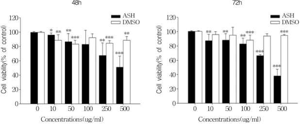
Figure 1. Effect of Acanthopanax senticosus Harms(ASH) on cell viability in AGS gastric cancer cells.
Cell viability was measured by MTT assay in AGS cells, in the presence of diverse ASH or DMSO concentrations after 48 and 72 h. 100% corresponds to cell viability under basal conditions(i.e., no ASH and DMSO for AGS cells). The data shown is a representative of three independent experiments which gave similar results. Bars, SD. (*p < 0.05, **p < 0.01, ***p < 0.001)
후 0.1% Tween 20이 포함된 1X-PBS(1X-
PBST)로 1시간 washing(10분⨉6회)을 하고 che- miluminescence detection system(Amersham- Pharmacia Biotech, Buckinghamshire, England) 을 사용하여 X-ray film으로 결과를 확인하였다.
8. 자료분석
본 실험에서 얻은 결과의 측정값을 Mean (평균)±S.E(표준오차)로 처리했으며, student t- testmethod(unpaired)로 분석하여 p값이 0.05 이하일 때 통계적 유의성을 인정하였다.
III. 연구결과
1. 가시오가피 에탄올 추출물의 위암세포주 증식 억제 효능
가시오가피가 AGS 위암세포주에서 항암효과
가 있는지 확인하기 위해 가시오가피에 대한 세포주의 증식억제여부를 MTT assay를 통하 여 확인하였다. 결과 DMSO군(가시오가피를 녹 인 negative․vehicle group)에 비해 가시오가피 를 농도별로(10, 50, 100, 250, 500 ㎍/㎖) 처리했 을 때, 위암 세포주의 증식이 억제되는 것을 확 인할 수 있었다. 또한 이러한 결과는 시간 의존 적으로 48시간 보다 72시간에서 가시오가피에 의한 세포의 증식 억제정도가 큰 것을 확인하 였다(Fig 1).
2. 가시오가피 에탄올 추출물에 의한 세포형태 변화 관찰
세포증식 억제에 효과가 있는 가시오가피 400
㎍/㎖ 농도를 AGS 세포주에 시간별(12, 24, 36 시간)로 처리하였을 때, 세포형태에 변화가 있 는지 현미경으로 관찰하였다(Fig 2). 결과 아무 것도 처리하지 않은 control군과 DMSO 0.2%
를 처리한 대조군에서는 36시간이 되었을 때 세
Con DMSO 0.2% ASH 400 ㎍/㎖
12h
24h
36h
⨉400
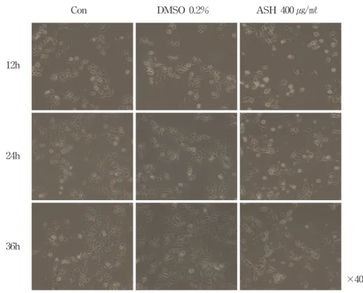
Figure 2. Acanthopanax senticosus Harms(ASH) changed the morphology of AGS gastric cancer cells.
AGS cells were exposed to 400 μg/ml of ASH or 0.2% DMSO for 12, 24 and 36 h. Morphology of cells were observed under light microscopy and photographed(x200). The data shown is a representative of three independent experiments which gave similar results(Con ; control, ASH ; Acanthopanax senticosus Harms).
포가 증식하였으며, 둥근 형태의 세포모양에도 변화가 없었다. 반면, 가시오가피 400 ㎍/㎖ 농 도를 처리한 실험군에서는 대조군과 비교했을 때, 12시간부터 생존세포 수에 차이가 보였으며, 36시간이 되었을 때는 생존세포 수 차이 뿐 아 니라 세포형태도 길쭉한 모양의 세포가 관찰 되었다.
3. 위암세포주에서 가시오가피 에탄올 추출물에 의한 세포 사멸조절 효과
가시오가피에 의한 위암 세포주의 증식억제
와 세포사멸과의 관련성을 확인하기 위해 AGS 세포주에 400 ㎍/㎖ 농도로 시간별(12, 24, 36시 간)로 처리 후, 유세포분석기를 이용하여 sub- G1의 DNA contents를 확인하였다(Fig 3A).
가시오가피가 AGS 세포주의 증식을 억제함에
도 불구하고, sub-G1은 아무것도 처리하지 않
은 그룹(Con)과 vehicle을 처리한 그룹(DMSO)
에 비해 그리 크게 증가하지 않았다. 또한 가
시오가피 농도에 의한 영향을 확인하기 위해
농도별(100, 200, 400 ㎍/㎖)로 24시간 동안 처
리 후, sub-G1의 DNA contents를 확인하였다
(Fig 3B). 결과 농도 의존적으로 sub-G1 약간
증가하기는 하지만, Con, DMSO에 비해 위암
DMSO ASH Time(h) C 12 24 12 24 DMSO ASH
Time(h) C 12 24 12 24 Fas
cleaved caspase-8 cleaved caspase-3 PARP
-actin C
Bas Bcl-2 procaspase-9 cleaved caspase-9 procaspase-3 cleaved caspase-3
-actin
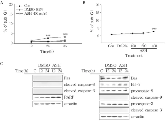
Figure 3. Effect of Acanthopanax senticosus Harms(ASH) on the apoptosis in AGS gastric cancer cells.
(A) AGS cells were exposed to 400 ㎍/㎖ of ASH or 0.2% DMSO for 12, 24, and 36 h
(B) AGS cells were exposed to various concentrations(100, 200, and 400 ㎍/㎖) of ASH for 24 h.
DNA content distribution was analyzed using a FACStar flow cytometer(Becton-Dickinson), and percentages of sub-G1 phase cells were determined based on a DNA content histogram.
The data shown is a representative of three independent experiments which gave similar results. Bars, SD (***p < 0.001).
(C) AGS cells were exposed to 400 ㎍/㎖ of ASH or 0.2% DMSO for 12 and 24 h. The cell lysates were separated by SDS-PAGE gel electrophoresis and Western blotting was performed with specific antibodies such as anti-Fas receptor, -procaspases(-9 and -3), -cleaved caspases(-9, -8 and -3), -PARP, -Bax, -Bcl-2 and -α-actin. The data shown is a representative of three independent experiments which gave similar results. (Con ; control, D ; DMSO, ASH ; Acanthopanax senticosus Harms)
B
A 20
15
10
5
0
20 15 10 5 0 Con
DMSO 0.2%
ASH 400 ㎍/㎖
***
*** ***
Con D 0.2% 100 200 400 12 24 36
Time(h) ASH Treatment
세포주의 세포사멸에 가시오가피에 의한 영향
이 경미함을 확인할 수 있었다(Fig 3B). 다음 으로는 가시오가피를 처리했을 때, 세포사멸과 관련된 단백질의 발현이 어떻게 변화하는지를 Western blotting으로 확인하였다(Fig. 3C). 세 포사멸의 경우 크게 Fas, caspase-8, -3, PARP
단백질을 통한 외재적경로(extrinsic pathway)
와 미토콘드리아 관련 단백질인 Bax와 Bcl-2,
그리고 caspase-9, -3 단백질들을 통한 내재적
경로(intrinsic pathway)로 나뉘는데, 이들 모
든 단백질에서 가시오가피에 의한 세포사멸로
사료되는 변화가 없었다.
4. 위암세포주에서 가시오가피 에탄올 추출물에 의한 세포 주기조절 효과
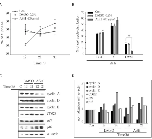
다음으로는 가시오가피에 대한 위암세포주의 증식억제와 세포주기와의 관련성을 확인하기 위해 AGS 세포주에 가시오가피를 400 ㎍/㎖ 농 도로 시간별(12, 24, 36시간)로 처리 한 후, 유 세포분석기를 이용하여 DNA contents histogram 을 측정하고, ModFit LT V2.0 프로그램을 사 용하여 세포주기를 분석하였다. 결과 시간 의존 적으로 가시오가피에 의해 위암세포주의 S기가 32.21%(12시간)에서 57.39%(36시간)로 증가하 여 세포주기를 억제시키는 것을 확인했다(Fig 4A and B). 또한 가시오가피를 농도별(100, 200, 400 ㎍/㎖)로 24시간동안 처리한 결과에서도 S 기의 증가가 관찰되었다(data not shown). 다 음으로는 가시오가피를 처리했을 때, 세포주기 와 관련된 단백질의 발현변화를 확인하기 위해 Western blotting을 수행하였다(Fig 4C and 4D).
그 결과 S기와 관련된 Cyclin A의 발현이 감 소함을 확인하였고, cyclin의존적 인산화효소 억 제제(Cyclin-dependent kinase inhibitor)로 알 려진 p27과 p16의 발현도 증가함을 확인할 수 있었다. 이러한 결과로 볼 때, 가시오가피 의한 AGS 위암 세포주의 증식억제는 세포주기억제 와 관련된다는 것을 알 수 있었다.
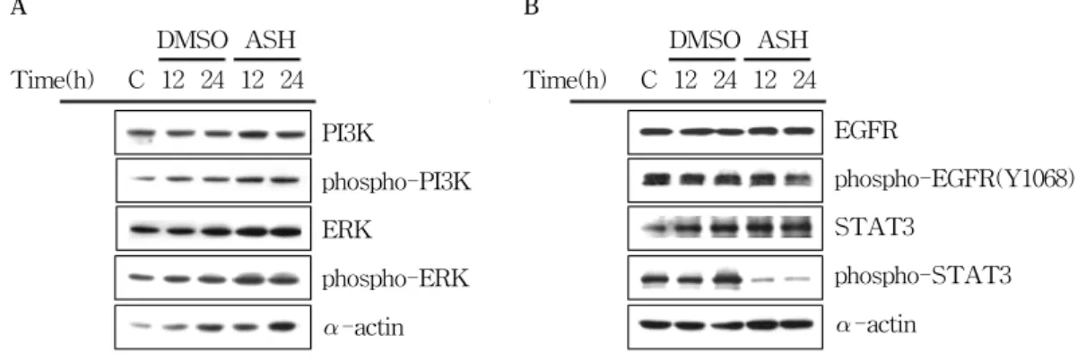
5. 위암세포주에서 가시오가피 에탄올 추출물에 의한 세포 증식관련경로 조절 효과
가시오가피가 세포 증식에 관련된 PI3K, ERK, EGFR pathway 단백질의 발현에도 영향을 미 치는지 확인한 결과, PI3K/phospho-PI3K와 ERK/phospho-ERK 단백질의 발현단계에는 아 무런 영향을 미치지 않는 것을 확인하였다(Fig 5A). 하지만 EGFR-STAT3 경로에서 가시오가
피가 EGFR과 STAT3의 인산화(phosphory- lation)를 억제시키는 것을 확인할 수 있었다(Fig 5B).
IV. 고 찰
오가피는 두릅나무과(Araliaceae)에 속하는 관목식물로 가시오가피, 두상오가피, 성오가피, 당오가피 등 여러 종류가 있지만 이중에서도 약효가 가장 좋다고 밝혀진 것이 ‘가시오가피’
이다. 가시오가피는 뿌리와 잎을 약용으로 사 용할 수 있으며 환경적응 약물로 육체적, 화학 적, 생물학적 스트레스 유발인자가 신체에 비 특이적 반응을 일으키게 되면 이러한 여러 인 자에 대항하여 신체 내부의 균형을 유지한다.
또한 항알레르기, 항산화, 항염증의 효능, 류마 티즘, 고혈압 등의 치료제로도 사용되고 있으 며, 항암 효능도 가지고 있는 것으로 알려져
있다.
8, 32)그러나 아직까지 위암에 대한 가시오
가피의 항암활성 기초 실험 연구가 많이 되어 있지 않아, 본 연구에서는 이러한 근거를 바탕 으로 위암 세포주를 사용하여 80% 에탄올 가 시오가피 추출물의 구체적인 항암효과 및 기전 을 확인하였다.
가시오가피 에탄올 추출물에 대한 항암작용
을 확인하기 위해 AGS 위암세포주에 가시오
가피를 처리한 결과 가시오가피가 위암 세포주
의 증식을 농도와 시간 의존적으로 억제시킨다
는 것을 확인하였다(Fig 1). 또한 현미경을 통한
세포형태 관찰 결과에서도 마찬가지로 가시오
가피 에탄올 추출물을 처리한 실험군에서 아무
것도 처리하지 않은 Control(Con) 그룹과 vehicle
(DMSO) 그룹에 비해 위암 세포주의 증식을 억
제시키는 것을 확인할 수 있었다(Fig 2). 다음
결과로부터 가시오가피 에탄올 추출물이 위암
세포에 대한 증식억제와 관련된 항암 효능을
나타내고, 결국 가시오가피는 위암과 관련하여
Figure 4. Acanthopanax senticosus Harms(ASH) induced S phase cell cycle arrest in AGS gastric cancer cells.
(A) AGS cells were exposed to 400 ㎍/㎖ of ASH or 0.2% DMSO for 12, 24, and 36 h, fixed in ethanol, and stained with propidium iodide, and cell cycle distribution was analyzed using a FACStar flow cytometer(Becton-Dickinson). The percentage of cells in each phase of the cell cycle was obtained by MODFIT software. The graph represents the percentage of ASH treated cells against untreated cells in S cell cycle. The data shown is a representative of three independent experiments which gave similar results. Bars, SD. **p < 0.05, ***p < 0.001.
(B) AGS cells were exposed to 400 ㎍/㎖ of ASH or 0.2% DMSO for 24 h.
(C) The cell lysates were separated by SDS-PAGE gel electrophoresis and Western blotting was performed with specific antibodies such as anti-cyclins(A, D and E), -CDK2, -p27, -p16 and -α-actin. The data shown is a representative of three independent experiments which gave similar results.
Con DMSO 0.2%
ASH 400 ㎍/㎖
12 24 36 Time(h) 70
60 50 40 30 20
Con DMSO 0.2%
ASH 400 ㎍/㎖
A B
G0/G1 S G2/M 70
60 50 40 30 20 10 0
24 h
**
DMSO ASH Time(h) C 12 24 12 24
cyclin A cyclin D cyclin E CDK2 p27 p16
-actin C
cyclin A cyclin D cyclin E CDK2 p27 p16 3
2
1
0
Con 12 24 12 24 DMSO ASH
Time(h) D
DMSO ASH Time(h) C 12 24 12 24
PI3K
phospho-PI3K ERK
phospho-ERK
-actin A
DMSO ASH Time(h) C 12 24 12 24
EGFR
phospho-EGFR(Y1068) STAT3
phospho-STAT3
-actin B
Figure 5. Effect of Acanthopanax senticosus Harms(ASH) on the EGFR-STAT3 pathway in AGS gastric cancer cells. (A, B) AGS cells were exposed to 400 μg/ml of ASH or 0.2% DMSO for 12 and 24 h. The cell lysates were separated by SDS-PAGE gel electrophoresis and Western blotting was performed with specific antibodies such as anti-PI3K, -ERK, -EGFR, -STAT3, -phospho forms(-PI3K, -ERK, -EGFR(Y1068) and -STAT3) and -α-actin. The data shown is a representative of three independent experiments which gave similar results.
증식억제 항암 효능을 타나낼 수 있는 약물로 써의 가능성을 확인하였다. 다음으로 이러한 증식억제 기전을 확인하기 위해 가시오가피에 대한 세포사멸과 세포주기를 확인하였다. 세포 사멸(apoptosis)의 경우 조직의 항상성을 유지 하거나 발생 과정에서 개체를 보호하기 위한 일 련의 진화적 과정으로 세포표면의 죽음 수용체 (death receptor)를 통하는 외재적 경로(extrinsic pathway)와 미토콘드리아를 경유하는 내재적 경로(intrinsic pathway)로 나뉘고 이들 두 경 로 모두 여러 종류의 caspase에 의해 조절된다.
또한 세포주기(cell cycle)는 cyclins과 CDKs
〔(cyclin-dependent kinases(serine/threonine protein))가 결합하여 G
1기를 거쳐 S기와 G
2기 를 지나 mitosis가 일어나는 과정으로, 성장한 하나의 세포가 두개의 세포로 분열되는 전 단 계를 의미한다. 암세포에 항암제를 처리하였을 때 인위적으로 세포사멸을 유도하거나, 세포주 기 중 일정한 단계에 머무르게 하면서 세포의 증식을 억제시키는 것을 볼 수 있는데 이런 현 상은 항암효과를 나타내는 대표적인 특징으로
알려져 있다. 가시오가피 에탄올 추출물의 세
포사멸과 관련된 증식억제 기능을 확인하기 위
해 가시오가피 400 ㎍/㎖를 시간별(12, 24, 36
h)로, 농도별로(100, 200, 400 ㎍/㎖) 처리한 결
과 sub-G
1fraction 비율은 아무것도 처리하지
않은 Control(Con) 그룹과 vehicle(DMSO) 그룹
에 비해 크게 증가되지 않은 것을 확인할 수 있
었고, 외재적 경로와 관련된 분자(Fas, caspase-
8)와 내재적 경로와 관련된 분자(Bax-Bcl2 ratio,
caspase-9), 또한 이들 두 경로와 모두 관련된
capase-3, PARP의 발현에 아무런 영향을 미
치지 못했음을 확인할 수 있었다. 이는 가시오
가피가 위암세포주에 대해 세포사멸을 유도하
지 않는 것으로 사료되었다(Fig 3). 반면에, 가
시오가피에 의한 세포주기 변화를 분석한 결과
가시오가피를 시간별로 처리하였을 때 S기가
증가하여 세포주기를 억제시키는 것을 확인하
였고, 400 ㎍/㎖ 농도에서 12, 24시간에는 S기
와 G
0/G
1기가 함께 증가하다가 36시간에 S기
가 약 60%에 이르는 증가를 보여 가시오가피
의 처리 농도와 시간에 따라 세포주기를 조절
하는 양상이 다름을 유추해 볼 수 있다. 또한 세포주기와 관련된 단백질의 발현을 확인해 본 결과, 가시오가피는 세포주기 각 단계에서 CDKs활성을 조절하는 세포내 CDKIs인 p27 또는 p16의 발현을 증가시키고 cyclin A의 발 현을 시간의존적으로 감소시켜 세포주기를 차단 하고 결국 위암세포주의 증식을 억제한다는 것 을 확인하였다(Fig 4). 또한 가시오가피에 의한 세포증식에 관련된 여러 경로들의 변화를 확인 해본 결과 PI3K/phospho-PI3K, ERK/phospho- ERK 단백질들의 발현에는 변화가 없었지만 (Fig 5A), EGFR-STAT3 경로에서 가시오가피 에 의해 EGFR과 STAT3의 인산화(phosphoryla- tion)을 감소시키는 것을 알 수 있었다(Fig 5B).
특히 EGFR/ErbB1은 receptor tyrosine kinases (RTKs)의 ErbB family에 속하는 것으로 인간 암세포에서 발현이 현저히 증가해 있는 것으로 알려져 있고,
33-36)이들 수용체(epithermal growth factor receptor, EGFR)에 성장인자(epithermal growth factor, EGF)가 결합하면 이 경로에 의 해 활성화 될 수 있는 타겟분자의 DNA에 STAT3 전사인자(transcription factor)가 결합하여 타켓 분자의 발현을 조절하게 된다.
37, 38)특히 EGFR에 의해 인산화 된 STAT3의 Tyr705잔기는 세포 주기를 진행시키는 유전자와 anti-apoptosis 기 능을 하는 유전자들의 발현을 조절한다고 알려 져 있다.
39-46)이러한 실험결과를 통해 가시오가 피가 EGFR과 STAT3 단백질의 인산화를 감소 시켜 세포주기와 관련된 유전자 발현을 조절함 을 통해 최종적으로 위암세포의 증식을 억제 하는 것으로 사료된다.
V. 결 론
위암세포주인 AGS에 80% 에탄올 가시오가 피 추출액을 처리하여 다음과 같은 결론을 얻 었다.
1. 가시오가피 에탄올 추출물이 위암세포주 의 증식 농도와 시간 의존적으로 억제시 켰다.
2. 가시오가피 에탄올 추출물의 위암세포주 증식억제기전과 관련하여 세포사멸과 상 관없이, cyclin A의 감소, p16 및 p27의 증가를 통해 세포주기의 이행을 억제시키 는 것을 확인하였다.
3. 가시오가피 에탄올 추출물은 세포증식에 관련된 EGFR-STAT3 경로에서 EGFR과 STAT3의 인산화(phosphorylation)을 억 제하였다
이러한 결과로부터 본 저자는 가시오가피에 의 한 위암세포주의 세포주기 이행 억제 및 EGFR- STAT3 억제를 통해 항암효과에 대한 가능성 을 발견하여 위암치료제나 개발에 가시오가피 가 활용될 수 있을 것으로 기대한다.
참고문헌